. 2010 Apr;75(4):914-22.
doi: 10.1016/j.urology.2009.10.031. Epub 2009 Dec 29.

2,8-dihydroxyadenine nephrolithiasis induces developmental stage-specific alterations in gene expression in mouse kidney

Affiliations

2,8-dihydroxyadenine nephrolithiasis induces developmental stage-specific alterations in gene expression in mouse kidney

Jianmin Chen et al. Urology. 2010 Apr.

Abstract

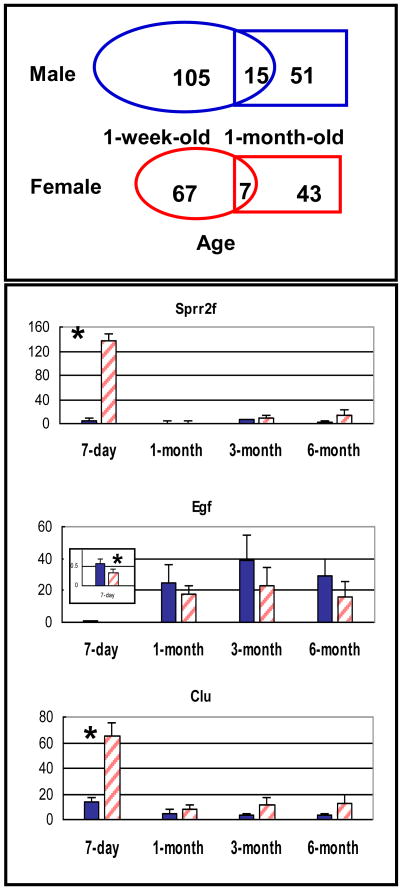
Objectives: To identify factors that may be crucial for the initiation and progression of stone-induced injury in the developing mouse kidney by a prospective observational study using microarray analysis. Kidney stone diseases are common in premature infants, but the underlying molecular and cellular mechanisms are not fully defined.

Methods: Mice with adenine phosphoribosyltransferase deficiency develop 2,8-dihydroxyadenine (DHA) nephrolithiasis. The gene expression changes between Aprt(-/-) and Aprt(+/+) kidneys from newborn and adult mice were compared using Affymetrix gene chips. Targets of interest were further analyzed by quantitative real-time polymerase chain reaction and immunohistochemistry.

Results: We identified a set of genes that were differentially expressed in the developing kidney in response to DHA-induced injury. In 1-week-old Aprt(-/-) mice, the expression of Sprr2f and Clu was highly augmented and that of Egf was significantly decreased. We also observed that maturation-related gene expression changes were delayed in developing Aprt(-/-) kidneys, and immature Aprt(-/-) kidneys contained large numbers of intercalated cells that were blocked from terminal differentiation.

Conclusions: This study presents a comprehensive picture of the transcriptional changes induced by DHA stone injury in the developing mouse kidney. Our findings help explain growth impairment in kidneys subject to injury during the early stages of development.


Figures

Figure 1
Figure 1. Pairwise overlap test and age-specific gene expression
Upper panel: Venn diagrams of pairwise overlap test. Numbers represent total number of genes differentially expressed in a specific age group or gender comparing Aprt-/- and wild type mice. Oval, 1-week-old; rectangle, 1-month-old. Blue, male; red, female. Lower panel: Age-specific gene expression. mRNA levels were measured by real-time PCR and normalized to Hprt expression. Bar color: blue (Aprt+/+), pink (Aprt-/-) Y-axis, the ratio between gene of interest and the internal control gene (Hprt). Insert is the zoomed-in image of 1-week-old kidneys which have very low Egf expression. * indicates p<0.05 (Welch's t-test).
Figure 2
Figure 2. Heatmap of hierarchical clustering of early response genes
The diagram shows reverse expression patterns of maturation-related and DHA injury-related genes. Sixty eight early response genes were clustered according to their temporal expression pattern using GeneSpring. Each vertical bar represents a gene and its average mRNA level in each age/genotype group (1W = 1-week-old; 1M = 1-month-old). Expression level color scheme is shown on the right: red >blue, dark red, high mRNA level; dark blue, no expression. Four 1st degree clusters were found. Clusters 1-3 represent genes with augmented expression in 1-week-old Aprt-/- mice whereas cluster 4 shows genes with reduced expression in these mice. Expression levels of these genes remain mostly unchanged when comparing 1-month-old Aprt-/- mice with age-matched controls, suggesting that the expression changes are unique to immature kidneys. Importantly, all genes in clusters 2 and 4 show reverse expression patterns when comparing 1-week-old to 1-month-old controls (kidney maturation) and 1-week-old Aprt-/- to 1-week-old controls (kidney injury), suggesting that injury delays maturation in developing kidneys.
Figure 3
Figure 3. LIF-sustained pluripotency markers
LIF-sustained pluripotency markers were significantly enriched among genes up-regulated in immature APRT-deficient kidneys. There were 9,239 genes after collapsing the 12,488 native probes present on the microarray. Upper panel: From left to right, the lines indicate the relative positions of the 9,239 genes within the rank order from the probe most up-regulated in Aprt-/- kidneys to the probe most down-regulated (Aprt-/- to Aprt+/+ log2 ratio from 4.4 to –1.7). The genes near the middle are unaffected by APRT deficiency, with Aprt-/- vs. Aprt+/+, log2 ratio equals 1. Lower panel: An example of indiscriminate distribution of a gene set with no bias toward Aprt-/- or Aprt+/+. In the upper panel, the LIF target genes are clearly enriched among genes up-regulated in immature Aprt-/- kidneys as evidenced by the increased density of black lines on the left side of the distribution, the positive enrichment score, and a nominal p = 0.007.
Figure 4
Figure 4. Sprr2, PCNA, and Ssea-1 immunostaining
Strong Sprr2 staining is detected only in collecting duct cells in immature APRT-deficient kidneys (B), but not in age-matched wild type kidneys (A). Sprr2 immuno-positive collecting duct cells (C, large arrow) in the developing mouse kidney are PCNA immuno-negative (D, large arrow), whereas Sprr2 immuno-negative cells are PCNA immuno-positive (C and D, small arrows). Sprr2 immuno-positive cells (green) are also Ssea-1 immuno-positive (red) (E). (F) is same as (E), but with higher magnification.

Similar articles

Cited by

References

    1. Verhulst A, Asselman M, De Naeyer S, et al. Preconditioning of the distal tubular epithelium of the human kidney precedes nephrocalcinosis. Kidney Int. 2005;68:1643–1647. - PubMed
    1. Verkoelen CF, van der Boom BG, Houtsmuller AB, et al. Increased calcium oxalate monohydrate crystal binding to injured renal tubular epithelial cells in culture. Am J Physiol. 1998;274:F958–965. - PubMed
    1. Asselman M, Verhulst A, Van Ballegooijen ES, et al. Hyaluronan is apically secreted and expressed by proliferating or regenerating renal tubular cells. Kidney Int. 2005;68:71–83. - PubMed
    1. Sahota AS, Tischfield JA, Kamatani N, Simmonds HA. Adenine phosphoribosyltransferase deficiency and 2,8-dihydroxyadenine lithiasis. In: Scriver CR, editor. The Metabolic and Molecular Bases of Inherited Disease. 8th. New York: McGraw-Hill; 2001. pp. 2571–2584.
    1. Evan AP, Lingeman JE, Coe FL, et al. Randall's plaque of patients with nephrolithiasis begins in basement membranes of thin loops of Henle. J Clin Invest. 2003;111:607–616. - PMC - PubMed