

Agreement for detecting glaucoma progression with the GDx guided progression analysis, automated perimetry, and optic disc photography
- PMID: 20036010
- PMCID: PMC2830299
- DOI: 10.1016/j.ophtha.2009.08.012
Agreement for detecting glaucoma progression with the GDx guided progression analysis, automated perimetry, and optic disc photography
Abstract
Purpose: To evaluate the ability of the GDx Variable Corneal Compensation (VCC) Guided Progression Analysis (GPA) software for detecting glaucomatous progression.
Design: Observational cohort study.
Participants: The study included 453 eyes from 252 individuals followed for an average of 46+/-14 months as part of the Diagnostic Innovations in Glaucoma Study. At baseline, 29% of the eyes were classified as glaucomatous, 67% of the eyes were classified as suspects, and 5% of the eyes were classified as healthy.
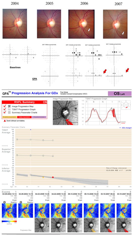
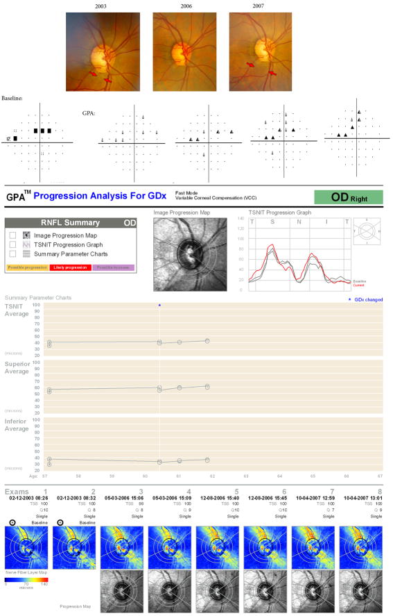
Methods: Images were obtained annually with the GDx VCC and analyzed for progression using the Fast Mode of the GDx GPA software. Progression using conventional methods was determined by the GPA software for standard automated achromatic perimetry (SAP) and by masked assessment of optic disc stereophotographs by expert graders.
Main outcome measures: Sensitivity, specificity, and likelihood ratios (LRs) for detection of glaucoma progression using the GDx GPA were calculated with SAP and optic disc stereophotographs used as reference standards. Agreement among the different methods was reported using the AC(1) coefficient.
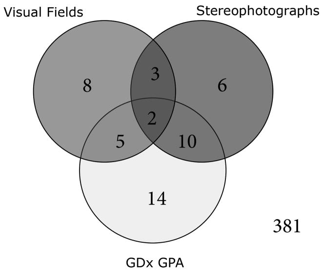
Results: Thirty-four of the 431 glaucoma and glaucoma suspect eyes (8%) showed progression by SAP or optic disc stereophotographs. The GDx GPA detected 17 of these eyes for a sensitivity of 50%. Fourteen eyes showed progression only by the GDx GPA with a specificity of 96%. Positive and negative LRs were 12.5 and 0.5, respectively. None of the healthy eyes showed progression by the GDx GPA, with a specificity of 100% in this group. Inter-method agreement (AC(1) coefficient and 95% confidence intervals) for non-progressing and progressing eyes was 0.96 (0.94-0.97) and 0.44 (0.28-0.61), respectively.
Conclusions: The GDx GPA detected glaucoma progression in a significant number of cases showing progression by conventional methods, with high specificity and high positive LRs. Estimates of the accuracy for detecting progression suggest that the GDx GPA could be used to complement clinical evaluation in the detection of longitudinal change in glaucoma.
Copyright 2010 American Academy of Ophthalmology. Published by Elsevier Inc. All rights reserved.
Conflict of interest statement
Figures
References
-
- Kass MA, Heuer DK, Higginbotham EJ, et al. Ocular Hypertension Treatment Study Group. The Ocular Hypertension Treatment Study: a randomized trial determines that topical ocular hypotensive medication delays or prevents the onset of primary open-angle glaucoma. Arch Ophthalmol. 2002;120:701–13. discussion 829–30. - PubMed
-
- Gordon MO, Beiser JA, Brandt JD, et al. Ocular Hypertension Treatment Study Group. The Ocular Hypertension Treatment Study: baseline factors that predict the onset of primary open-angle glaucoma. Arch Ophthalmol. 2002;120:714–20. discussion 829–30. - PubMed
-
- Tuulonen A, Airaksinen PJ. Initial glaucomatous optic disk and retinal nerve fiber layer abnormalities and their progression. Am J Ophthalmol. 1991;111:485–90. - PubMed
-
- European Glaucoma Prevention Study (EGPS) Group. Results of the European Glaucoma Prevention Study. Ophthalmology. 2005;112:366–75. - PubMed
-
- Sommer A, Katz J, Quigley HA, et al. Clinically detectable nerve fiber atrophy precedes the onset of glaucomatous field loss. Arch Ophthalmol. 1991;109:77–83. - PubMed
Publication types
MeSH terms
Grants and funding
LinkOut - more resources
Full Text Sources
Other Literature Sources
Medical
Miscellaneous
