

Effect of pro-inflammatory mediators on membrane-associated mucins expressed by human ocular surface epithelial cells
- PMID: 20036239
- PMCID: PMC2880853
- DOI: 10.1016/j.exer.2009.12.009
Effect of pro-inflammatory mediators on membrane-associated mucins expressed by human ocular surface epithelial cells
Abstract
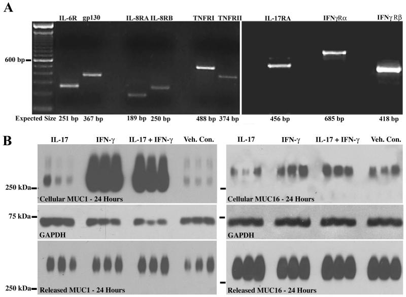
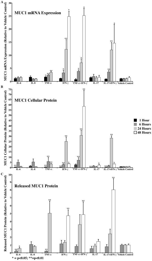
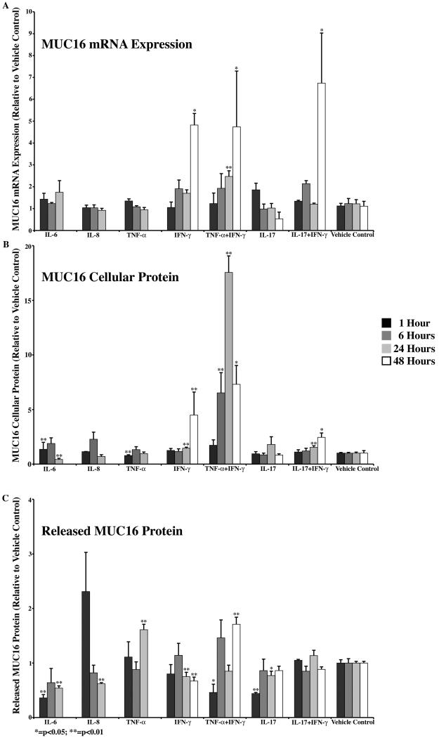
Membrane-associated mucins are altered on the ocular surface in non-Sjögren's dry eye. This study sought to determine if inflammatory mediators, present in tears of dry eye patients, regulate membrane-associated mucins MUC1 and -16 at the level of gene expression, protein biosynthesis and/or ectodomain release. A human corneal limbal epithelial cell line (HCLE), which produces membrane-associated mucins, was used. Cells were treated with interleukin (IL)-6, -8, or -17, tumor necrosis factor-alpha (TNF-alpha), and Interferon-gamma (IFN-gamma), or a combination of TNF-alpha and IFN-gamma, or IFN-gamma and IL-17, for 1, 6, 24, or 48 h. Presence of receptors for these mediators was verified by RT-PCR. Effects of the cytokines on expression levels of MUC1 and -16 were determined by real-time PCR, and on mucin protein biosynthesis and ectodomain release in cell lysates and culture media, respectively, by immunoblot analysis. TNF-alpha and IFN-gamma each significantly induced MUC1 expression, cellular protein content and ectodomain release over time. Combined treatment with the two cytokines was not additive. By comparison, one of the inflammatory mediators, IFN-gamma, affected all three parameters-gene expression, cellular protein, and ectodomain release-for MUC16. Combined treatment with TNF-alpha and IFN-gamma showed effects similar to IFN-gamma alone, except that ectodomain release followed that of TNF-alpha, which induced MUC16 ectodomain release. In conclusion, inflammatory mediators present in tears of dry eye patients can affect MUC1 and -16 on corneal epithelial cells and may be responsible for alterations of surface mucins in dry eye.
Figures
References
-
- Afonso AA, Monroy D, Stern ME, Feuer WJ, Tseng SC, Pflugfelder SC. Correlation of tear fluorescein clearance and Schirmer test scores with ocular irritation symptoms. Ophthalmology. 1999;106:803–810. - PubMed
-
- Argueso P, Spurr-Michaud S, Russo CL, Tisdale A, Gipson IK. MUC16 mucin is expressed by the human ocular surface epithelia and carries the H185 carbohydrate epitope. Invest. Ophthalmol. Vis. Sci. 2003;44:2487–2495. - PubMed
-
- Blalock T, Spurr-Michaud S, Tisdale A, Heimer S, Gilmore M, Ramesh V, Gipson I. Functions of MUC16 in corneal epithelial cells. Invest. Ophthalmol. Vis. Sci. 2007;48:4509–4518. - PubMed
Publication types
MeSH terms
Substances
Grants and funding
LinkOut - more resources
Full Text Sources
Other Literature Sources
Research Materials
Miscellaneous
