. 2010 Mar;222(1):93-107.
doi: 10.1016/j.expneurol.2009.12.011. Epub 2009 Dec 28.

The effects of sympathetic outflow on upregulation of vanilloid receptors TRPV(1) in primary afferent neurons evoked by intradermal capsaicin

Affiliations

The effects of sympathetic outflow on upregulation of vanilloid receptors TRPV(1) in primary afferent neurons evoked by intradermal capsaicin

Xijin Xu et al. Exp Neurol. 2010 Mar.

Abstract

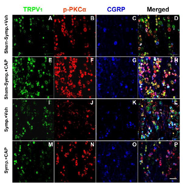
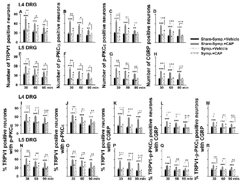
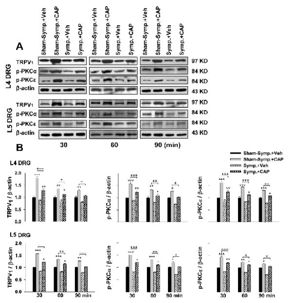
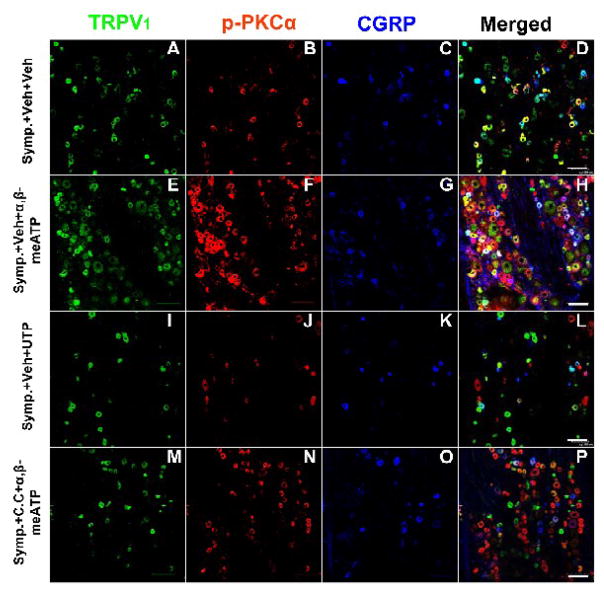
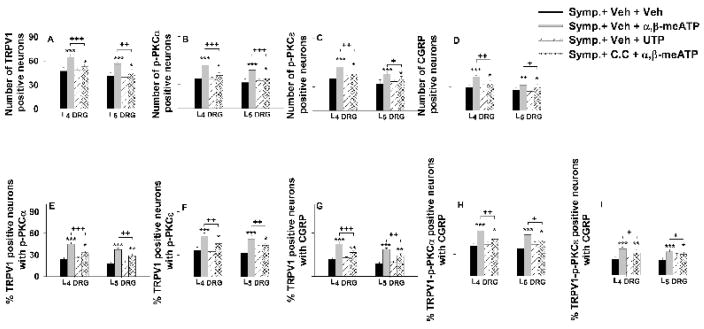
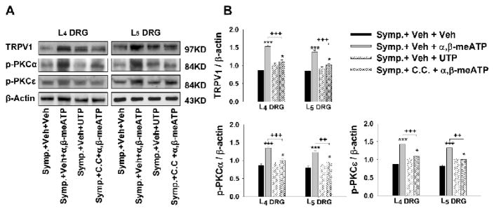
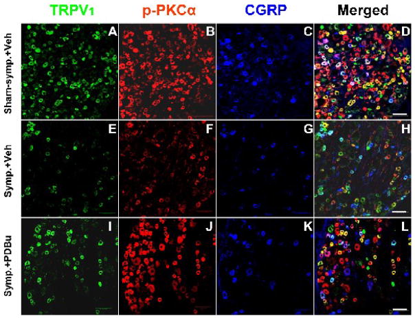
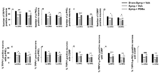
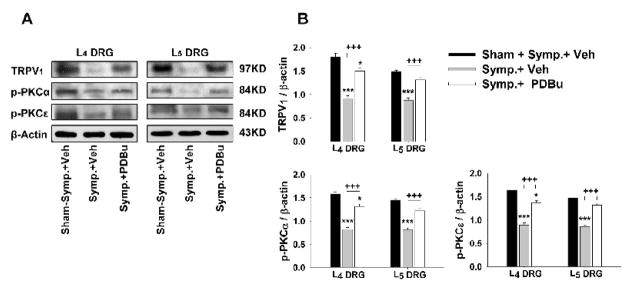
The vanilloid receptor TRPV(1) is a key nociceptive molecule located in primary afferent nociceptive neurons in dorsal root ganglia (DRG) for initiating neurogenic inflammation and pain. Our recent study demonstrates that up-regulation of TRPV(1) receptors by intradermal injection of capsaicin is modulated by activation of the protein kinase C (PKC) cascade. Neurogenic inflammation and pain resulting from capsaicin injection are sympathetically dependent, responding to norepinephrine, adenosine 5'-triphosphate (ATP) and/or neuropeptide Y released from sympathetic efferents. In a rat model of acute neurogenic inflammatory pain produced by capsaicin injection, we used immunofluorescence and Western blots combined with pharmacology and surgical sympathectomies to analyze whether the capsaicin-evoked up-regulation of TRPV(1) in DRG neurons is affected by sympathetic outflow by way of activating the PKC cascade. Sympathetic denervation reduced significantly the capsaicin-evoked expressions of TRPV(1), calcitonin gene-related peptide and/or phosphorylated PKC and their co-expression. These reductions could be restored by exogenous pretreatment with an analog of ATP, alpha,beta-methylene ATP. Inhibition of PKC with chelerythrine chloride prevented the ATP effect. Consistent results were obtained from experiments in which capsaicin-evoked changes in cutaneous inflammation (vasodilation and edema) were examined after sympathetic denervation, and the effects of the above pharmacological manipulations were evaluated. Our findings suggest that the capsaicin-evoked up-regulation of TRPV(1) receptors in DRG neurons is modulated sympathetically by the action of ATP released from sympathetic efferents to activate the PKC cascade. Thus, this study proposes a potential new mechanism of sympathetic modulation of neurogenic inflammation.


Figures

Figure 1
Figure 1
Confocal immunofluorescence images showing single labeling of TRPV1, p-PKCα and CGRP, and double or triple labeling of these three molecules in L4 DRG on the side ipsilateral to intradermal injection of CAP at 30 min after intradermal injection of CAP or vehicle in sham-sympathectomized (Sham-symp.+Veh, Sham-symp.+CAP) and sympathectomized (Symp.+Veh, Symp.+CAP) rats. Bar=100 μm.
Figure 2
Figure 2
Grouped data summarizing changes in the number of TRPV1-, p-PKC (α and β subunits)-, and CGRP-positive neurons (A–H) and in percent TRPV1 positive neurons with p-PKC double staining, percent TRPV1 positive neurons with CGRP double staining, and percent TRPV1-p-PKC positive neurons with CGRP triple staining (I–R) in L4–5 DRGs after CAP injection in sympathetically intact (Sham-symp.+CAP) and sympathectomized (Symp.+CAP) groups. DRGs were sampled at 30, 60 and 90 min after vehicle or CAP injection. Statistical analysis (*, P<0.05, **, P<0.01; ***, P<0.001) was made of changes due to CAP injection vs. those due to vehicle injection at the same time point in the group having sham-sympathectomy or sympathectomy, and of the CAP-induced effects between the groups of sham-sympathectomy and sympathectomy at the same time point (+, P<0.05; ++, P<0.01; + ++, P<0.001).
Figure 3
Figure 3
Western blot analyses of the evoked changes in the relative density of TRPV1, p-PKCα and p-PKCε in L4 and L5 DRG tissue following CAP injection in sham-sympathectomized and sympathectomized rats at 30, 60 and 90 min after unilateral injection of CAP. Immunoreactivity of TRPV1, p-PKCα and p-PKCε was normalized to β-actin. A. Representative Western blots for TRPV1, p-PKCα and p-PKCε to CAP injection at 30, 60 and 90 min after injection in the sham-sympathectomized and sympathectomized rats. B. Grouped data of changes in these molecules. * P<0.05, ** P<0.01, *** P<0.001, compared with vehicle injection at the same time point in the group of sham-sympathectomy or sympathectomy. +, P<0.05, ++ P<0.01, +++ P<0.001, compared with the CAP-induced effects under sham-sympathectomized conditions.
Figure 4
Figure 4
Confocal immunofluorescence images showing the restoration by activation of peripheral P2X (not P2Y) receptors of the reduction in the CAP-evoked single staining for TRPV1, p-PKC, and CGRP and double or triple staining for these molecules in L4 DRG on the side ipsilateral to CAP injection after sympathectomy and the effects of PKC inhibition. Data were sampled at 30 min after CAP injection. Symp.+Veh+Veh: CAP-evoked expression in a sympathectomized rat with vehicle pretreatments (used for dissolving the P2X or P2Y agonist and C.C.); Symp.+Veh+α,β-meATP: CAP-evoked expression in a sympathectomized rat without C.C. and with α,β-meATP pretreatments; Symp.+Veh+UTP: CAP-evoked expression in a sympathectomized rat without C.C. and with UTP pretreatments; Symp.+C.C.+α,β-meATP: CAP-evoked expression with C.C. and α,β-meATP pretreatments. Scale bars=100 μm.
Figure 5
Figure 5
Grouped data summarizing the restoration by activation of peripheral P2X (not P2Y) receptors on the reduction in the CAP-evoked single staining for TRPV1, p-PKC, and CGRP (A–D) and the reduction in percentage TRPV1 positive neurons with p-PKC double staining (E and F), percentage TRPV1 positive neurons with CGRP double staining (G), and percentage TRPV1-p-PKC positive neurons with CGRP triple staining (H and I) in L4 and L5 DRGs on the side ipsilateral to CAP injection after sympathectomy and the effects of PKC inhibition. *, **, and ***: P<0.05, P<0.01 and P<0.001, compared to the group of sympathectomy with vehicle pretreatment (Symp.+Veh+Veh). +, ++, and +++: P<0.05, P<0.01 and P<0.001, comparison between the groups of sympathectomy without C.C. and with α,β-meATP pretreatments (Symp.+Veh+α,β-meATP) and of sympathectomy with C.C. and α,β-meATP pretreatments (Symp.+C.C.+α,β-meATP).
Figure 6
Figure 6
Western blot analyses of the restoration by activation of peripheral P2X (not P2Y) receptors on the reduction in the CAP-evoked expressions of TRPV1, p-PKCα and p-PKCε proteins in L4 and L5 DRGs after sympathectomy and the effects of PKC inhibition. A: Representative Western blots for TRPV1, p-PKCα and p-PKCε in DRGs ipsilateral to CAP injection at 30 min after CAP injection in the sympathectomized rats pretreated with and without α,β-meATP or UTP when PKC was and was not inhibited by chelerythrine chloride. B: Grouped data of showing changes in these molecules. * and ***: P<0.05 and P<0.001, compared to the group with symapthectomy and vehicle pretreatments (Symp.+Veh+Veh). ++ and +++: P<0.01 and P<0.001, comparison between the groups of sympathectomy without C.C. and with α,β-meATP pretreatments (Symp.+Veh+α,β-meATP) and sympathectomy with C.C. and α,β-meATP pretreatments (Symp.+C.C.+ α,β-meATP).
Figure 7
Figure 7
Confocal immunofluorescence images showing changes in the CAP-evoked single labeling of TRPV1, p-PKCα, and CGRP and double or triple labeling of these molecules in neurons of the L4 DRG on the side ipsilateral to CAP injection after sympathectomy (E–H, Symp.+Veh) compared to the CAP-evoked changes in the sham-sympathectomized (A–D, Sham-symp.+Veh) and the effects of PKC activation by pretreatment with PDBu (I–L, Symp.+PDBu). Data were sampled at 30 min after CAP injection. Scale bar=100 μm.
Figure 8
Figure 8
Grouped data summarizing changes in the CAP-evoked single labeling for TRPV1, p-PKC (α and ε subunits), and CGRP (A–D) and changes in the CAP-evoked percentage of TRPV1 positive neurons with p-PKC double staining (E and F), percentage of TRPV1 positive neurons with CGRP double staining (G), and percentage of TRPV1-p-PKC positive neurons with CGRP triple staining (H and I) in L4 and L5 DRGs on the side ipsilateral to CAP injection after sympathectomy and the effects of PKC activation by pretreatment with PDBu (Symp.+PDBu). Data were sampled at 30 min after CAP injection. *, **, and ***, P<0.05, P<0.01, and P<0.001, compared with the group having sham-sympathectomy with vehicle pretreatment (Sham-symp.+Veh). +, ++, and +++: P<0.05, P<0.01, and P<0.001, comparison between the groups of sympathectomy with vehicle pretreatment (Symp.+Veh) and of sympathectomy with PDBu pretreatment (Symp.+PDBu).
Figure 9
Figure 9
Western blot analyses of the CAP-evoked changes in the relative density of TRPV1, p-PKCα and p-PKCε in L4 and L5 DRG tissue after sympathectomy and the effects of PKC activation by pretreatment with PDBu. Data were sampled at 30 min after CAP injection. A: Representative Western blots for TRPV1, p-PKCα and p-PKCε in DRG tissue ipsilateral to CAP injection at 30 min after CAP injection in the sham-sympathectomized and sympathectomized rats with and without PDBu pretreatments. B: Grouped data of changes in these molecules. * and ***: P<0.05 and P<0.001, compared to the group of sham-sympathectomy with vehicle pretreatment. +++: P<0.001, comparison between groups of sympathectomy with vehicle pretreatment and sympathectomy with PDBu pretreatment.
Figure 10
Figure 10
A: Restorative effect of activation of peripheral P2X (not P2Y) receptors on the reduction in the CAP-induced enhancement of vasodilation (left panel) and edema (right panel) due to sympathectomy and the effect of PKC inhibition. Chelerythrine chloride (C.C., a PKC inhibitor) was given 20 min prior to CAP injection. α,β-meATP (a P2X agonist) or UTP (a P2Y agonist) was injected 10 min prior to CAP injection. **: P<0.01, compared to the group of sympathectomy with vehicle pretreatment/without C.C. pretreatment (Symp. +Vehicle+Vehicle). +: P<0.05, compared to the group of sympathectomy with α,β-meATP pretreatment/without C.C. pretreatment (Symp.+Vehicle+α,β-meATP). B: Restorative effect of activation of PKC by intra-arterial injection of PDBu (a PKC activator) on the reduction in the CAP-induced enhancement of vasodilation (left panel) and edema (right panel) due to sympathectomy (Symp.). PDBu was given 10 min prior to CAP injection. **: P<0.01, compared to the group of sympathectomy with vehicle pretreatment (Symp. +Vehicle). +: P<0.05, compared to the group of sham-sympathectomy with vehicle pretreatment (Sham-symp.+Vehicle).

Similar articles

Cited by

References

    1. Ahern GP, Premkumar LS. Voltage-dependent priming of rat vanilloid receptor: effects of agonist and protein kinase C activation. J Physiol (Lond) 2002;545:441–451. - PMC - PubMed
    1. Aoki Y, Ohtori S, Takahashi K, Ino H, Douya H, Ozawa T, Saito T, Moriya H. Expression and Co-expression of VR1, CGRP, and IB-4-binding glycoprotein in dorsal root ganglion neurons in rats: differences between the disc afferents and the cutaneous afferents. Spine. 2005;30:1496–1500. - PubMed
    1. Bhave G, Hu HJ, Glauner KS, Zhu W, Wang H, Brasier DJ, Oxford GS, Gereau RW. Protein kinase C phosphorylation sensitizes but does not activate the capsaicin receptor transient receptor potential vanilloid 1 (TRPV1) Proc Natl Acad Sci. 2003;100:12480–12485. - PMC - PubMed
    1. Birder LA, Perl ER. Expression of alpha2-adrenergic receptors in rat primary afferent neurones after peripheral nerve injury or inflammation. J Physiol (Lond) 1999;515:533–542. - PMC - PubMed
    1. Burnstock G. Purine-mediated signaling in pain and visceral perception. Trends Pharmacol Sci. 2001;22:182–188. - PubMed

Publication types

MeSH terms