

N-myristoyltransferase from Leishmania donovani: structural and functional characterisation of a potential drug target for visceral leishmaniasis
- PMID: 20036251
- PMCID: PMC2829124
- DOI: 10.1016/j.jmb.2009.12.032
N-myristoyltransferase from Leishmania donovani: structural and functional characterisation of a potential drug target for visceral leishmaniasis
Abstract
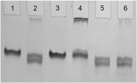
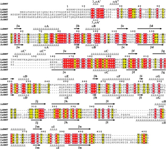
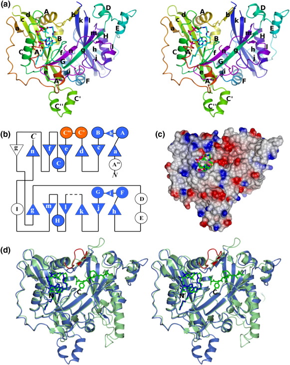
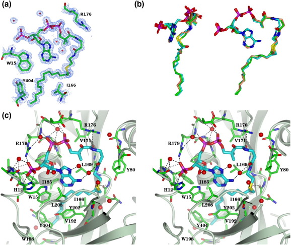
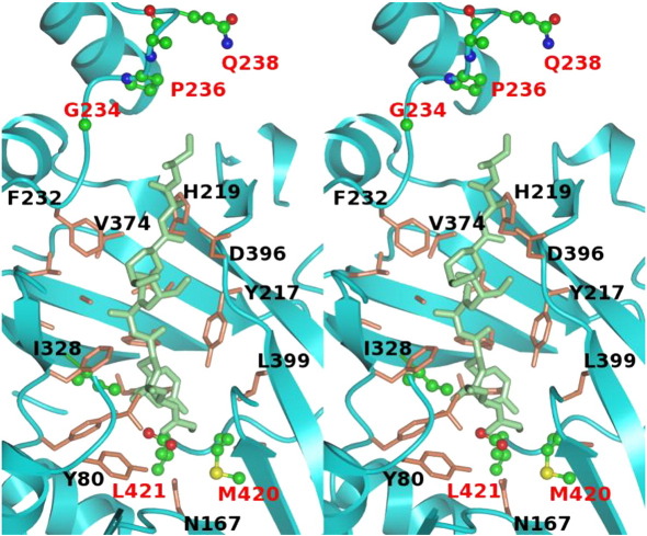
N-Myristoyltransferase (NMT) catalyses the attachment of the 14-carbon saturated fatty acid, myristate, to the amino-terminal glycine residue of a subset of eukaryotic proteins that function in multiple cellular processes, including vesicular protein trafficking and signal transduction. In these pathways, N-myristoylation facilitates association of substrate proteins with membranes or the hydrophobic domains of other partner peptides. NMT function is essential for viability in all cell types tested to date, demonstrating that this enzyme has potential as a target for drug development. Here, we provide genetic evidence that NMT is likely to be essential for viability in insect stages of the pathogenic protozoan parasite, Leishmania donovani, causative agent of the tropical infectious disease, visceral leishmaniasis. The open reading frame of L. donovani NMT has been amplified and used to overproduce active recombinant enzyme in Escherichia coli, as demonstrated by gel mobility shift assays of ligand binding and peptide-myristoylation activity in scintillation proximity assays. The purified protein has been crystallized in complex with the non-hydrolysable substrate analogue S-(2-oxo)pentadecyl-CoA, and its structure was solved by molecular replacement at 1.4 A resolution. The structure has as its defining feature a 14-stranded twisted beta-sheet on which helices are packed so as to form an extended and curved substrate-binding groove running across two protein lobes. The fatty acyl-CoA is largely buried in the N-terminal lobe, its binding leading to the loosening of a flap, which in unliganded NMT structures, occludes the protein substrate binding site in the carboxy-terminal lobe. These studies validate L. donovani NMT as a potential target for development of new therapeutic agents against visceral leishmaniasis.
(c) 2009 Elsevier Ltd. All rights reserved.
Figures

Similar articles
-
Structure of N-myristoyltransferase from Aspergillus fumigatus.Acta Crystallogr D Biol Crystallogr. 2015 Apr;71(Pt 4):754-61. doi: 10.1107/S1399004715000401. Epub 2015 Mar 26. Acta Crystallogr D Biol Crystallogr. 2015. PMID: 25849386
-
Pharmacological Validation of N-Myristoyltransferase as a Drug Target in Leishmania donovani.ACS Infect Dis. 2019 Jan 11;5(1):111-122. doi: 10.1021/acsinfecdis.8b00226. Epub 2018 Nov 12. ACS Infect Dis. 2019. PMID: 30380837 Free PMC article.
-
Studies of the catalytic activities and substrate specificities of Saccharomyces cerevisiae myristoyl-coenzyme A: protein N-myristoyltransferase deletion mutants and human/yeast Nmt chimeras in Escherichia coli and S. cerevisiae.J Biol Chem. 1992 Nov 25;267(33):23852-61. J Biol Chem. 1992. PMID: 1429724
-
Antiparasitic chemotherapy: tinkering with the purine salvage pathway.Adv Exp Med Biol. 2008;625:116-32. doi: 10.1007/978-0-387-77570-8_10. Adv Exp Med Biol. 2008. PMID: 18365663 Review.
-
Drug discovery in leishmaniasis using protein lipidation as a target.Biophys Rev. 2021 Nov 4;13(6):1139-1146. doi: 10.1007/s12551-021-00855-0. eCollection 2021 Dec. Biophys Rev. 2021. PMID: 35035594 Free PMC article. Review.
Cited by
-
Identification of and Structural Insights into Hit Compounds Targeting N-Myristoyltransferase for Cryptosporidium Drug Development.ACS Infect Dis. 2023 Oct 13;9(10):1821-1833. doi: 10.1021/acsinfecdis.3c00151. Epub 2023 Sep 18. ACS Infect Dis. 2023. PMID: 37722671 Free PMC article.
-
Association of NMT2 with the acyl-CoA carrier ACBD6 protects the N-myristoyltransferase reaction from palmitoyl-CoA.J Lipid Res. 2016 Feb;57(2):288-98. doi: 10.1194/jlr.M065003. Epub 2015 Nov 30. J Lipid Res. 2016. PMID: 26621918 Free PMC article.
-
Structure-guided optimization of quinoline inhibitors of Plasmodium N-myristoyltransferase.Medchemcomm. 2017 Jan 1;8(1):191-197. doi: 10.1039/c6md00531d. Epub 2016 Nov 11. Medchemcomm. 2017. PMID: 28626547 Free PMC article.
-
Functional analysis of Leishmania cyclopropane fatty acid synthetase.PLoS One. 2012;7(12):e51300. doi: 10.1371/journal.pone.0051300. Epub 2012 Dec 10. PLoS One. 2012. PMID: 23251490 Free PMC article.
-
Design and synthesis of high affinity inhibitors of Plasmodium falciparum and Plasmodium vivax N-myristoyltransferases directed by ligand efficiency dependent lipophilicity (LELP).J Med Chem. 2014 Mar 27;57(6):2773-88. doi: 10.1021/jm500066b. Epub 2014 Mar 18. J Med Chem. 2014. PMID: 24641010 Free PMC article.
References
-
- Chappuis F., Sundar S., Hailu A., Ghalib H., Rijal S., Peeling R.W. Visceral leishmaniasis: what are the needs for diagnosis, treatment and control? Nat. Rev. Microbiol. 2007;5:873–882. - PubMed
-
- Shaw J. The leishmaniases-survival and expansion in a changing world. A mini-review. Mem. Inst. Oswaldo Cruz. 2007;102:541–547. - PubMed
-
- Croft S.L., Seifert K., Yardley V. Current scenario of drug development for leishmaniasis. Indian J. Med. Res. 2006;123:399–410. - PubMed
Publication types
MeSH terms
Substances
Associated data
- Actions
- Actions
Grants and funding
LinkOut - more resources
Full Text Sources
Other Literature Sources
