

Backbone model of an aquareovirus virion by cryo-electron microscopy and bioinformatics
- PMID: 20036256
- PMCID: PMC2900198
- DOI: 10.1016/j.jmb.2009.12.027
Backbone model of an aquareovirus virion by cryo-electron microscopy and bioinformatics
Abstract
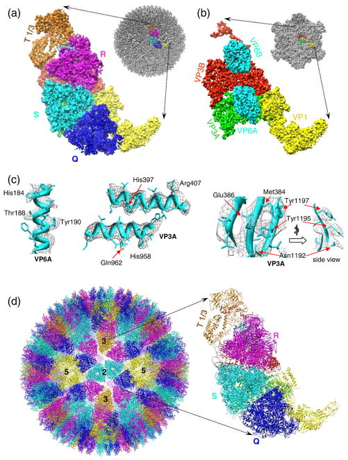
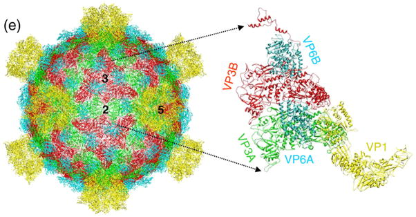
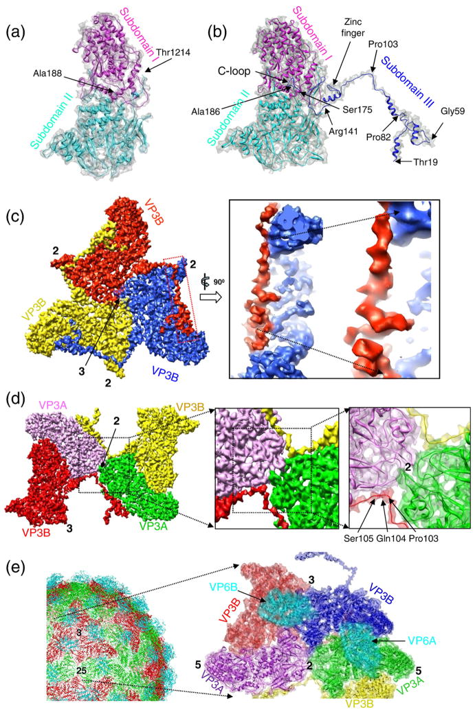
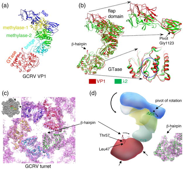
Grass carp reovirus (GCRV) is a member of the aquareovirus genus in the Reoviridae family and has a capsid with two shells-a transcription-competent core surrounded by a coat. We report a near-atomic-resolution reconstruction of the GCRV virion by cryo-electron microscopy and single-particle reconstruction. A backbone model of the GCRV virion, including seven conformers of the five capsid proteins making up the 1500 molecules in both the core and the coat, was derived using cryo-electron microscopy density-map-constrained homology modeling and refinement. Our structure clearly showed that the amino-terminal segment of core protein VP3B forms an approximately 120-A-long alpha-helix-rich extension bridging across the icosahedral 2-fold-symmetry-related molecular interface. The presence of this unique structure across this interface and the lack of an external cementing molecule at this location in GCRV suggest a stabilizing role of this extended amino-terminal density. Moreover, part of this amino-terminal extension becomes invisible in the reconstruction of transcription-competent core particles, suggesting its involvement in endogenous viral RNA transcription. Our structure of the VP1 turret represents its open state, and comparison with its related structures at the closed state suggests hinge-like domain movements associated with the mRNA-capping machinery. Overall, this first backbone model of an aquareovirus virion provides a wealth of structural information for understanding the structural basis of GCRV assembly and transcription.
Copyright 2009. Published by Elsevier Ltd.
Figures

Similar articles
-
Subnanometer-resolution structures of the grass carp reovirus core and virion.J Mol Biol. 2008 Sep 26;382(1):213-22. doi: 10.1016/j.jmb.2008.06.075. Epub 2008 Jul 3. J Mol Biol. 2008. PMID: 18625243 Free PMC article.
-
3D reconstruction and capsid protein characterization of grass carp reovirus.Sci China C Life Sci. 2005 Dec;48(6):593-600. doi: 10.1360/062004-105. Sci China C Life Sci. 2005. PMID: 16483138
-
Building and refining protein models within cryo-electron microscopy density maps based on homology modeling and multiscale structure refinement.J Mol Biol. 2010 Apr 2;397(3):835-51. doi: 10.1016/j.jmb.2010.01.041. Epub 2010 Jan 28. J Mol Biol. 2010. PMID: 20109465 Free PMC article.
-
High-resolution 3D structures reveal the biological functions of reoviruses.Virol Sin. 2013 Dec;28(6):318-25. doi: 10.1007/s12250-013-3341-6. Epub 2013 Nov 6. Virol Sin. 2013. PMID: 24254888 Free PMC article. Review.
-
Structural studies on orbivirus proteins and particles.Curr Top Microbiol Immunol. 2006;309:221-44. doi: 10.1007/3-540-30773-7_8. Curr Top Microbiol Immunol. 2006. PMID: 16909901 Review.
Cited by
-
Gorgon and pathwalking: macromolecular modeling tools for subnanometer resolution density maps.Biopolymers. 2012 Sep;97(9):655-68. doi: 10.1002/bip.22065. Biopolymers. 2012. PMID: 22696403 Free PMC article.
-
Modeling protein structure at near atomic resolutions with Gorgon.J Struct Biol. 2011 May;174(2):360-73. doi: 10.1016/j.jsb.2011.01.015. Epub 2011 Feb 4. J Struct Biol. 2011. PMID: 21296162 Free PMC article.
-
N-Terminal Myristoylated VP5 is Required for Penetrating Cell Membrane and Promoting Infectivity in Aquareoviruses.Virol Sin. 2018 Jun;33(3):287-290. doi: 10.1007/s12250-018-0036-z. Epub 2018 Jun 5. Virol Sin. 2018. PMID: 29869748 Free PMC article. No abstract available.
-
Inhibitor analysis revealed that clathrin-mediated endocytosis is involed in cellular entry of type III grass carp reovirus.Virol J. 2018 May 24;15(1):92. doi: 10.1186/s12985-018-0993-8. Virol J. 2018. PMID: 29793525 Free PMC article.
-
Functional investigation of grass carp reovirus nonstructural protein NS80.Virol J. 2011 Apr 14;8:168. doi: 10.1186/1743-422X-8-168. Virol J. 2011. PMID: 21489306 Free PMC article.
References
-
- Reinisch KM. The dsRNA viridae and their catalytic capsids. Nat Struct Biol. 2002;9:714–716. - PubMed
-
- Mertens P. The dsRNA viruses. Virus Res. 2004;101:3–13. - PubMed
-
- Caston JR, Luque D, Trus BL, Rivas G, Alfonso C, Gonzalez JM, et al. Three-dimensional structure and stoichiometry of Helmintosporium victoriae 190S totivirus. Virology. 2006;347:323–332. - PubMed
-
- Naitow H, Tang J, Canady M, Wickner RB, Johnson JE. L-A virus at 3.4 Å resolution reveals particle architecture and mRNA decapping mechanism. Nat Struct Biol. 2002;9:725–728. - PubMed
-
- Huiskonen JT, De Haas F, Bubeck D, Bamford DH, Fuller SD, Butcher SJ. Structure of the bacteriophage phi6 nucleocapsid suggests a mechanism for sequential RNA packaging. Structure. 2006;14:1039–1048. - PubMed
Publication types
MeSH terms
Substances
Associated data
- Actions
Grants and funding
LinkOut - more resources
Full Text Sources
Research Materials
