. 2010 Mar 5;396(4):967-84.
doi: 10.1016/j.jmb.2009.12.030. Epub 2009 Dec 28.

Molecular and structural insight into proNGF engagement of p75NTR and sortilin

Affiliations

Molecular and structural insight into proNGF engagement of p75NTR and sortilin

Dan Feng et al. J Mol Biol. .

Abstract

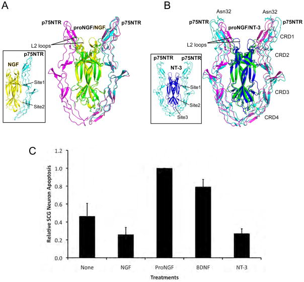
Nerve growth factor (NGF) is initially synthesized as a precursor, proNGF, that is cleaved to release its C-terminal mature form. Recent studies suggested that proNGF is not an inactive precursor but acts as a signaling ligand distinct from its mature counterpart. proNGF and mature NGF initiate opposing biological responses by utilizing both distinct and shared receptor components. In this study, we carried out structural and biochemical characterization of proNGF interactions with p75NTR and sortilin. We crystallized proNGF complexed to p75NTR and present the structure at 3.75-A resolution. The structure reveals a 2:2 symmetric binding mode, as compared with the asymmetric structure of a previously reported crystal structure of mature NGF complexed to p75NTR and the 2:2 symmetric complex of neurotrophin-3 (NT-3) and p75NTR. Here, we discuss the possible origins and implications of the different stoichiometries. In the proNGF-p75NTR complex, the pro regions of proNGF are mostly disordered and two hairpin loops (loop 2) at the top of the NGF dimer have undergone conformational changes in comparison with mature NT structures, suggesting possible interactions with the propeptide. We further explored the binding characteristics of proNGF to sortilin using surface plasmon resonance and cell-based assays and determined that calcium ions promote the formation of a stable ternary complex of proNGF-sortilin-p75NTR. These results, together with those of previous structural and mechanistic studies of NT-receptor interactions, suggest the potential for distinct signaling activities through p75NTR mediated by different NT-induced conformational changes.


Figures

Figure 1
Figure 1. Characterization of recombinant proNGF activity
(A). Schematic diagram of proNGF primary structure. Signal peptide was colored in green, pro-peptide was colored in white and mature NGF was colored in gray. Three furin cleavage sites located at positions -1, -2, -40, -41, -70, and -71 were mutated to alanine residues. There are two predicted N-glycosylation sites located in pro-peptide domain positions -8 and -53. (B). Western blot analysis of receptor expression in HT1080 cell line that was transfected with p75NTR, or sortilin or both, with wild type HT1080 cells as control. (C). Uptake of Alexa-conjugated proNGF triple mutant by HT1080 cells expressing different receptors: (1) none (WT), (2) sortilin, (3) p75NTR and (4) p75NTR/sortilin. (D). Quantitation of uptake of proNGF triple mutant. Results are representative of five independent experiments.
Figure 2
Figure 2. Biochemical and crystallographic analysis of proNGF/p75NTR complex
(A) Chromatographic profiles on a Superdex 200 HR 10/30 size-exclusion column. Curves were shown in different colors: Blue dashed line for p75NTR Asn32 mutant; Orange dashed line for proNGF; Magenta dashed line for proNGF + p75NTR wild type; Black solid line for proNGF + p75NTR Asn32 mutant. The column molecular weight calibration with globular protein standards is shown at the top. (B). SDS-PAGE gel corresponding to proNGF/p75NTRmut (Asn32 mutant) purification (black solid curve in panel (A)). Molecular weight standards are shown at the left side. Lanes are labeled as eluting volume from gel filtration column. ProNGF migrated at ~32 kDa and p75NTR migrated at ~20 kDa, as indicated by arrows at the right side of gel. (C). Crystal gel for proNGF/p75NTR crystal. Samples were the starting material used for crystallization, and then only from washed crystals. Crystals were washed by reservoir solution several times to eliminate carry over of starting material. ProNGF appears at ~32 kDa, indicating it is in an intact state in crystal. (D). Composite omit electron density in the asymmetric unit. Map is shown at contour level 1.2 σ. All molecules in the asymmetric unit are shown in ribbon representation. Mature NGF is shown in green and p75NTR molecules are shown in magenta. Symmetry related molecules are grey. All structural representations in this work were generated with PyMOL (DeLano Scientific LLC). (E). Molecular packing in the crystal lattice is mediated by p75NTR. Mature NGF is in green and p75NTR is in magenta. Crystal lattice in other directions is shown in Supplementary Figure 2. (F). Crystal structure of proNGF/p75NTR complex. Mature regions of the two proNGF molecules are colored in green and the two p75NTR molecules are colored in magenta. N and C terminus of mature NGF in proNGF were labeled as green.
Figure 3
Figure 3. Symmetric and asymmetric binding in neurotrophin complexes with p75NTR
(A). Superposition of proNGF/p75NTR structure with mature NGF/p75NTR structure (PDB code: 1SG1) using mature NGF as the superposition template. Both structures are represented as ribbons. For proNGF/p75NTR complex, mature NGF is green and p75NTR is magenta. For NGF/p75NTR structure, NGF is yellow and p75NTR is cyan. L2 loops of one NGF molecule in each structure are indicated. The inset shows the asymmetric NGF/p75NTR structure. (B). Superposition of proNGF/p75NTR structure and mature NT-3/p75NTR structure (PDB code: 3BUK). ProNGF/p75NTR structure is colored as in panel (A). For NT-3/p75NTR structure, NT-3 is blue and p75NTR is cyan. N-Glycans at Asn32 are highlighted as cyan sticks. The inset shows the NT-3/p75NTR structure. (C) Cell apoptosis assay for NGF, proNGF, BDNF, and NT-3. Replica cultures of SCG neurons (DIV 9) were washed free of NGF and were treated with no additive (None), 10 ng/ml NGF, 5 ng/ml proNGF, 100 ng/ml BDNF, or 100 ng/ml recombinant NT-3 in the presence of 12.5 mM KCl. Thirty-six hours later, cultures were processed and scored for apoptotic neurons as described (Materials and Methods). The data were normalized to the number of dying neurons under 12.5 mM KCl treatment. Results were summarized from three independently conducted experiments. Vertical error bars represent S.E.M.
Figure 4
Figure 4. Molecular masses of NGF and proNGF alone and in complexes with p75NTR
Molar masses of NGF (A), proNGF (B), p75NTR (C), NGF/p75NTR (D), and proNGF/p75NTR (E) are measured with multi-angle light scattering, and are plotted against elution time from Superdex 200 gel filtration column. Peaks are visualized as unitless relative refractive index measurements. Grey lines represent the linear fitting of the molar mass measurements.
Figure 5
Figure 5. Conformational changes of L2 loops in mature neurotrophins versus proNGF
Top view of molecular surfaces of (A) NGF/p75NTR, (B) NT-3/p75NTR and (C) proNGF/p75NTR. p75NTR is magenta in all structures. Neurotrophins are colored in light grey, except for L2 loops. L2 loops are yellow for NGF/p75NTR in (A), blue for NT-3/p75NTR in (B) and green for proNGF/p75NTR in (C). Composite omit map electron densities between L2 loops of proNGF are shown at contour level 1.2 σ. Tryptophan residues at position 99 are shown as green sticks. (D). Comparison of L2 loops in proNGF/p75NTR structure with L2 loops in uncomplexed structures of neurotrophins. L2 loops in proNGF/p75NTR structure are green, with composite omit map of this region at contour level 1.2 σ. L2 in other structures include unliganded NGF (1BET) in gray, NGF complexed to p75NTR (1SG1) in yellow, NGF complexed to TrkA-D5 (1WWW) in magenta, NGF complexed to TrkA (D1-D5) (2IFG) in cyan, NT-4 (1B98) in slate, brain-derived neurotrophic factor (1B8M) in orange, and NT-3 complexed to p75NTR in blue (3BUK). Distance between the L2’s of the proNGF dimer is measured as 14 Å and indicated by black dashed line. (E) Side view of L2 loop region of proNGF. Composite omit map densities were shown for the region around L2 loops. For comparison, L2 loops of NGF and NT-3 are also shown in yellow and blue respectively. Trp99 is shown in green sticks.
Figure 6
Figure 6. Dissecting the assembly of the proNGF/sortilin/p75NTR complex by surface plasmon resonance
Sortilin was immobilized on a streptavidin (SA) chip. Different ligands were flowed over the chip surface including (A) proNGF, (C) NGF, and (E) proNGF/p75NTR complex. proNGF used is the triple-mutant form; other forms of proNGF are not stable enough for SPR. Experiments were performed over a series of concentrations as shown in each panel. Competition experiments were done by adding increasing amounts of the specific antagonist neurotensin to proNGF (B), NGF (D), and proNGF/p75NTR complex (F).
Figure 7
Figure 7. Calcium-dependent association of proNGF and sortilin
(A) The three components (proNGF, p75NTR and sortilin) were mixed and applied to a Superose 6 gel filtration column with 1 mM Calcium included both in the sample mixture and in the running buffer. Inset shows the SDS gel for fractions from 11 ml to 18 ml as labeled at the top of gel. Column calibration with globular protein molecular weight standards is shown at top of the panel. In panel (B), fractions from 11 ml to 14 ml in panel (A) were pooled, and then divided into two equal aliquots which were re-applied to the Superose 6 column with Calcium (B) or without Calcium (C) included in running buffer. Gels for fractions 11 ml to 18 ml are shown as insets in each panel. For comparison, profiles for individual components are also shown in panel (C) with green dash for sortilin, orange dash for proNGF and blue dash for p75NTR. HEK-293 cells were transfected with proNGF and sortilin, and cell lysates (D) or culture media (E) were immunoprecipitated with sortilin antibody in calcium-supplemented or depleted conditions followed by Western blot analysis. (F) HT-1080P/S cells were treated with proNGF (500 ng/ml), and cell lysates were subjected to immunoprecipitation with p75NTR antibody in calcium-supplemented or depleted conditions. Lysates, 10 μg/lane.

Similar articles

Cited by

References

    1. Levi-Montalcini R. The nerve growth factor 35 years later. Science. 1987;237:1154–62. - PubMed
    1. Snider WD. Functions of the neurotrophins during nervous system development: what the knockouts are teaching us. Cell. 1994;77:627–38. - PubMed
    1. Edwards RH, Selby MJ, Garcia PD, Rutter WJ. Processing of the native nerve growth factor precursor to form biologically active nerve growth factor. J Biol Chem. 1988;263:6810–5. - PubMed
    1. Rattenholl A, Lilie H, Grossmann A, Stern A, Schwarz E, Rudolph R. The pro-sequence facilitates folding of human nerve growth factor from Escherichia coli inclusion bodies. Eur J Biochem. 2001;268:3296–303. - PubMed
    1. Farhadi HF, Mowla SJ, Petrecca K, Morris SJ, Seidah NG, Murphy RA. Neurotrophin-3 sorts to the constitutive secretory pathway of hippocampal neurons and is diverted to the regulated secretory pathway by coexpression with brain-derived neurotrophic factor. J Neurosci. 2000;20:4059–68. - PMC - PubMed

Publication types

MeSH terms

Associated data