

Ionic storm in hypoxic/ischemic stress: can opioid receptors subside it?
- PMID: 20036308
- PMCID: PMC2843769
- DOI: 10.1016/j.pneurobio.2009.12.007
Ionic storm in hypoxic/ischemic stress: can opioid receptors subside it?
Abstract
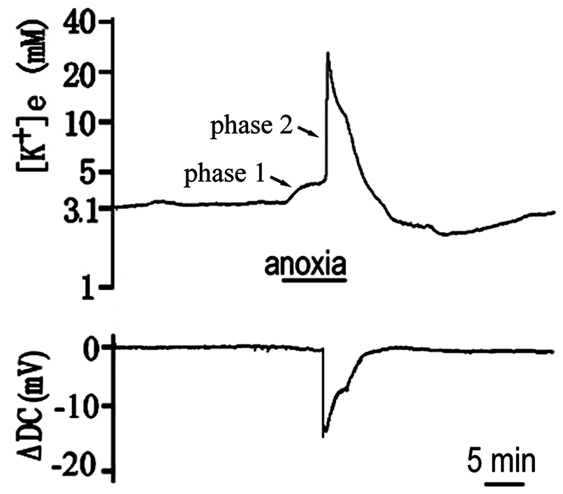
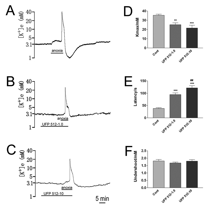
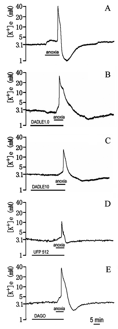
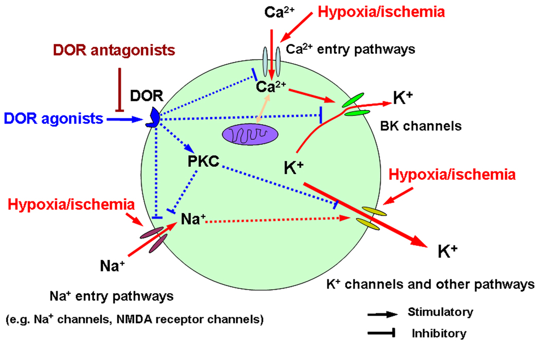
Neurons in the mammalian central nervous system are extremely vulnerable to oxygen deprivation and blood supply insufficiency. Indeed, hypoxic/ischemic stress triggers multiple pathophysiological changes in the brain, forming the basis of hypoxic/ischemic encephalopathy. One of the initial and crucial events induced by hypoxia/ischemia is the disruption of ionic homeostasis characterized by enhanced K(+) efflux and Na(+)-, Ca(2+)- and Cl(-)-influx, which causes neuronal injury or even death. Recent data from our laboratory and those of others have shown that activation of opioid receptors, particularly delta-opioid receptors (DOR), is neuroprotective against hypoxic/ischemic insult. This protective mechanism may be one of the key factors that determine neuronal survival under hypoxic/ischemic condition. An important aspect of the DOR-mediated neuroprotection is its action against hypoxic/ischemic disruption of ionic homeostasis. Specially, DOR signal inhibits Na(+) influx through the membrane and reduces the increase in intracellular Ca(2+), thus decreasing the excessive leakage of intracellular K(+). Such protection is dependent on a PKC-dependent and PKA-independent signaling pathway. Furthermore, our novel exploration shows that DOR attenuates hypoxic/ischemic disruption of ionic homeostasis through the inhibitory regulation of Na(+) channels. In this review, we will first update current information regarding the process and features of hypoxic/ischemic disruption of ionic homeostasis and then discuss the opioid-mediated regulation of ionic homeostasis, especially in hypoxic/ischemic condition, and the underlying mechanisms.
Keywords: Ca2+ channels; K+ channels; Na+ channels; hypoxia/ischemia; ionic homeostasis; neuroprotection; opioids; δ-opioid receptor.
Copyright 2010 Elsevier Ltd. All rights reserved.
Figures
Similar articles
-
Attenuating Ischemic Disruption of K+ Homeostasis in the Cortex of Hypoxic-Ischemic Neonatal Rats: DOR Activation vs. Acupuncture Treatment.Mol Neurobiol. 2016 Dec;53(10):7213-7227. doi: 10.1007/s12035-015-9621-4. Epub 2015 Dec 19. Mol Neurobiol. 2016. PMID: 26687186
-
DOR activation inhibits anoxic/ischemic Na+ influx through Na+ channels via PKC mechanisms in the cortex.Exp Neurol. 2012 Aug;236(2):228-39. doi: 10.1016/j.expneurol.2012.05.006. Epub 2012 May 15. Exp Neurol. 2012. PMID: 22609332 Free PMC article.
-
Cortical delta-opioid receptors potentiate K+ homeostasis during anoxia and oxygen-glucose deprivation.J Cereb Blood Flow Metab. 2007 Feb;27(2):356-68. doi: 10.1038/sj.jcbfm.9600352. Epub 2006 Jun 14. J Cereb Blood Flow Metab. 2007. PMID: 16773140
-
Neuroprotection Against Hypoxic/Ischemic Injury: δ-Opioid Receptors and BDNF-TrkB Pathway.Cell Physiol Biochem. 2018;47(1):302-315. doi: 10.1159/000489808. Epub 2018 May 11. Cell Physiol Biochem. 2018. PMID: 29768254 Review.
-
Neuroprotection against hypoxia/ischemia: δ-opioid receptor-mediated cellular/molecular events.Cell Mol Life Sci. 2013 Jul;70(13):2291-303. doi: 10.1007/s00018-012-1167-2. Epub 2012 Sep 27. Cell Mol Life Sci. 2013. PMID: 23014992 Free PMC article. Review.
Cited by
-
Effect of electroacupuncture on rat ischemic brain injury: importance of stimulation duration.Evid Based Complement Alternat Med. 2013;2013:878521. doi: 10.1155/2013/878521. Epub 2013 May 13. Evid Based Complement Alternat Med. 2013. PMID: 23737851 Free PMC article.
-
δ-Opioid receptor activation rescues the functional TrkB receptor and protects the brain from ischemia-reperfusion injury in the rat.PLoS One. 2013 Jul 2;8(7):e69252. doi: 10.1371/journal.pone.0069252. Print 2013. PLoS One. 2013. PMID: 23844255 Free PMC article.
-
Effect of δ-opioid receptor activation on BDNF-TrkB vs. TNF-α in the mouse cortex exposed to prolonged hypoxia.Int J Mol Sci. 2013 Jul 31;14(8):15959-76. doi: 10.3390/ijms140815959. Int J Mol Sci. 2013. PMID: 23912236 Free PMC article.
-
Force spectroscopy measurements show that cortical neurons exposed to excitotoxic agonists stiffen before showing evidence of bleb damage.PLoS One. 2013 Aug 30;8(8):e73499. doi: 10.1371/journal.pone.0073499. eCollection 2013. PLoS One. 2013. PMID: 24023686 Free PMC article.
-
Effects of inhibitory amino acids on expression of GABAA Rα and glycine Rα1 in hypoxic rat cortical neurons during development.Brain Res. 2011 Nov 24;1425:1-12. doi: 10.1016/j.brainres.2011.09.045. Epub 2011 Sep 29. Brain Res. 2011. PMID: 22018691 Free PMC article.
References
-
- Aarts M, Iihara K, Wei WL, Xiong ZG, Arundine M, Cerwinski W, MacDonald JF, Tymianski M. A key role for TRPM7 channels in anoxic neuronal death. Cell. 2003;115:863–877. - PubMed
-
- Adams DJ, Trequattrini C. Opioid receptor-mediated inhibition of ω-conotoxin GVIA-sensitive calcium channel currents in rat intracardiac neurons. J. Neurophysiol. 1998;79:753–762. - PubMed
-
- Adams HP, Jr, Olinger CP, Barsan WG, Butler MJ, Graff-Radford NR, Brott TG, Biller J, Damasio H, Tomsick T, Goldberg M. A dose-escalation study of large doses of naloxone for treatment of patients with acute cerebral ischemia. Stroke. 1986;17:404–409. - PubMed
-
- Agostinho P, Duarte CB, Oliveira CR. Intracellular free Na+ concentration increases in cultured retinal cells under oxidative stress conditions. Neurosci. Res. 1996;25:343–351. - PubMed
Publication types
MeSH terms
Substances
Grants and funding
LinkOut - more resources
Full Text Sources
Miscellaneous
