. 2010;261(2):134-43.
doi: 10.1016/j.cellimm.2009.11.011. Epub 2009 Dec 5.

Cyclophosphamide induces bone marrow to yield higher numbers of precursor dendritic cells in vitro capable of functional antigen presentation to T cells in vivo

Affiliations

Cyclophosphamide induces bone marrow to yield higher numbers of precursor dendritic cells in vitro capable of functional antigen presentation to T cells in vivo

Mohamed L Salem et al. Cell Immunol. 2010.

Abstract

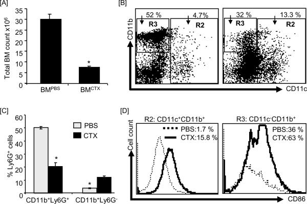
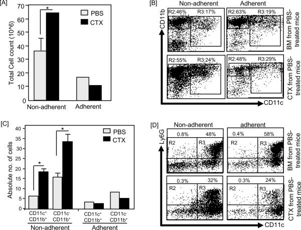
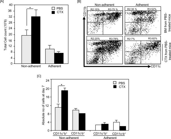
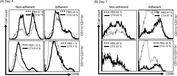
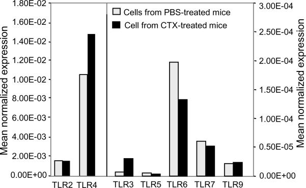
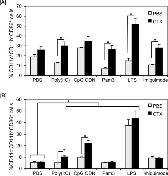
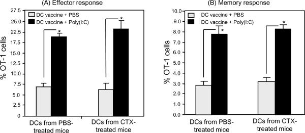
We have shown recently that cyclophosphamide (CTX) treatment induced a marked increase in the numbers of immature dendritic cells (DCs) in blood, coinciding with enhanced antigen-specific responses of the adoptively transferred CD8(+) T cells. Because this DC expansion was preceded by DC proliferation in bone marrow (BM), we tested whether BM post CTX treatment can generate higher numbers of functional DCs. BM was harvested three days after treatment of C57BL/6 mice with PBS or CTX and cultured with GM-CSF/IL-4 in vitro. Compared with control, BM from CTX-treated mice showed faster generation and yielded higher numbers of DCs with superior activation in response to toll-like receptor (TLR) agonists. Vaccination with peptide-pulsed DCs generated from BM from CTX-treated mice induced comparable adjuvant effects to those induced by control DCs. Taken together, post CTX BM harbors higher numbers of DC precursors capable of differentiating into functional DCs, which be targeted to create host microenvironment riches in activated DCs upon treatment with TLR agonists.


Figures

Figure 1
Figure 1. CTX treatment induced an increased level of DCs in fresh BM
C57BL/6 mice were i.p. treated with PBS (n=3 mice/group) or 4mg/mouse CTX (n=6 mice/group) and BM was harvested 3 days after treatments. BM single cell suspensions were prepared and stained with anti-CD11c, anti-CD11b, anti-Ly6G (Gr-1), and anti-CD86 mAbs. (A) Shows the total cell number of BM cells. (B) Shows the expression levels of CD11c and CD11b in the total BM. Cells are gated on CD11c+CD11b+ and CD11c+CD11b (R2) populations and on CD11cCD11b+ population (R3). (C) Shows the expression level of Ly6G in the total BM cells. (D) Shows the expression levels of CD86 on cells gated on R2 (gated on CD11c+CD11b+ + CD11cCD11b+) and R3 (gated on CD11cCD11b+). *, Statistically significant (P<0.05).
Figure 2
Figure 2. Non-adherent population harvested on day-4 culture of BM from CTX-treated mice showed higher number of CD11c+ cells
C57BL/6 mice were i.p. treated with PBS (n=3 mice/group) or 4mg/mouse CTX (n=6 mice/group) and BM was harvested 3 days after treatments. BM single cell suspensions were prepared and cultured with 10ng/mL of GM-CSF and IL-4. On day 4, the non-adherent and adherent populations were harvested, counted and their phenotypic characterization was analyzed by flow cytometry after staining with anti-CD11c, anti-CD11b, and anti-Ly6G mAbs. (A) Shows the total cell number of the non-adherent and adherent populations. (B) Shows the expression levels of CD11b and CD11c in the non-adherent (left panel) and adherent (right panel) cells in BM from PBS-treated mice (upper panel) and BM from CTX-treated mice (lower panel). (C) Shows the absolute numbers of CD11c+ and CD11b+ cells in the non-adherent and adherent populations from BM from PBS-treated mice (upper panel) and BM from CTX-treated mice (lower panel). The absolute number of cells was calculated by multiplying the % cells with a certain phenotype by the total number of the non-adherent and adherent cells. (D) Shows the expression levels of CD11c and Ly6G in in the non-adherent (left panel) and adherent (right panel) in BM from PBS-treated mice (upper panel) and BM from CTX-treated mice (lower panel). *, Statistically significant (P<0.05).
Figure 3
Figure 3. Non-adherent population of day 7 BM from CTX-treated mice culture showed higher number of CD11c+ cells
C57BL/6 mice were i.p. treated with PBS (n=3 mice/group) or 4mg/mouse CTX (n=6 mice/group) and BM was harvested 3 days after treatments. BM single cell suspensions were prepared and cultured with 10ng/mL of GM-CSF and IL-4. On day 4, the non-adherent and adherent cells were harvested, counted and their phenotypic characterization was analyzed by flow cytometry. (A) Shows the total cell number of the non-adherent and adherent populations of BM from PBS-treated mice and BM from CTX-treated mice. (B) Shows the expression levels of CD11b and CD11c in the non-adherent (left panel) and adherent (right panel) cells of BM from PBS-treated mice (upper panel) and BM from CTX-treated mice (lower panel). (C) Shows the absolute numbers of CD11c+ and CD11b+ subsets in the non-adherent and adherent populations of BM from PBS-treated mice and BM from CTX-treated mice harvested on day 7. The absolute number of cells was calculated by multiplying the % cells with a certain phenotype by the total number of non-adherent and adherent cells. *, Statistically significant (P<0.05).
Figure 4
Figure 4. Non-adherent cells from BM from CTX-treated mice showed higher activation phenotype
C57BL/6 mice were i.p. treated with PBS (n=3 mice/group) or 4mg/mouse CTX (n=6 mice/group) and BM was harvested 3 days after treatments. BM single cell suspensions were prepared and cultured with 10ng/mL of GM-CSF and IL-4. On day 4, (A) and 7 (B), the non-adherent (left panel) and adherent (right panel) cells from BM of PBS- and CTX-treated mice were harvested and stained with anti-CD11c, anti-CD11b, and anti-CD86 mAbs and the expression level of the activation marker CD86 was analyzed by 3-color flow cytometry in CD11c+CD11b+ (upper panel) and CD11cCD11b+ (lower panel) cells. *, Statistically significant (P<0.05).
Figure 5
Figure 5. DCs generated from BM from CTX-treated mice culture express simlar levels of different TLRs
C57BL/6 mice were i.p. treated with PBS (n=3 mice/group) or 4mg/mouse CTX (n=6 mice/group) and BM was harvested 3 days after treatments. BM single cell suspensions were prepared and cultured with 10ng/mL of GM-CSF and IL-4. On day 7 of culture, the non-adherent cells were harvested and processed for the real-time PCR analysis. Results are expressed as normalized mean expression.
Figure 6
Figure 6. DCs generated from BM from CTX-treated mice can respond to stimulation with TLR agonists
C57BL/6 mice were i.p. treated with PBS (n=3 mice/group) or 4mg/mouse CTX (n=6 mice/group) and BM was harvested 3 days after treatments. BM single cell suspensions were prepared and cultured with 10ng/mL of GM-CSF and IL-4. The non-adherent cells were harvested on day 7, counted, re-plated and treated for 24 hours with pam3CysK4 (a TLR2/6 agonist; 50μg/mL), poly(I:C) (a TLR3 agonist; 25μg/mL); LPS (a TLR4 agonist, 10ng/mL), imiquimode (a TLR7/8 agonist; 10μg/mL), or CpG ODN (a TLR9 agonist; 4μg/mL). (A) Shows the expression level of the activation marker CD86 on CD11c+CD11b+ and (B) shows the percentage of CD86 expression on CD11cCD11b+ cells. *, Statistically significant (P<0.05).
Figure 7
Figure 7. DCs generated from BM from CTX-treated mice can prime T cells in vivo
B6 mice (on Ly5.2 background) were treated with PBS and CTX as described in Figure 1 legend. After 3 days of treatment, BM was harvested and cultured with 10ng/mL of GM-CSF and IL-4. On day 7 of the culture, the non-adherent cells were harvested, counted and activated with 10ng/mL LPS for 24 hours. Cells were washed and pulsed with 5μg/mL of SIINFEKL peptide for 2 hours. Cells were then washed and injected through lateral tail vein into recipient B6 mice (on Ly5.2 background; n=4/group) adoptively transferred 1 day before with 1.5 × 10⁶ cells of naïve OT-1 cells harvested from lymph nodes and spleen of B6 mice on Ly5.1 background. (A) Shows the numbers of effector OT-1 cells in the peripheral blood harvested 7 days after vaccination with DCs. (B) Shows the number of memory OT-1 cells in the peripheral blood harvested 3 days after s.c. revaccination with 100μg/mouse SIINFEKL 40 days after priming. *, Statistically significant (P<0.05).

References

    1. Jurado JM, Sanchez A, Pajares B, Perez E, Alonso L, Alba E. Combined oral cyclophosphamide and bevacizumab in heavily pre-treated ovarian cancer. Clin Transl Oncol. 2008;10:583–6. - PubMed
    1. Rosell R, Moreno I, Maestre J, Olazabal A, Carles J, Barnadas A, Abad-Esteve A, Ribelles N, Canela M. Cyclophosphamide and ifosfamide combination as neoadjuvant chemotherapy for locally advanced nonsmall-cell lung cancer: a meta-analytic review. J Surg Oncol. 1990;45:124–30. - PubMed
    1. Saxton ML, Longo DL, Wetzel HE, Tribble H, Alvord WG, Kwak LW, Leonard AS, Ullmann CD, Curti BD, Ochoa AC. Adoptive transfer of anti-CD3-activated CD4+ T cells plus cyclophosphamide and liposome-encapsulated interleukin-2 cure murine MC-38 and 3LL tumors and establish tumor-specific immunity. Blood. 1997;89:2529–36. - PubMed
    1. Curti BD, Ochoa AC, Powers GC, Kopp WC, Alvord WG, Janik JE, Gause BL, Dunn B, Kopreski MS, Fenton R, Zea A, Dansky-Ullmann C, Strobl S, Harvey L, Nelson E, Sznol M, Longo DL. Phase I trial of anti-CD3-stimulated CD4+ T cells, infusional interleukin-2, and cyclophosphamide in patients with advanced cancer. J Clin Oncol. 1998;16:2752–60. - PubMed
    1. Bracci L, Moschella F, Sestili P, La Sorsa V, Valentini M, Canini I, Baccarini S, Maccari S, Ramoni C, Belardelli F, Proietti E. Cyclophosphamide enhances the antitumor efficacy of adoptively transferred immune cells through the induction of cytokine expression, B-cell and T-cell homeostatic proliferation, and specific tumor infiltration. Clin Cancer Res. 2007;13:644–53. - PubMed

Publication types

MeSH terms