

Role of myoepithelial cells in breast tumor progression
- PMID: 20036817
- PMCID: PMC2904307
- DOI: 10.2741/3617
Role of myoepithelial cells in breast tumor progression
Abstract
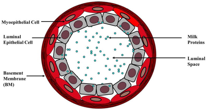
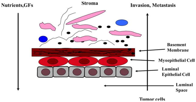
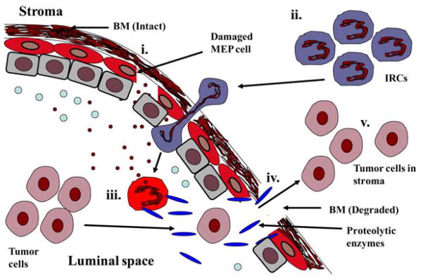
Myoepithelial cells form a semi-continuous protective sheet separating the human breast epithelium and the surrounding stroma. They suppress stromal invasion of tumor cells by the secretion of various anti-angiogenic and anti-invasive factors. The disruption of this cell layer results in the release of the growth factors, angiogenic factors, and reactive oxygen species causing an alteration in the microenvironment. This helps in the proliferation of surrounding cells and increases the invasiveness of tumor cells. Two theories are proposed for the mechanism of tumor epithelial cells progression from in situ to invasive stage. According to the first theory, tumor cell invasion is triggered by the overproduction of proteolytic enzymes by myoepithelial cells and surrounding tumor cells. The second theory states that tumor invasion is a multistep process, the interactions between damaged myoepithelial cells and the immunoreactive cells trigger the release of basement membrane degrading enzymes causing tumor progression. Further studies in understanding of molecular mechanism of myoepithelial cell functions in tumor suppression may lead to the identification of novel therapeutic targets for breast cancer.
Figures
Similar articles
-
Myoepithelial cell-specific expression of stefin A as a suppressor of early breast cancer invasion.J Pathol. 2017 Dec;243(4):496-509. doi: 10.1002/path.4990. Epub 2017 Oct 31. J Pathol. 2017. PMID: 29086922
-
Do myoepithelial cells hold the key for breast tumor progression?J Mammary Gland Biol Neoplasia. 2005 Jul;10(3):231-47. doi: 10.1007/s10911-005-9584-6. J Mammary Gland Biol Neoplasia. 2005. PMID: 16807803 Review.
-
Focal degeneration of aged or injured myoepithelial cells and the resultant auto-immunoreactions are trigger factors for breast tumor invasion.Med Hypotheses. 2007;69(6):1340-57. doi: 10.1016/j.mehy.2007.02.031. Epub 2007 May 9. Med Hypotheses. 2007. PMID: 17493765
-
Myoepithelial molecular markers in human breast carcinoma PMC42-LA cells are induced by extracellular matrix and stromal cells.In Vitro Cell Dev Biol Anim. 2006 Nov-Dec;42(10):298-307. doi: 10.1290/0601004.1. In Vitro Cell Dev Biol Anim. 2006. PMID: 17316063
-
Myoepithelial cells: autocrine and paracrine suppressors of breast cancer progression.J Mammary Gland Biol Neoplasia. 2005 Jul;10(3):249-60. doi: 10.1007/s10911-005-9585-5. J Mammary Gland Biol Neoplasia. 2005. PMID: 16807804 Review.
Cited by
-
Comparative Analysis of P63, Maspin and Matrix Metalloproteinase 2 Expression in Mucoepidermoid Carcinoma and Adenoid Cystic Carcinoma of Salivary Glands.J Dent (Shiraz). 2020 Jun;21(2):95-101. doi: 10.30476/DENTJODS.2019.77868.0. J Dent (Shiraz). 2020. PMID: 32582823 Free PMC article.
-
Tumor-infiltrating immune cells promoting tumor invasion and metastasis: existing theories.J Cancer. 2013;4(1):84-95. doi: 10.7150/jca.5482. Epub 2013 Jan 5. J Cancer. 2013. PMID: 23386907 Free PMC article.
-
The obese inflammatory microenvironment may promote breast DCIS progression.Front Immunol. 2024 Jul 12;15:1384354. doi: 10.3389/fimmu.2024.1384354. eCollection 2024. Front Immunol. 2024. PMID: 39072314 Free PMC article.
-
Re-evaluation of the myoepithelial cells roles in the breast cancer progression.Cancer Cell Int. 2022 Dec 12;22(1):403. doi: 10.1186/s12935-022-02829-y. Cancer Cell Int. 2022. PMID: 36510219 Free PMC article. Review.
-
Inhibition of the transition of ductal carcinoma in situ to invasive ductal carcinoma by a Gemini vitamin D analog.Cancer Prev Res (Phila). 2014 Jun;7(6):617-26. doi: 10.1158/1940-6207.CAPR-13-0362. Epub 2014 Apr 1. Cancer Prev Res (Phila). 2014. PMID: 24691501 Free PMC article.
References
-
- Murrell TGC. The potential for oxytocin (OT) to prevent breast cancer: a hypothesis. Breast Cancer Res Trea. 1995;35:225–229. - PubMed
-
- Man YG, Sang QX. Significance of focal myoepithelial cell layer disruptions in human breast tumor invasion: a paradigm shift from the “protease-centered” hypothesis. Exp Cell Res. 2004;301(2):103–18. - PubMed
-
- Clarke C, Sandle J, Lakhani SR. Myoepithelial cells: pathology, cell separation and markers of myoepithelial differentiation. J Mammary Gland Biol Neoplasia. 2005;(3):273–80. - PubMed
Publication types
MeSH terms
Substances
Grants and funding
LinkOut - more resources
Full Text Sources
Medical
