Comparative Study
. 2010 May;31(5):868-73.
doi: 10.3174/ajnr.A1937. Epub 2009 Dec 24.

Comparison of the added value of contrast-enhanced 3D fluid-attenuated inversion recovery and magnetization-prepared rapid acquisition of gradient echo sequences in relation to conventional postcontrast T1-weighted images for the evaluation of leptomeningeal diseases at 3T

Affiliations
Comparative Study

Comparison of the added value of contrast-enhanced 3D fluid-attenuated inversion recovery and magnetization-prepared rapid acquisition of gradient echo sequences in relation to conventional postcontrast T1-weighted images for the evaluation of leptomeningeal diseases at 3T

H Fukuoka et al. AJNR Am J Neuroradiol. 2010 May.

Abstract

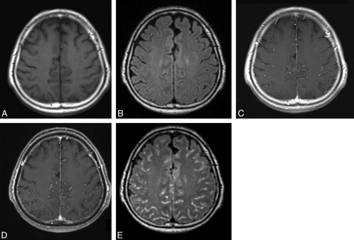
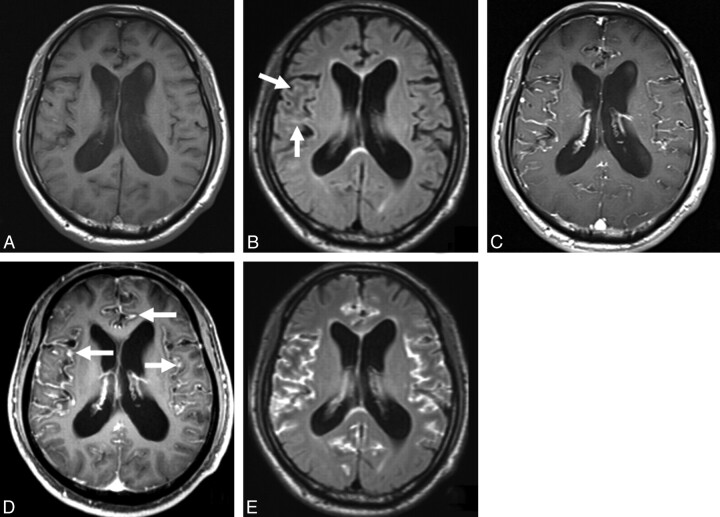
Background and purpose: The usefulness of contrast-enhanced 3D T2-FLAIR MR imaging for the evaluation of leptomeningeal diseases has not been systematically investigated. The purpose of this study was to assess the value added by contrast-enhanced 3D T2-FLAIR and MPRAGE sequences to conventional postcontrast T1-weighted images in the evaluation of leptomeningeal diseases. We also undertook in vitro studies in attempts to understand the consequences of our patient study.

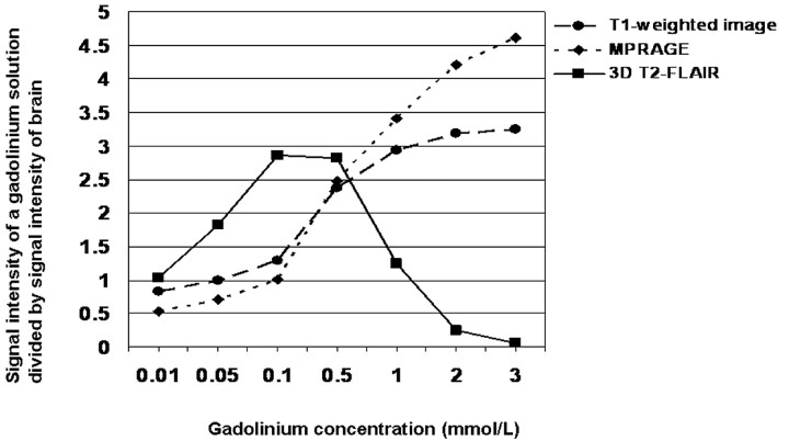
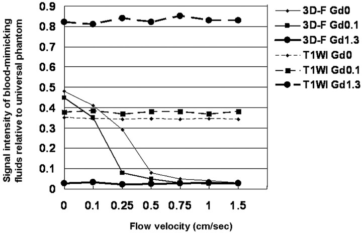
Materials and methods: Twelve patients with confirmed leptomeningeal diseases underwent postcontrast T1-weighted, MPRAGE, and 3D T2-FLAIR imaging at 3T. Two radiologists independently assessed the presence of additional information on postcontrast 3D MR images compared with postcontrast T1-weighted images. The effect of different Gd concentrations and flow velocities on the signal intensity on 3D T2-FLAIR images was investigated in vitro.

Results: According to both reviewers, 3D T2-FLAIR images yielded significantly more information than did MPRAGE images (P < .05 and P < .01, respectively). In the in vitro study, 3D T2-FLAIR was more highly sensitive to low Gd concentrations and less sensitive to high Gd concentrations than were T1-weighted or MPRAGE sequences. On 3D T2-FLAIR sequences, at a flow velocity exceeding 1.0 cm/s, the signal intensity of blood-mimicking fluids at concentrations of 0 and 0.1 mmol/L was as low as at 1.3 mmol/L.

Conclusions: For the depiction of leptomeningeal diseases, postcontrast 3D T2-FLAIR provides more additional information than postcontrast MPRAGE imaging. The superiority of the 3D T2-FLAIR sequence is associated with its high sensitivity to flow.


Figures

Fig 1.
Fig 1.
Diagram depicting the experimental setup of our flow velocity study. In the in vitro study, a 5-mm-diameter tube filled with a blood-mimicking fluid containing 3 different concentrations of gadopentetate dimeglumine (0, 0.1, and 1.3 mmol/L) was used. A continuous nonpulsatile flow (velocity, 0, 0.1, 0.25, 0.5, 0.75, 1.0, and 1.5 cm/s) was created in the tube with syringe pumps. A universal phantom and the tube were scanned simultaneously.
Fig 2.
Fig 2.
MR images in a 66-year-old man (case 7) with viral meningitis treated with an antiviral agent. A and B, Precontrast T1-weighted (A) and 3D T2-FLAIR (B) images show no apparent leptomeningeal abnormality. C and D, Postcontrast T1-weighted (C) and MPRAGE (D) images depict enhancement in the sulci, corresponding to the vessels. Compared with the postcontrast T1-weighted image (C), the postcontrast MPRAGE image does not provide additional information. Both reviewers ranked the MPRAGE sequence as grade 0. E, Postcontrast 3D T2-FLAIR image shows abnormal leptomeningeal enhancement in the sulci. Compared with the postcontrast T1-weighted image (C), postcontrast 3D T2-FLAIR provided additional information about abnormal leptomeningeal enhancement. Both reviewers scored the 3D T2-FLAIR sequence as grade 3.
Fig 3.
Fig 3.
MR images in a 65-year-old man (case 10) with leptomeningeal metastasis from melanoma. A, Precontrast T1-weighted image shows no apparent leptomeningeal abnormality. B, Precontrast 3D T2-FLAIR image demonstrates slight hyperintense areas in the sulci of the right frontal operculum (arrows). C, Postcontrast T1-weighted image depicts enhancement in the bilateral Sylvian fissures. It is difficult to discriminate between the vessels and the leptomeningeal lesions. D, Compared with the postcontrast T1-weighted image (C), the postcontrast MPRAGE image shows probable thickened enhanced areas in the sulci and Sylvian fissures (arrows). Both reviewers judged that the MPRAGE sequence provided additional information (grade 2). E, Postcontrast 3D T2-FLAIR image reveals markedly enhanced areas in the sulci and Sylvian fissures. As the cortical veins are not enhanced, it is easy to differentiate the vessels from leptomeningeal lesions. Compared with the postcontrast T1-weighted image (C), the postcontrast 3D T2-FLAIR image provides additional information about definite leptomeningeal lesion. Both reviewers scored the 3D T2-FLAIR sequence as grade 3.
Fig 4.
Fig 4.
Comparison of the relative signal intensity of a Gd solution among various pulse sequences. Three-dimensional T2-FLAIR sequences exhibited higher sensitivity to low Gd concentrations and lower sensitivity to high Gd concentrations than did T1-weighted or MPRAGE sequences.
Fig 5.
Fig 5.
Comparison of the signal intensity of blood-mimicking fluids relative to the universal phantom at various flow velocities on T1-weighted and 3D T2-FLAIR images. On T1-weighted images, the SI ratio at the 3 Gd concentrations is almost constant regardless of flow velocity. At no flow of the blood-mimicking fluids at concentrations of 0 and 0.1 mmol/L, the SI ratio is higher on 3D T2-FLAIR than on T1-weighted sequences. On 3D T2-FLAIR images at a Gd concentration of 1.3 mmol/L the SI ratio is extremely low regardless of flow velocity. At the concentrations of 0 and 0.1 mmol/L the SI ratio decreases rapidly with increasing velocity; at a flow velocity >1.0 cm/s, the SI ratio of blood-mimicking fluids at the concentrations of 0 and 0.1 mmol/L is as low as at 1.3 mmol/L. 3D-F Gd0 = 3D T2-FLAIR at 0 mmol/L of gadopentetate dimeglumine (Gd); 3D-F Gd0.1 = 3D T2-FLAIR at 0.1 mmol/L Gd; 3D-F Gd1.3 = 3D T2-FLAIR at 1.3 mmol/L Gd; T1WI Gd0 = T1-weighted imaging at 0 mmol/L Gd; T1WI Gd0.1 = T1-weighted imaging at 0.1 mmol/L Gd; T1WI Gd1.3 = T1-weighted imaging at 1.3 mmol/L of Gd.

References

    1. Mathews VP, Caldemeyer KS, Lowe MJ, et al. . Brain: gadolinium-enhanced fast fluid-attenuated inversion-recovery MR imaging. Radiology 1999;211:257–63 - PubMed
    1. Tsuchiya K, Katase S, Yoshino A, et al. . FLAIR MR imaging for diagnosing intracranial meningeal carcinomatosis. AJR Am J Roentgenol 2001;176:1585–88 - PubMed
    1. Griffiths PD, Coley SC, Romanowski CAJ, et al. . Contrast-enhanced fluid-attenuated inversion recovery imaging for leptomeningeal disease in children. AJNR Am J Neuroradiol 2003;24:719–23 - PMC - PubMed
    1. Splendiani A, Puglielli E, Amicis RD, et al. . Contrast-enhanced FLAIR in the early diagnosis of infectious meningitis. Neuroradiology 2005;47:591–98 - PubMed
    1. Kallmes DF, Hui FK, Mugler JP, 3rd. Suppression of cerebrospinal fluid and blood flow artifacts in FLAIR MR imaging with a single-slab three-dimensional pulse sequence: initial experience. Radiology 2001;221:251–55 - PubMed