. 2010 Feb 26;285(9):6275-84.
doi: 10.1074/jbc.M109.046151. Epub 2009 Dec 25.

The human carnitine transporter SLC22A16 mediates high affinity uptake of the anticancer polyamine analogue bleomycin-A5

Affiliations

The human carnitine transporter SLC22A16 mediates high affinity uptake of the anticancer polyamine analogue bleomycin-A5

Mustapha Aouida et al. J Biol Chem. .

Abstract

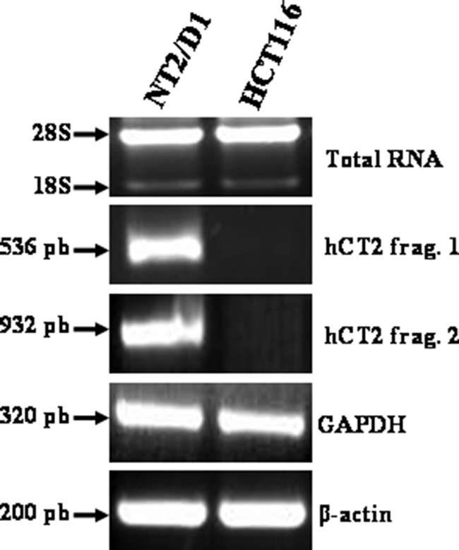
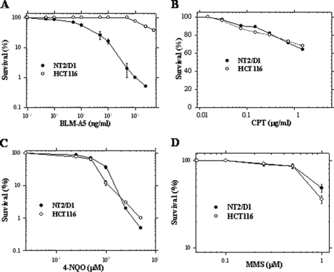
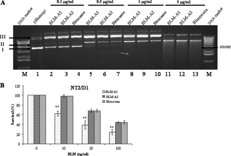
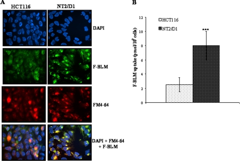
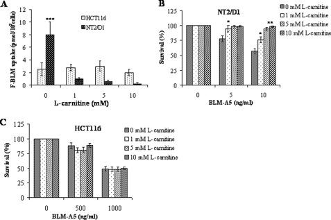
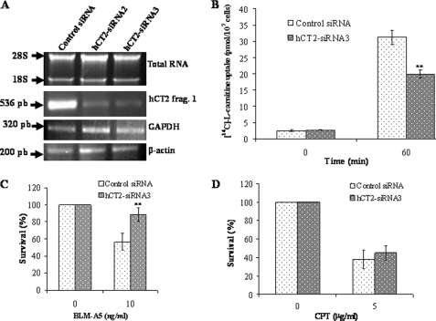
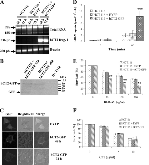
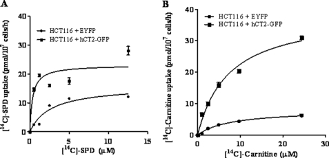
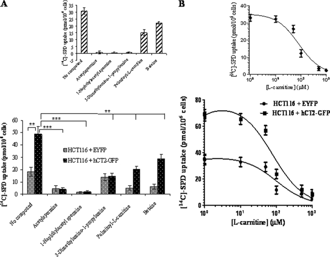
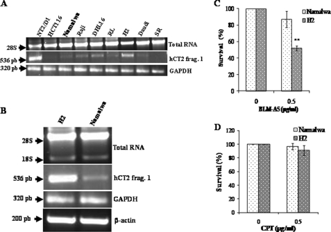
Bleomycin is used in combination with other antineoplastic agents to effectively treat lymphomas, testicular carcinomas, and squamous cell carcinomas of the cervix, head, and neck. However, resistance to bleomycin remains a persistent limitation in exploiting the full therapeutic benefit of the drug with other types of cancers. Previously, we documented that the Saccharomyces cerevisiae L-carnitine transporter Agp2 is responsible for the high affinity uptake of polyamines and of the polyamine analogue bleomycin-A5. Herein, we document that the human L-carnitine transporter hCT2 encoded by the SLC22A16 gene is involved in bleomycin-A5 uptake, as well as polyamines. We show that NT2/D1 human testicular cancer cells, which highly express hCT2, are extremely sensitive to bleomycin-A5, whereas HCT116 human colon carcinoma cells devoid of detectable hCT2 expression or MCF-7 human breast cancer cells that only weakly express the permease showed striking resistance to the drug. NT2/D1 cells accumulated fluorescein-labeled bleomycin-A5 to substantially higher levels than HCT116 cells. Moreover, L-carnitine protected NT2/D1 cells from the lethal effects of bleomycin-A5 by preventing its influx, and siRNA targeted to hCT2 induced resistance to bleomycin-A5-dependent genotoxicity. Furthermore, hCT2 overexpression induced by transient transfection of a functional hCT2-GFP fusion protein sensitized HCT116 cells to bleomycin-A5. Collectively, our data strongly suggest that hCT2 can mediate bleomycin-A5 and polyamine uptake, and that the rate of bleomycin-A5 accumulation may account for the differential response to the drug in patients.


Figures

FIGURE 1.
FIGURE 1.
RT-PCR analysis of hCT2 expression in testicular and colon cancer cell lines. Briefly, total RNA was isolated from the cancer cell lines, and 5 μg were used to synthesize cDNA using Superscript First-Strand Synthesis Systems for RT-PCR with random primers. PCR products were separated by electrophoresis on a 1% agarose gel and detected with ethidium bromide. The GAPDH and β-actin genes were used as controls.
FIGURE 2.
FIGURE 2.
Sensitivity of NT2/D1 and HCT116 cells toward various genotoxic agents. NT2/D1 and HCT116 cancer cells were treated with the indicated concentrations of bleomycin A5 (BLM-A5) (A), cisplatin (CPT) (B), 4-nitroquinoline-1-oxide (4-NQO) (C), and methyl methane sulfonate (MMS) (D) for 1 h and monitored for viability using clonogenic assay. Results are expressed as the mean ± S.D. from three separate experiments.
FIGURE 3.
FIGURE 3.
DNA damage potential by BLM-A5, BLM-A2, and blenoxane and cytotoxicity on NT2/D1 cells. A, damage to plasmid DNA by increasing concentrations of bleomycin species. Fixed amount of plasmid DNA (pCMV-hCT2-HA) was incubated at room temperature for 15 min with increasing concentrations of BLM-A5, BLM-A2, and blenoxane. Damaged to the DNA was assessed by monitoring the conversion of the covalently closed circular superhelical form (Form I) to the nicked form (Form II) or to the linear form (Form III). The DNA forms were analyzed on an agarose gel (1%) stained by ethidium bromide. Lane M, DNA size standard. B, NT2/D1 cells are hypersensitive to BLM-A5, as opposed to BLM-A2 or blenoxane. Cells were treated with the indicated concentrations of BLM for 1 h and monitor for viability using clonogenic assay. Results are expressed as the mean ± S.D. from three separate experiments with triplicate.
FIGURE 4.
FIGURE 4.
Increased accumulation of F-BLM in NT2/D1 cells. A, fluorescence analysis of F-BLM subcellular distribution in HCT116 and NT2/D1 cells. Cells were incubated with F-BLM (1.2 μm) for 1 h and prepared for microscopy as described under “Experimental Procedures.” DAPI and FM4–64 were used for nuclear and lysosomal staining, respectively. B, quantification of F-BLM levels in NT2/D1 and HCT116 cells. ***, p < 0.001.
FIGURE 5.
FIGURE 5.
l-carnitine blocks F-BLM uptake in NT2/D1, but not in HCT116 cells and protects against the cytotoxicity of BLM-A5. A, quantification of F-BLM uptake in NT2/D1 and HCT116 upon coincubation with l-carnitine. Cells were coincubated with increasing concentrations of l-carnitine (0–10 mm) and with F-BLM (1.2 μm) for 1 h, and the amount of internalized F-BLM measured using a spectrofluorometer. B and C, l-carnitine protects NT2/D1, but not HCT116, cells from the cytotoxic effects of BLM-A5. Cells were coincubated with l-carnitine and 1.2 μm BLM-A5 for 1 h and then monitored for survival using clonogenic assay. Results are expressed as the mean ± S.D. from three separate experiments. *, p < 0.05; **, p < 0.01; ***, p < 0.001.
FIGURE 6.
FIGURE 6.
hCT2 siRNA knockdown in NT2/D1 cells protects against BLM-A5 toxicity. A, RT-PCR analysis showing siRNA knockdown of hCT2 expression in NT2/D1 cells. Cells were transfected with 70 nm of either the scrambled control siRNA (lane 1), siRNA2 derived from the middle portion (lane 2), or siRNA3 against the C terminus of hCT2 cDNA (lane 3), and 5 h later, growth medium was added to the transfected cells followed by incubation for another 60 h. Total RNA was extracted and RT-PCR was performed using specific primers for either hCT2, GAPDH, or β-actin (ACTB). PCR products were detected as in Fig. 1. B, hCT2-siRNA3 knockdown of hCT2 decreases l-carnitine uptake. Cells were transfected with the hCT2-siRNA3 as in panel A, harvested, washed, and resuspended in DMEM medium without serum. Uptake was monitored at the indicated times following the addition of l-[14C]carnitine (10 μm). Qualitatively similar results were obtained using siRNA2 (not shown). C and D, siRNA knockdown of hCT2 protects NT2/D1 cells against BLM-A5-, but not cisplatin-induced cytotoxicity. Cells were transfected with siRNA as in panel A and cells were treated with either BLM-A5 (C) or cisplatin (CPT) (D) for 1 h and then monitored for survival by a clonogenic assay as in Fig. 4.
FIGURE 7.
FIGURE 7.
hCT2 overexpression sensitizes HCT116 human colon cancer cells to BLM-A5. A, RT-PCR analysis for the transient expression of hCT2-EYFP in HCT116 cells. Cells (60–70% confluent) were transfected without or with either the empty vector pEYFP or the plasmid pCMV-hCT2-GFP and cells harvested at the indicated time to assess for hCT2-GFP expression. B, overexpression of hCT2-GFP fusion protein in transiently transfected HCT116 cells. Cells were transfected as in A, and total extracts (50 μg) were analyzed for hCT2-EYFP expression by Western blot probed with anti-GFP monoclonal antibody. The result is representative of three independent analyses. Molecular mass standards in kDa are indicated by arrows. C, plasma membrane localization of the hCT2-EYFP fusion protein in HCT116 cells. Cells were transiently transfected for 48 and 72 h with either the empty vector pEYFP or the plasmid pCMV-hCT2-GFP, and fixed for confocal microscopy. D, overexpression of hCT2-GFP enhances F-BLM uptake in HCT116 cells. Cells were transfected with either the EYFP or the pCMV-hCT2-GFP vectors for 48 h, followed by incubation with a fixed amount of F-BLM (2.3 μg/ml) for 1 h, and the level of drug uptake determined using a spectrofluorometer. E and F, hCT2 overexpression sensitizes HCT116 cells to BLM-A5, but not cisplatin toxicity. Cells were transiently transfected for 48 h with either the empty vector pEYFP or the pCMV-hCT2-GFP plasmid, followed by exposure to increasing concentrations of either BLM-A5 (E) or cisplatin (CPT) (F) for 1 h, and then monitored for survival by clonogenic assay. Results are expressed as the mean ± S.D. from three separate experiments.
FIGURE 8.
FIGURE 8.
hCT2 expression stimulates [14C]spermidine and l-carnitine uptake into HCT116 cells. A and B, concentration dependence and kinetic analysis of [14C]spermidine and l-carnitine uptake. Cells were incubated with increasing concentrations of radiolabeled spermidine or l-carnitine for 1 h, before determination of intracellular radioactivity. The Vmax and Km were determined by Lineweaver-Burke analysis and plotted using the GraphPad software Prism 5. The plot shows the real effect of hCT2-GFP.
FIGURE 9.
FIGURE 9.
Polyamine- and l-carnitine-related compounds compete for hCT2-mediated uptake of [14C]spermidine uptake. A, the uptake levels of [14C]spermidine (1.5 μm) by HCT116 cells overexpressing hCT2 was determined in the absence or presence of the indicated compounds inhibitor at 50 μm for 30 min. Results are expressed as the mean ± S.D. from two separate experiments. B, concentration-dependent inhibition of [14C]spermidine (1.5 μm) by unlabeled l-carnitine using HCT116 cells overexpressing hCT2. The uptake was monitored after 30 min of incubation and the experiment was done twice. The inhibition constant was determined using the GraphPad Software Prism 5.
FIGURE 10.
FIGURE 10.
BLM-A5 sensitivity of lymphoma cell lines with different levels of hCT2 expression. A and B, RT-PCR analysis showing the levels of hCT2 expression in several lymphoma cell lines. RT-PCR analysis was performed on cDNA derived from total RNA and the PCR products assessed as in Fig. 1. C and D, the high-level hCT2-expressing cells H2 exhibit hypersensitivity to BLM-A5, but not to cisplatin. Cells were treated with 0.5 μg/ml of either BLM-A5 (C) or cisplatin (CPT) (D) for 1 h and then monitored for survival using clonogenic assay.

Similar articles

Cited by

References

    1. Umezawa H. (1965) Antimicrobial Agents Chemother. 5, 1079–1085 - PubMed
    1. Umezawa H., Maeda K., Takeuchi T., Okami Y. (1966) J. Antibiot. 19, 200–209 - PubMed
    1. Umezawa H. (1971) Pure. Appl. Chem. 28, 665–680 - PubMed
    1. Giloni L., Takeshita M., Johnson F., Iden C., Grollman A. P. (1981) J. Biol. Chem. 256, 8608–8615 - PubMed
    1. Steighner R. J., Povirk L. F. (1990) Mutat. Res. 240, 93–100 - PubMed

Publication types