. 2010 Feb 26;285(9):6316-26.
doi: 10.1074/jbc.M109.073601. Epub 2009 Dec 25.

Inhibition of ERK1/2 and activation of liver X receptor synergistically induce macrophage ABCA1 expression and cholesterol efflux

Affiliations

Inhibition of ERK1/2 and activation of liver X receptor synergistically induce macrophage ABCA1 expression and cholesterol efflux

Xiaoye Zhou et al. J Biol Chem. .

Abstract

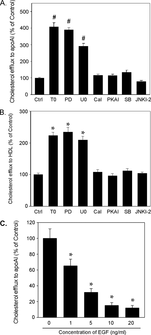
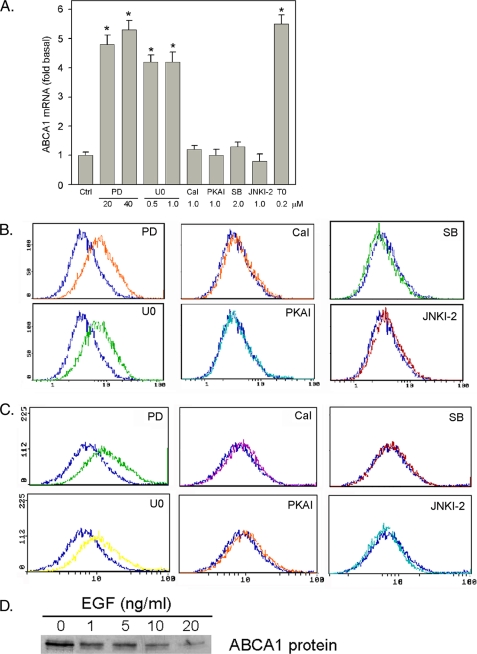
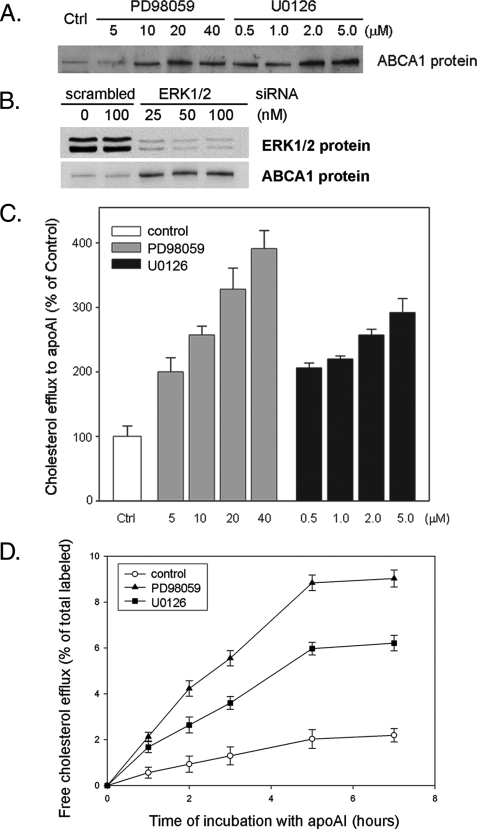
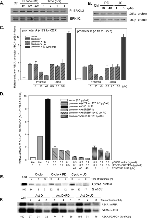
ATP-binding cassette transporter A1 (ABCA1), a molecule mediating free cholesterol efflux from peripheral tissues to apoAI and high density lipoprotein (HDL), inhibits the formation of lipid-laden macrophage/foam cells and the development of atherosclerosis. ERK1/2 are important signaling molecules regulating cellular growth and differentiation. The ERK1/2 signaling pathway is implicated in cardiac development and hypertrophy. However, the role of ERK1/2 in the development of atherosclerosis, particularly in macrophage cholesterol homeostasis, is unknown. In this study, we investigated the effects of ERK1/2 activity on macrophage ABCA1 expression and cholesterol efflux. Compared with a minor effect by inhibition of other kinases, inhibition of ERK1/2 significantly increased macrophage cholesterol efflux to apoAI and HDL. In contrast, activation of ERK1/2 reduced macrophage cholesterol efflux and ABCA1 expression. The increased cholesterol efflux by ERK1/2 inhibitors was associated with the increased ABCA1 levels and the binding of apoAI to cells. The increased ABCA1 by ERK1/2 inhibitors was due to increased ABCA1 mRNA and protein stability. The induction of ABCA1 expression and cholesterol efflux by ERK1/2 inhibitors was concentration-dependent. The mechanism study indicated that activation of liver X receptor (LXR) had little effect on ERK1/2 expression and activation. ERK1/2 inhibitors had no effect on macrophage LXRalpha/beta expression, whereas they did not influence the activation or the inhibition of the ABCA1 promoter by LXR or sterol regulatory element-binding protein (SREBP). However, inhibition of ERK1/2 and activation of LXR synergistically induced macrophage cholesterol efflux and ABCA1 expression. Our data suggest that ERK1/2 activity can play an important role in macrophage cholesterol trafficking.


Figures

FIGURE 1.
FIGURE 1.
Regulation of macrophage free cholesterol efflux by ERK1/2 activity. A and B, inhibition of ERK1/2 increases macrophage free cholesterol efflux to apoAI and HDL, respectively. RAW macrophages in 12-well plates were pre-labeled as described under “Experimental Procedures” and then received treatment overnight. Free cholesterol efflux was performed by incubating the treated cells in serum-free medium containing apoAI (10 μg/ml) (A) or HDL (15 μg/ml) (B) for 5 h at 37 °C. Radioactivity in medium was determined and normalized by cellular protein content. Treatments were as follows: ERK1/2 inhibitors, PD98059 (PD, 40 μm) and U0126 (2 μm); protein kinase C inhibitor, calphostin C (Cal, 1 μm); PKA inhibitor, myristoylated PKA inhibitor amide 14–22 (PKAI, 1 μm); p38 MAPK inhibitor, SB203580 (SB, 2 μm), and JNKs inhibitor, JNK inhibitor II (JNKI-2, 2 μm). LXR ligand, T0901317 (T0, 200 nm), was used as a positive control. # and *, significantly different from the corresponding controls at p < 0.05 by Student's t test (n = 4). C, EGF decreases macrophage free cholesterol efflux to apoAI. Pre-labeled RAW cells were treated with EGF at the indicated concentrations overnight followed by determination of free cholesterol efflux to apoAI (10 μg/ml). *, significantly different from control at p < 0.05 by Student's t test (n = 4).
FIGURE 2.
FIGURE 2.
Inhibition of ERK1/2 induces macrophage ABCA1 expression and the binding of apoAI to cells. A, RAW macrophages in serum-free medium were treated with inhibitors for various kinases as indicated overnight. LXR ligand, T0901317, was used as a positive control. Total cellular RNA was extracted and used to determine expression of ABCA1 mRNA by quantitative real-time PCR as described under “Experimental Procedures.” *, significantly different from control at p < 0.05 by Student t test (n = 4). B, RAW cells in serum-free medium were treated with inhibitors for various kinases (40 μm PD98059 (PD), 1 μm U0126 (U0), 1 μm calphostin C (Cal), 1 μm PKA inhibitor (PKAI), 2 μm SB203580 (SB), and 2 μm JNK inhibitor II (JNKI-2)) overnight. After washing with PBS, surface ABCA1 protein levels were determined for cells by FACS assay as described under “Experimental Procedures.” Blue line, control cells. C, macrophages received same treatments as in Fig. 2B. The binding of apoAI to treated cells was completed as described under “Experimental Procedures” and assessed by FACS assay. Blue line, control cells. D, RAW macrophages were treated with EGF at the indicated concentrations overnight. Expression of ABCA1 protein was determined by Western blot as described under “Experimental Procedures.”
FIGURE 3.
FIGURE 3.
ERK1/2 inhibitors induce macrophage ABCA1 expression and free cholesterol efflux in a concentration- and time-dependent manner. A, peritoneal macrophages were isolated from thioglycolate-elicited wild-type mice and cultured in complete RMPI medium for 2 days. Cells were then treated with ERK1/2 inhibitors, PD98059 and U0126, at the indicated concentrations in serum-free medium overnight. Total cellular proteins were extracted and used to determine ABCA1 protein expression by Western blot as described under “Experimental Procedures.” B, RAW macrophages in a 6-well plate were transfected with scrambled siRNA or the mixed siRNA against ERK1 and ERK2 (equal amount of each) at the indicated concentrations for 24 h as described under “Experimental Procedures.” Cells were used to extract total protein and determined the expression of ERK1/2 and ABCA1 by Western blot. C, pre-labeled RAW macrophages were treated with ERK1/2 inhibitors at the indicated concentrations overnight followed by determination of free cholesterol efflux to apoAI (10 μg/ml, 5 h) as described under “Experimental Procedures.” D, pre-labeled RAW macrophages were treated with PD98059 (40 μm) and U0126 (2 μm) overnight. After washing with PBS, cells were incubated in serum-free medium containing 10 μg/ml apoAI for indicated times, and the radioactivity in medium was determined. Free cholesterol efflux was calculated as the percentage of total labeled free cholesterol.
FIGURE 4.
FIGURE 4.
Mechanisms by which ERK1/2 inhibitors induce macrophage ABCA1 expression. A, LXR ligand has no effect on ERK1/2 expression and phosphorylation. Macrophages in serum-free medium were treated with T0901317 at the indicated concentrations for 2 h or with 200 nm T0901317 for indicated times. Levels of total and phosphorylated ERK1/2 (Pi-ERK1/2) were determined by Western blot. B, ERK1/2 inhibitors do not affect LXR expression. Macrophages were treated with ERK1/2 inhibitors, PD98059 and U0126, at the indicated concentrations overnight. Nuclear proteins were extracted and used to determine expression of LXRα and LXRβ proteins by Western blot as described under “Experimental Procedures.” C, the effects of ERK1/2 inhibitors on ABCA1 promoter activity. 293 cells in 24-well plates were transfected with ABCA1 promoter DNA (0.2 μg/well) and Renilla luciferase DNA as described under “Experimental Procedures” and received the indicated treatment overnight. Activity of firefly or Renilla luciferase in cellular lysate was determined by using the Dual-Luciferase Reporter Assay System (n = 4). D, the effects of ERK1/2 inhibitors on SREBP1a-inhibited ABCA1 promoter activity. 293 cells in 24-well plates were transfected with DNA for ABCA1 promoter and nSREBP1a at the indicated concentrations. Cells were then treated with ERK1/2 inhibitors at the indicated concentrations overnight. Activity of ABCA1 promoter was determined as described above. E, ERK1/2 inhibitors increase macrophage ABCA1 protein stability. Macrophages were treated with 5 μm cycloheximide (Cycle) or cycloheximide plus 40 μm PD98059 (PD) or 1 μm U0126 (U0) for the indicated times. Total cellular proteins were extracted and used to determine ABCA1 protein by Western blot as described under “Experimental Procedures.” F, ERK1/2 inhibitors increase macrophage ABCA1 mRNA stability. Macrophages were treated with 2 μm actinomycin D (Act D) or actinomycin D plus 40 μm PD98059 (PD) or 1 μm U0126 (U0) for the indicated times. Total cellular RNA was extracted and used to determine ABCA1 mRNA levels by Northern blot as described under “Experimental Procedures.” The same blot was hybridized with 32P-labeled glyceraldehyde-3-phosphate dehydrogenase (GAPDH) probe.
FIGURE 5.
FIGURE 5.
ERK1/2 inhibitors and LXR ligand synergistically induce macrophage free cholesterol efflux and ABCA1 expression. A, pre-labeled macrophages were treated with ERK1/2 inhibitor (20 μm PD98059 or 2 μm U0126), or LXR ligand (T0901317), or ERK1/2 inhibitor plus T0901317 at the indicated concentrations overnight. Free cholesterol efflux from macrophages was conducted by incubating treated cells in serum-free medium containing 10 μg/ml apoAI for 5 h at 37 °C. Radioactivity in medium was determined and normalized by protein content. B, pre-labeled macrophages were treated with 50 nm T0901317, or ERK1/2 inhibitor (PD98059 or U0126) at the indicated concentrations, or the combination of 50 nm T0901317 with an ERK1/2 inhibitor at the indicated concentrations overnight followed by determination of cholesterol efflux to apoAI (10 μg/ml). C, macrophages in serum-free medium were treated with PD98059 (20 μm) or U0126 (2 μm) or T0901713 at the indicated concentrations or the combination of PD98059 or U0126 with T0901317 at the indicated concentrations overnight. Changes in ABCA1 protein were determined by Western blot as described under “Experimental Procedures.” D, macrophages in serum-free medium were treated with 5 nm T0901317, or PD98059 or U0126 at the indicated concentrations, or the combination of 5 nm T0901317 with PD98059 or U0126 at the indicated concentrations overnight. Expression of ABCA1 protein in response to treatment was determined by Western blot as described under “Experimental Procedures.”

Similar articles

Cited by

References

    1. Arora S., Nicholls S. J. (2008) Drugs Today 44, 711–718 - PubMed
    1. de Villiers W. J., Smart E. J. (1999) J. Leukocyte Biol. 66, 740–746 - PubMed
    1. Febbraio M., Podrez E. A., Smith J. D., Hajjar D. P., Hazen S. L., Hoff H. F., Sharma K., Silverstein R. L. (2000) J. Clin. Invest. 105, 1049–1056 - PMC - PubMed
    1. Suzuki H., Kurihara Y., Takeya M., Kamada N., Kataoka M., Jishage K., Ueda O., Sakaguchi H., Higashi T., Suzuki T., Takashima Y., Kawabe Y., Cynshi O., Wada Y., Honda M., Kurihara H., Aburatani H., Doi T., Matsumoto A., Azuma S., Noda T., Toyoda Y., Itakura H., Yazaki Y., Kodama T. (1997) Nature 386, 292–296 - PubMed
    1. Braun A., Trigatti B. L., Post M. J., Sato K., Simons M., Edelberg J. M., Rosenberg R. D., Schrenzel M., Krieger M. (2002) Circ. Res. 90, 270–276 - PubMed

Publication types

MeSH terms