. 2010 Feb;13(2):253-60.
doi: 10.1038/nn.2466. Epub 2009 Dec 27.

The descending corticocollicular pathway mediates learning-induced auditory plasticity

Affiliations

The descending corticocollicular pathway mediates learning-induced auditory plasticity

Victoria M Bajo et al. Nat Neurosci. 2010 Feb.

Abstract

Descending projections from sensory areas of the cerebral cortex are among the largest pathways in the brain, suggesting that they are important for subcortical processing. Although corticofugal inputs have been shown to modulate neuronal responses in the thalamus and midbrain, the behavioral importance of these changes remains unknown. In the auditory system, one of the major descending pathways is from cortical layer V pyramidal cells to the inferior colliculus in the midbrain. We examined the role of these neurons in experience-dependent recalibration of sound localization in adult ferrets by selectively killing the neurons using chromophore-targeted laser photolysis. When provided with appropriate training, animals normally relearn to localize sound accurately after altering the spatial cues available by reversibly occluding one ear. However, this ability was lost after eliminating corticocollicular neurons, whereas normal sound-localization accuracy was unaffected. The integrity of this descending pathway is therefore critical for learning-induced localization plasticity.


Figures
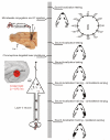
Figure 1
Figure 1. Experimental design
The vertical gray bar represents the chronological order with behavioral measurements on the right and surgical procedures on the left. After obtaining baseline data from two blocks of testing on a 12-speaker sound-localization task, we gave the ferrets multiple injections of fluorescent microspheres conjugated with chlorine e6 monoethylene diamine disodium (chlorine e6) in the left inferior colliculus. Two more blocks of behavioral testing were followed by ablation of retrograde-labeled layer V corticocollicular neurons by illumination of the ipsilateral auditory cortex with near-infrared light. After re-testing the ability of the animals to localize sound, we examined their capacity to relearn to localize sound after altering the spatial cues available. This was done by providing sound localization training over a 2-week period while the ferrets wore a unilateral earplug, first in the right ear, contralateral to the corticocollicular pathway lesion, then again in the right ear, and finally in the left ear. A, anterior; AAF, anterior auditory field; A1, primary auditory cortex; IC, inferior colliculus; M, medial. Calibration bar represents 5 mm.
Figure 2
Figure 2. Effect of unilateral auditory corticocollicular lesions on sound-localization accuracy
(a,b,c) Percentage correct scores at each of the 12 loudspeakers positioned at equal intervals in the horizontal plane. 0° is directly in front of the ferret and negative speaker angles denote stimulus locations on the ferret’s left. Accuracy is shown at three different stimulus sound durations, 1,000 ms (a), 200 ms (b) and 40 ms (c). (d) Cumulative response time in the auditory-localization approach-to-target task, subdivided by whether the ferrets approached the correct reward spout (solid lines) or not (dotted lines). (e,f) The initial sound-evoked head-orienting responses were recorded in the same trials, from which we derived the mean final head bearing for each stimulus location (e) and the latency of those movements (f). The gray bands correspond to 1 s.d. on either side of the mean values achieved by control ferrets and the different colors represent the mean values for the ferrets with corticocollicular lesions before and after each stage of the chromophore-targeted laser photolysis procedure. AC, auditory cortex.
Figure 3
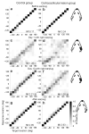
Figure 3. Effect of monaural occlusion on auditory-localization accuracy by the control ferrets (left column) and the ferrets with left auditory corticocollicular lesions (right column)
Each plot shows the distribution of the conditional probabilities of the response. (a,b) Data from the last session before inserting an earplug. (c,d) Data from the first day with an earplug in the right ear. (e,f) Data from the last (tenth) day with an earplug in the right ear. (g,h) Data from the first session after the plug was removed. Stimulus (target) location is plotted along the abscissa and response location along the ordinate of each panel, with negative numbers representing the left hemifield. Gray scale represents the conditional probability of the response location selected by the ferrets for each target location. The mutual information (MI) in bits between response and stimulus locations is given in each panel.
Figure 4
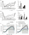
Figure 4. Effect of occluding the right ear on sound-localization accuracy
(a) Percentage correct scores (averaged across all speaker locations) in the session before the right ear was plugged (Preplug), on each of the 10 d over which the plug was worn (day 1–10) and in the session following its removal (Postplug). Data from control animals are shown in gray and from the ferrets with corticocollicular lesions in black; the symbols represent different animals and the lines show the mean scores. The mean (± s.d.) scores on the first (D1) and tenth (D10) day of monaural occlusion are shown for each group on the right. (b) Percentage correct scores for the left and right speaker locations are plotted separately, as indicated. Prior to insertion of the earplug, all of the ferrets scored ≥90% correct, but performed poorly when the right ear was first plugged. Daily training with the earplug in place led to a recovery in localization accuracy, except on the right side of space for the ferrets with corticocollicular lesions. *P < 0.05, **P < 0.01. (c) Accuracy of the head-orienting responses made by the same ferrets at the start of each trial, shown by plotting final head bearing against target location, with negative numbers indicating locations on the ferrets’ left. Mean (± s.d.) preplug and postplug data are shown in black, whereas data averaged over the first three (D1–3) and last two (D9–10) days are shown by the green and blue lines, respectively. The gray background indicates the side on which the earplug was worn. The corresponding mutual information value between target location and final head bearing is shown in each panel. Note the lack of recovery in the accuracy of acoustic orientation behavior in the ferrets with corticocollicular lesions.
Figure 5
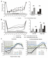
Figure 5. Effect of occluding the left ear on sound-localization accuracy
(a) Percentage correct scores (averaged across all speaker locations) in the session before the left ear was plugged (Preplug), on each of the 10 d over which the plug was worn (day 1-10) and in the session following its removal (Postplug). Data from control ferrets are shown in gray and from the ferrets with corticocollicular lesions in black. (b) Percentage correct scores for the left and right speaker locations are plotted separately, as indicated. (c) Accuracy of the head-orienting responses made by the same ferrets, shown by plotting final head bearing against target location. Data are otherwise presented as described in Figure 4. The previous experience of wearing an earplug in the right ear resulted in a larger deficit when the left earplug was first inserted. As with occlusion of the right ear, more complete recovery was seen in the control ferrets than in the ferrets with corticocollicular lesions. *P < 0.05, **P < 0.01.
Figure 6
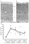
Figure 6. Reduction in density of layer V neurons in the primary auditory cortex in the corticocollicular lesion group
(a,b) We used antibody to NeuN as a neuronal marker and found that fewer large pyramidal cells (arrows) were present in layer V of the MEG in the left (lesioned) hemisphere (a) than in the right hemisphere (b). Calibration bars represent 0.1 mm. (c) Using the optical fractionator as a stereological estimator of the number of neurons in each layer of the MEG, we calculated and plotted the neuronal density across cortical layers in each of the ferrets that received corticocollicular lesions. The symbols indicate different ferrets and the lines are the mean values, plotted in black for the right MEG and in gray for the left (lesioned) side. The density of neurons in layer V was lower in the left MEG than in the right (t2 = 4.483, *P < 0.05), with no significant differences between the two hemispheres in other layers. LC, left cortex; RC, right cortex.
Figure 7
Figure 7. Reduction in the number of layer V pyramidal neurons in the primary auditory cortex following corticocollicular lesions
(a-d) Staining with the SMI32 antibody, a marker of layer III and layer V pyramidal cortical neurons, was sparser on the left (lesioned) side of the MEG, resulting in a less distinct bilaminar appearance (a) than on the right side (b), whereas no differences between the hemispheres were found in other parts of the auditory cortex, such as the adjacent anterior ectosylvian gyrus (AEG) (c,d). Calibration bar represents 0.1 mm. (e) The mean (± s.d.) number of SMI32-positive cells was plotted at different antero-posterior levels of the MEG, counting from the posterior corner of the suprasylvian sulcus (antero-posterior = 0). The number of SMI32-positive cells in layer V was significantly lower (P < 0.01) in the left MEG than on the right side, whereas no differences were found for layer III. The inset indicates the antero-posterior location of the sections used for quantifying SMI32-positive staining. The sparser staining in layer III of the left MEG in a was a result of there being fewer apical dendrites extending from layer V neurons. PEG, posterior ectosylvian gyrus.
Figure 8
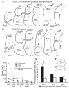
Figure 8. (a,b) Two examples of the distribution of retrograde-labeled fluorescent cells in the left auditory cortex for a ferret from the corticocollicular lesion group (a) and one from the control group (b)
Coronal sections of the ectosylvian gyrus were arranged from anterior to posterior and numbered starting from its posterior extent (e). Calibration bar represents 1 mm. (c) Density of retrograde-labeled fluorescent cells plotted for the different regions of the auditory cortex (MEG, PEG and AEG) in each hemisphere. Three ferrets with left corticocollicular lesions (filled symbols and black line showing the average density) were compared with the control case F0702 (open symbols and gray line) in which beads were injected in the left inferior colliculus, but the cortex was not exposed to the laser. (d) Mean (± s.d.) percentage of retrograde-labeled cells in the right hemisphere, contralateral to the inferior colliculus injection sites, for the different regions of the right auditory cortex. The significant increase (**P < 0.01) in this percentage in the MEG of the animals in the lesion group (black bars) indicates a substantial loss of left corticocollicular neurons compared with the controls (open bars). D, dorsal; P, posterior; pss, pseudosylvian sulcus; sss, suprasylvian sulcus.

Similar articles

Cited by

References

    1. Gilbert CD, Li W, Piech V. Perceptual learning and adult cortical plasticity. J. Physiol. 2009;587:2743–2751. - PMC - PubMed
    1. Dahmen JC, King AJ. Learning to hear: plasticity of auditory cortical processing. Curr. Opin. Neurobiol. 2007;17:456–464. - PubMed
    1. Edeline JM, Weinberger NM. Thalamic short-term plasticity in the auditory system: associative returning of receptive fields in the ventral medial geniculate body. Behav. Neurosci. 1991;105:618–639. - PubMed
    1. Tzounopoulos T, Kraus N. Learning to encode timing: mechanisms of plasticity in the auditory brainstem. Neuron. 2009;62:463–469. - PMC - PubMed
    1. Suga N, Ma X. Multiparametric corticofugal modulation and plasticity in the auditory system. Nat. Rev. Neurosci. 2003;4:783–794. - PubMed

Publication types