

doi: 10.1038/nmeth.1413.
Epub 2009 Dec 27.
Two-color, two-photon uncaging of glutamate and GABA
Affiliations
- PMID: 20037590
- PMCID: PMC4135702
- DOI: 10.1038/nmeth.1413
Item in Clipboard
Two-color, two-photon uncaging of glutamate and GABA
Nat Methods.
2010 Feb.
Abstract
We developed a caged GABA (gamma-aminobutyric acid), which, when combined with an appropriate caged glutamate, allows bimodal control of neuronal membrane potential with subcellular resolution using optically independent two-photon uncaging of each neurotransmitter. We used two-color, two-photon uncaging to fire and block action potentials from rat hippocampal CA1 neurons in brain slices with 720-nm and 830-nm light, respectively. Our method should be generalizable to other chemical messenger pairs.
Figures
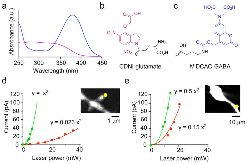
Absorption spectra of chromophores for two-color photostimulation. (a) UV-visible absorption spectra of CDNI-Glu (b) and N-DCAC-GABA (c). (d) Power dependence of the evoked current from uncaging CDNI-Glu (5 mM) at 720 nm (green squares) and at 830 nm (red squares) near a spine head (inset) on a CA1 pyramidal neuron in an acutely isolated brain slices from the hippocampus of a 13-day old rat. (e) Power dependence of the evoked current from uncaging N-DCAC-GABA (4 mM) at 720 nm (green squares) and at 830 nm (red squares) at the proximal dendrites (inset). The lines drawn through the points were fitted to the equation y = k×x2 in (d) and (e). The ratio of constants (k) revealed that CDNI-Glu and N-DCAC-GABA were 38 and 3.3 times, respectively, more photosensitive at 720 nm compared with 830 nm.
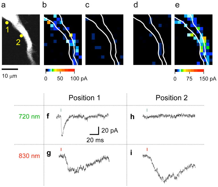
Optical independent two-color, 2P mapping of functional AMPA and GABA-A receptors on a CA1 pyramidal neuron. This experiment shows representative data from fice similar experiments in which CDNI-Glu (2 mM) and N-DCAC-GABA (4 mM) were co-applied to an acutely isolated brain slice from the rat hippocampus. The neuron was visualized by perfusion through a patch pipette with a CsCl-based solution containing Alexa-594. The proximal apical dendrite (a) was subjected to functional mapping by photolysis at 720 and 830 nm under voltage clamp by pseudo-random scanning of each pixel as shown in (b)–(e). For uncaging at 720 nm (b, c) 8.7 mW was applied for 1 ms (green bars in f and h), and at 830 nm (d, e) 20.6 mW for 2 ms (red bars in g and i). The evoked currents are represented on a pseudo-color scale: 0–100 pA for (b–d), and 0–150 pA for (e). Two time-windows for current integration [2–3 ms (b, d) and 30–35 ms (c, e)] after uncaging show the AMPA-receptor and GABA-A receptors are activated with optical independence at 720 nm and 830 nm, respectively. (f–i) Examples of current traces. The current trace from pixel 1 in (a) shows an AMPA current at 720 nm (f) but not 830 nm, and a GABA current at 830 nm but not 720 nm (g) whereas pixel 2 in (a) shows only a GABA current at 830 nm (i), and no current evoked by irradiation at 720 nm (h), showing only 830 nm light uncaged GABA. The membrane potential was held at −70 mV.
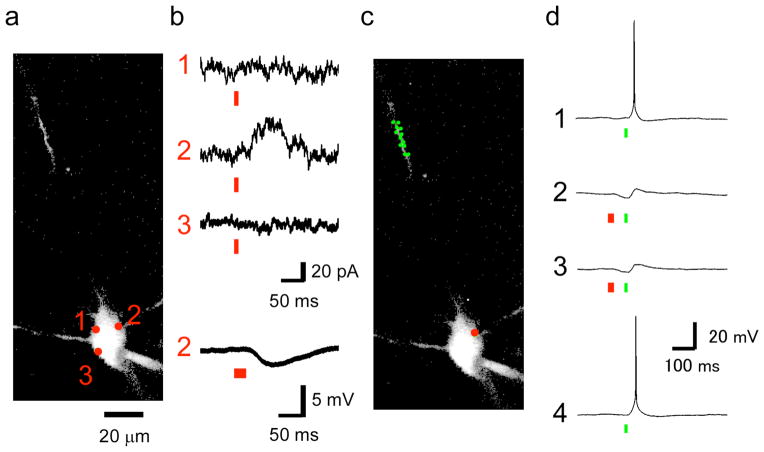
Two-color, 2P uncaging of Glu and GABA fired and blocked action potentials. This figure shows representative data from seven similar experiments when CDNI-Glu (2 mM) and N-DCAC-GABA (4 mM) were co-applied to an acutely isolated brain slice from the rat hippocampus. (a) The neuron was visualized by perfusion through a patch pipette with a K-gluconate-based solution containing Alexa-594, and 2P imaging was performed at 900 nm. A two-dimensional projection of a three-dimensional z-stack is shown (the proximal dendrite was outside of the imaging volume). (b) Functional mapping of currents in voltage clamp of the soma at −20 mV. Two-photon photolysis at 830 nm and 55 mW for 9 ms (red bars) revealed a GABA-A receptor hotspot at position 2. The lowest trace indicates 2pIPSP induced by sequential uncaging of three points adjacent to the position 2 (red bars, 27 ms in total). (c) The somatic position of uncaging at 830 nm (red circle) and dendritic twelve positions (green circles) for uncaging at 720 nm are shown. (d) Four consecutive voltage traces where elicited with an interval of 5 s during the current clamp experiments in which membrane potential was held at −48 mV. Action potentials were elicited by sequential uncaging at 720 nm of the twelve positions in the dendrites (c) each with the laser power of 9 mW for 1 ms (green bars, 12 ms in total). 2pIPSCs were induced as shown in (b). Action potential was suppressed when 2pIPSP was induced.
References
Publication types
MeSH terms
Substances
Grants and funding
LinkOut - more resources
Full Text Sources
Other Literature Sources
Miscellaneous
