

Anatomy and physical examination of the knee menisci: a narrative review of the orthopedic literature
- PMID: 20037697
- PMCID: PMC2796951
Anatomy and physical examination of the knee menisci: a narrative review of the orthopedic literature
Abstract
Objective: The objective of this study was to review the physical examination tests available to a practitioner in order to arrive at a clinical diagnosis or suspicion of a meniscal lesion.
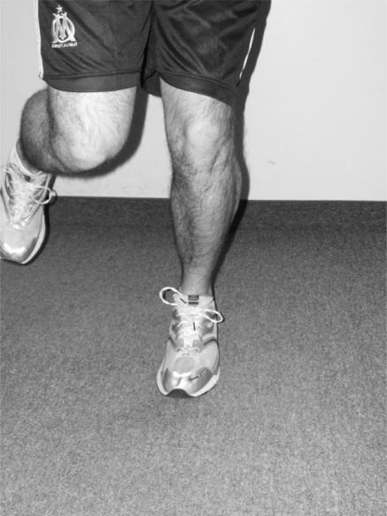
Background: The menisci transmit weight bearing forces and increase stability of the knee. The menisci also facilitate nutrition, provide lubrication and shock absorption for the articular cartilage and promote knee proprioception. The combinations of torsional and axial loading appear to be the cause of most meniscal injuries. Diagnosis of acute knee injuries has long been a topic for discussion throughout the orthopedic literature. Many clinical tests and diagnostic studies have been developed to increase the clinician's ability to accurately diagnose these types of disorders of the knee.
Conclusion: The accuracy of all diagnostic tests is thought to be dependant upon the skill of the examiner, and the severity and location of the injury. The multitude of tests described to assess meniscal lesions suggests that none are consistently reliable. However, recent research has focused on a composite score to accurately predict meniscus lesions. The combination of a comprehensive history, multiple physical tests and diagnostic imaging for confirmation is typical for a clinical meniscal lesion diagnosis while the gold standard remains the arthroscopic procedure itself.
Objectif :: L’objectif de cette étude était de passer en revue les examens physiques que peut faire passer un praticien pour en arriver à un diagnostic clinique ou un soupçon de lésion méniscale.
Contexte :: Le ménisque transmet les forces de charge et accroît la stabilité du genou. Le ménisque facilite également la nutrition et fournit de la lubrification et une absorption des chocs pour le cartilage articulaire, en plus de promouvoir la proprioception du genou. Les combinaisons de charge de torsion et axiale semblent être la cause de la plupart des blessures méniscales. Le diagnostic d’une blessure aiguë au genou est depuis longtemps un sujet de discussion dans la littérature orthopédique. Bon nombre de tests cliniques et d’études diagnostiques ont été créés pour accroître la capacité d’un clinicien à diagnostiquer avec exactitude ces types de troubles du genou.
Conclusion :: On croit que l’exactitude des tests diagnostiques dépend de l’aptitude de l’examinateur, ainsi que de la gravité et l’endroit de la blessure. La multitude de tests décrits pour évaluer les lésions méniscales suggère qu’il n’en existe pas qui soit fiable de façon constante. Cependant, la recherche récente a mis l’accent sur un résultat combiné pour prédire avec exactitude les lésions méniscales. La combinaison d’antécédents détaillés, de nombreux tests physiques et d’imagerie diagnostique aux fins de confirmation est typique pour le diagnostic clinique d’une lésion méniscale, bien que la norme d’excellence reste la technique arthroscopique.
Keywords: knee; meniscus; orthopedic; tests.
Figures
References
-
- Levangie P, Norkin C. Joint Structure and Function. FA Davis Company; 2001.
-
- Lee J, Fu F. The meniscus: basic science and clinical applications. Operative Techniques in Orthopaedics. 2000;10:162–168.
-
- Lento P, Akuthota V. Meniscal injuries: a critical review. J Back Musculoskel Rehabil. 2000;15:55–62. - PubMed
LinkOut - more resources
Full Text Sources