

Improved method for in vitro secondary amastigogenesis of Trypanosoma cruzi: morphometrical and molecular analysis of intermediate developmental forms
- PMID: 20037731
- PMCID: PMC2796335
- DOI: 10.1155/2010/283842
Improved method for in vitro secondary amastigogenesis of Trypanosoma cruzi: morphometrical and molecular analysis of intermediate developmental forms
Abstract
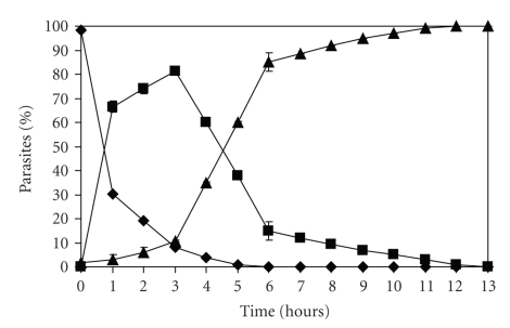
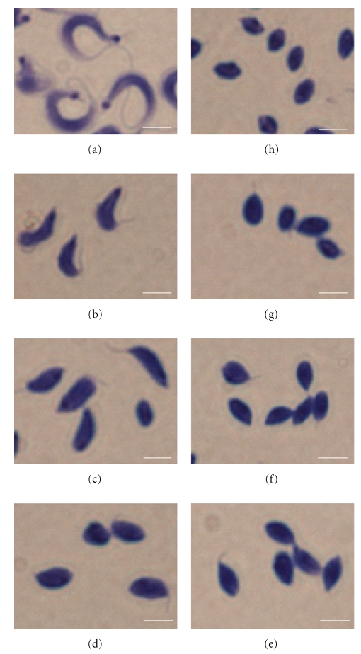
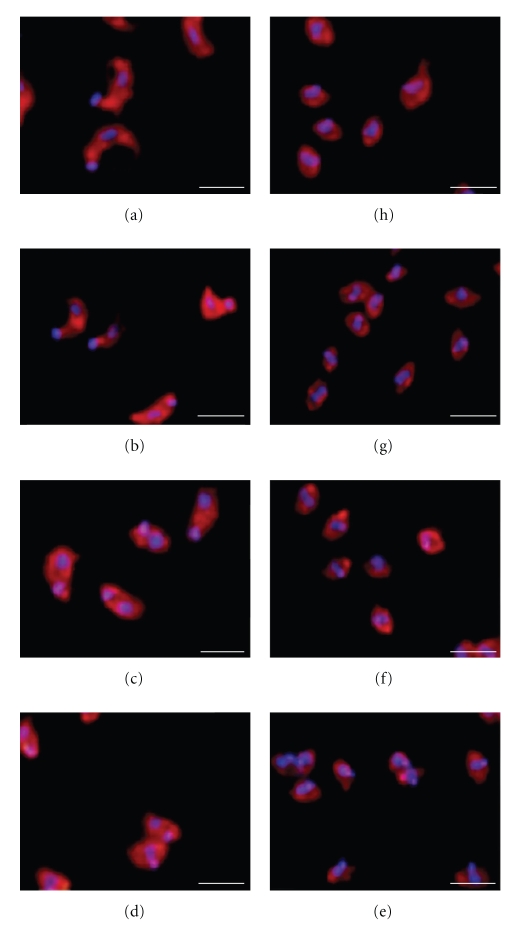
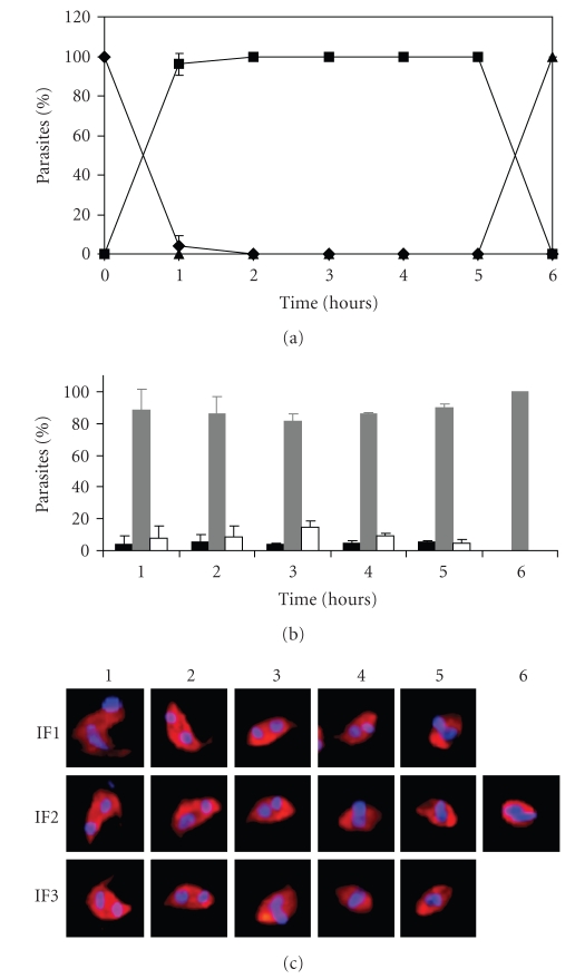
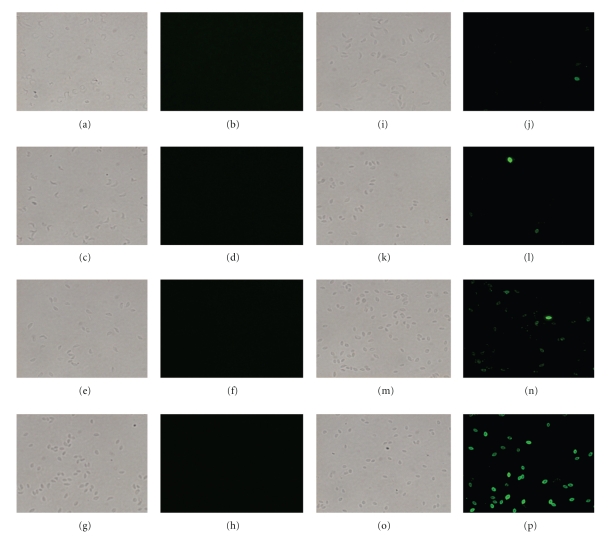
Trypanosoma cruzi undergoes a biphasic life cycle that consists of four alternate developmental stages. In vitro conditions to obtain a synchronic transformation and efficient rates of pure intermediate forms (IFs), which are indispensable for further biochemical, biological, and molecular studies, have not been reported. In the present study, we established an improved method to obtain IFs from secondary amastigogenesis. During the transformation kinetics, we observed progressive decreases in the size of the parasite body, undulating membrane and flagellum that were concomitant with nucleus remodeling and kinetoplast displacement. In addition, a gradual reduction in parasite movement and acquisition of the amastigote-specific Ssp4 antigen were observed. Therefore, our results showed that the in vitro conditions used obtained large quantities of highly synchronous and pure IFs that were clearly distinguished by morphometrical and molecular analyses. Obtaining these IFs represents the first step towards an understanding of the molecular mechanisms involved in amastigogenesis.
Figures
Similar articles
-
Morphological comparison of axenic amastigogenesis of trypomastigotes and metacyclic forms of Trypanosoma cruzi.Mem Inst Oswaldo Cruz. 2003 Jan;98(1):83-91. doi: 10.1590/s0074-02762003000100012. Epub 2003 Apr 9. Mem Inst Oswaldo Cruz. 2003. PMID: 12700866
-
Quantitative proteomic and phosphoproteomic analysis of Trypanosoma cruzi amastigogenesis.Mol Cell Proteomics. 2014 Dec;13(12):3457-72. doi: 10.1074/mcp.M114.040329. Epub 2014 Sep 15. Mol Cell Proteomics. 2014. PMID: 25225356 Free PMC article.
-
Exoproteome profiling of Trypanosoma cruzi during amastigogenesis early stages.PLoS One. 2019 Nov 22;14(11):e0225386. doi: 10.1371/journal.pone.0225386. eCollection 2019. PLoS One. 2019. PMID: 31756194 Free PMC article.
-
The life cycle of Trypanosoma cruzi revisited.Int J Parasitol. 2001 May 1;31(5-6):472-81. doi: 10.1016/s0020-7519(01)00153-9. Int J Parasitol. 2001. PMID: 11334932 Review.
-
Basic cell biology of Trypanosoma cruzi.Curr Pharm Des. 2002;8(4):269-85. doi: 10.2174/1381612023396276. Curr Pharm Des. 2002. PMID: 11860366 Review.
Cited by
-
The Kinetoplastid-Specific Protein TcCAL1 Plays Different Roles During In Vitro Differentiation and Host-Cell Invasion in Trypanosoma cruzi.Front Cell Infect Microbiol. 2022 Jun 30;12:901880. doi: 10.3389/fcimb.2022.901880. eCollection 2022. Front Cell Infect Microbiol. 2022. PMID: 35846750 Free PMC article.
-
The Potent Trypanocidal Effect of LQB303, a Novel Redox-Active Phenyl-Tert-Butyl-Nitrone Derivate That Causes Mitochondrial Collapse in Trypanosoma cruzi.Front Microbiol. 2021 Apr 15;12:617504. doi: 10.3389/fmicb.2021.617504. eCollection 2021. Front Microbiol. 2021. PMID: 33935988 Free PMC article.
-
Differential Expression of Matrix Metalloproteinases 2, 9 and Cytokines by Neutrophils and Monocytes in the Clinical Forms of Chagas Disease.PLoS Negl Trop Dis. 2017 Jan 24;11(1):e0005284. doi: 10.1371/journal.pntd.0005284. eCollection 2017 Jan. PLoS Negl Trop Dis. 2017. PMID: 28118356 Free PMC article.
-
Metacyclogenesis defects and gene expression hallmarks of histone deacetylase 4-deficient Trypanosoma cruzi cells.Sci Rep. 2021 Nov 4;11(1):21671. doi: 10.1038/s41598-021-01080-1. Sci Rep. 2021. PMID: 34737385 Free PMC article.
-
In vitro characterization of Trypanosoma cruzi infection dynamics in skeletal and cardiac myotubes models suggests a potential cell-to-cell transmission in mediating cardiac pathology.PLoS Negl Trop Dis. 2024 Jun 24;18(6):e0012288. doi: 10.1371/journal.pntd.0012288. eCollection 2024 Jun. PLoS Negl Trop Dis. 2024. PMID: 38913744 Free PMC article.
References
-
- Almeida-De-Faria M, Freymuller E, Colli W, Alves MJ. Trypanosoma cruzi: characterization of an intracellular epimastigote-like form. Experimental Parasitology. 1999;92(4):263–274. - PubMed
-
- Tyler KM, Engman DM. The life cycle of Trypanosoma cruzi revisited. International Journal for Parasitology. 2001;31(5-6):472–481. - PubMed
-
- Andrews NW, Hong KS, Robbins ES, Nussenzweig V. Stage-specific surface antigens expressed during the morphogenesis of vertebrate forms of Trypanosoma cruzi. Experimental Parasitology. 1987;64(3):474–484. - PubMed
-
- Avila AR, Dallagiovanna B, Yamada-Ogatta SF, et al. Stage-specific gene expression during Trypanosoma cruzi metacyclogenesis. Genetics and Molecular Research. 2003;2(1):159–168. - PubMed
-
- Navarro MC, De Lima AR, Askue J, Contreras VT. Morphological comparison of axenic amastigogenesis of trypomastigotes and metacyclic forms of Trypanosoma cruzi. Memórias do Instituto Oswaldo Cruz. 2003;98(1):83–91. - PubMed
Publication types
MeSH terms
Substances
LinkOut - more resources
Full Text Sources
