. 2009:2009:789526.
doi: 10.1155/2009/789526. Epub 2009 Dec 16.

High harvest yield, high expansion, and phenotype stability of CD146 mesenchymal stromal cells from whole primitive human umbilical cord tissue

Affiliations

High harvest yield, high expansion, and phenotype stability of CD146 mesenchymal stromal cells from whole primitive human umbilical cord tissue

Rebecca C Schugar et al. J Biomed Biotechnol. 2009.

Abstract

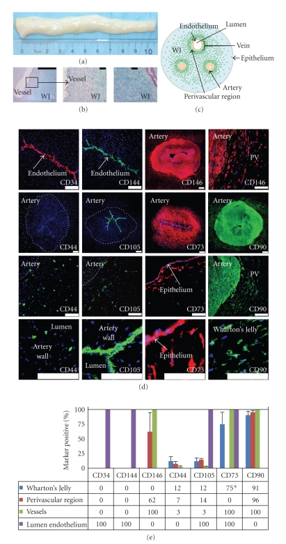
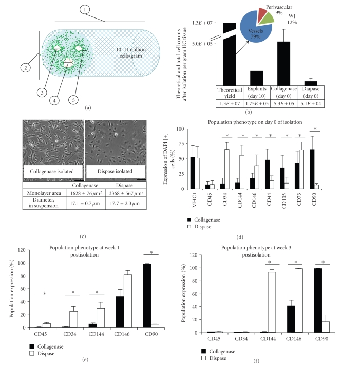
Human umbilical cord blood is an excellent primitive source of noncontroversial stem cells for treatment of hematologic disorders; meanwhile, new stem cell candidates in the umbilical cord (UC) tissue could provide therapeutic cells for nonhematologic disorders. We show novel in situ characterization to identify and localize a panel of some markers expressed by mesenchymal stromal cells (MSCs; CD44, CD105, CD73, CD90) and CD146 in the UC. We describe enzymatic isolation and purification methods of different UC cell populations that do not require manual separation of the vessels and stroma of the coiled, helical-like UC tissue. Unique quantitation of in situ cell frequency and stromal cell counts upon harvest illustrate the potential to obtain high numerical yields with these methods. UC stromal cells can differentiate to the osteogenic and chondrogenic lineages and, under specific culturing conditions, they exhibit high expandability with unique long-term stability of their phenotype. The remarkable stability of the phenotype represents a novel finding for human MSCs, from any source, and supports the use of these cells as highly accessible stromal cells for both basic studies and potentially therapeutic applications such as allogeneic clinical use for musculoskeletal disorders.


Figures

Figure 1
Figure 1
In situ examination of human umbilical cords. (a) UCs range in length up to 60 cm; shown is a cut 10 cm portion with UC blood removed. (b) Histology of 10 micron thick cryosections stained with Alcian blue. (c) Schematic of UC cross-section. Green dots represent cells; all other structures are as indicated. (d) Detection of CD34, CD144, CD146, CD44, CD105, CD73, and CD90. CD34 and CD144 are expressed only in the endothelial lumen of the UC vessels. CD146 expression is present in both the vascular and perivascular regions, but not in the bulk of the WJ. CD44- and CD105-positive cells are found in the WJ, perivascular region, and in the vessels. CD105 is also in the lumen. A low level of CD73 expression is found throughout the vessels and high in the region just beneath the epithelium (*75% positivity only in the region just below epithelium). CD90 positivity was detected in all regions, except the endothelium. Scale bars on whole vessel images represent 200 μm; all others represent 100 μm. (e) The percentage of cells expressing these markers was determined by immunostaining: (PV: perivascular region).
Figure 2
Figure 2
Tissue processing and method of isolation affect total cell yield and population phenotype. (a) Physical properties of the UC tissue provide an estimate that 10-11 million cells per gram can be isolated from the tissue (also see Table 1 and Table 1 in Supplementary Material available online at doi:10.1155/2009/789526). (b) The theoretical yield of 10-11 million cells/UC gram includes 79% vascular cells, 9% perivascular cells, and 12% WJ cells. Distinct phenotypes based on enzymatic method are evident by both morphology (c) and molecular phenotype (d)–(f). (c) Cell area or spreading in 2D monolayer is significantly larger for dispase-isolated cells versus collagenase-isolated cells (mean ± s.e.m., P < .001), although the cells have similar diameter or size when measured prior to surface adhesion. (d) Mean surface marker expression on day 0 of isolation. Collagenase-isolated cell populations have a significantly higher level of expression of CD44 (P = .005), CD105 (P = .014), and CD90 (P < .001), as compared to dispase-isolated cells that exhibit a significantly higher level of endothelial marker expression, CD34 (P < .001), CD144 (P < .001), and CD146 (P = .049), and CD73 (P < .001) which was also detected in the endothelium. (e) Marker expression at week 1 postisolation. Significant differences were detected for expression of CD45 (P = .01), CD34 (P < .001), CD144 (P = .006), and CD90 (P = .003). (f) Marker expression at week 3 postisolation. Significant differences were detected for expression of CD144 (P = .017), CD146 (P = .008), and CD90 (P = .001). The use of collagenase enzyme yields a cell population with significantly higher MSC marker expression levels in comparison to both dispase enzyme digestion and mechanical dissociation.The latter two methods, however, yield populations with significantly higher endothelial cell marker expression levels than the collagenase enzyme digest.
Figure 3
Figure 3
UCSCs demonstrate a high level of ex vivo expansion and retain MSC surface marker phenotype for up to 10 passages, or 5 weeks in culture. (a) UCSCs showed a significantly higher level of proliferation in EGM2 growth medium as compared to DMEM (72 hours, P = .038, 96 hours, P = .10). (b) Cells cultured in the different media were distinct morphologically; DMEM-cultured cells were significantly larger in 2D as compared to cells grown in EGM2 (mean ± s.e.m.). (c) We expanded UCSCs for more than 55 population doublings, PDs. Thirteen UCSC populations were expanded for 10 passages, and 3 of these UCSC populations were continued for another 10 passages. The theoretical expansion yield is 1020 cells from an initial 2 × 10⁴ cells. (d) Mean population doubling time of the UCSCs was 24 ± 1.1 hours for the populations at 0–10 PDs, 23 ± 1.7 hours at 10–20 PDs, which are significantly faster than the PDT of 28 ± 1.3 hours for UCSCs at 20–30 PDs (P < .05, bars represent mean ± s.e.m.). (e) We observed no significant changes in cellular division times which was similar for cells at 0–10 PDs (14.8 ± 0.4 hours), 10–20 PDs (15.1 ± 0.7 hours), and 20–30 PDs (15.9 ± 0.8 hours). (f) Histograms are representative of UCSCs surface marker expression 7 days postisolation. The population is homogeneously positive for mesenchymal cell markers CD44, CD105, CD73, and CD90, and ~50% positive for endothelial cell marker CD146, and negative for hematopoietic cell markers CD34 and CD45, and endothelial cell marker CD144. (g) Surface marker expression levels through 10 passages. UCSCs have steady but moderate CD146 expression levels and remain negative for CD45, CD34, and CD144 expression. The cells are consistently highly positive (≥97%) for all mesenchymal markers analyzed throughout the five-week period and support the reproducibility of the isolation method and stability of the MSC-like phenotype. (h) Representative PCR analysis of low-passage and high-passage UCScs confirmed the presence of mRNA transcripts for MSC genes. Lane 1: UCSCs at passage 2; Lane 2: UCSCs at passage 8; Lane 3: human vascular endothelial cells (HUVEC); Lane 4: no DNA.
Figure 3
Figure 3
UCSCs demonstrate a high level of ex vivo expansion and retain MSC surface marker phenotype for up to 10 passages, or 5 weeks in culture. (a) UCSCs showed a significantly higher level of proliferation in EGM2 growth medium as compared to DMEM (72 hours, P = .038, 96 hours, P = .10). (b) Cells cultured in the different media were distinct morphologically; DMEM-cultured cells were significantly larger in 2D as compared to cells grown in EGM2 (mean ± s.e.m.). (c) We expanded UCSCs for more than 55 population doublings, PDs. Thirteen UCSC populations were expanded for 10 passages, and 3 of these UCSC populations were continued for another 10 passages. The theoretical expansion yield is 1020 cells from an initial 2 × 10⁴ cells. (d) Mean population doubling time of the UCSCs was 24 ± 1.1 hours for the populations at 0–10 PDs, 23 ± 1.7 hours at 10–20 PDs, which are significantly faster than the PDT of 28 ± 1.3 hours for UCSCs at 20–30 PDs (P < .05, bars represent mean ± s.e.m.). (e) We observed no significant changes in cellular division times which was similar for cells at 0–10 PDs (14.8 ± 0.4 hours), 10–20 PDs (15.1 ± 0.7 hours), and 20–30 PDs (15.9 ± 0.8 hours). (f) Histograms are representative of UCSCs surface marker expression 7 days postisolation. The population is homogeneously positive for mesenchymal cell markers CD44, CD105, CD73, and CD90, and ~50% positive for endothelial cell marker CD146, and negative for hematopoietic cell markers CD34 and CD45, and endothelial cell marker CD144. (g) Surface marker expression levels through 10 passages. UCSCs have steady but moderate CD146 expression levels and remain negative for CD45, CD34, and CD144 expression. The cells are consistently highly positive (≥97%) for all mesenchymal markers analyzed throughout the five-week period and support the reproducibility of the isolation method and stability of the MSC-like phenotype. (h) Representative PCR analysis of low-passage and high-passage UCScs confirmed the presence of mRNA transcripts for MSC genes. Lane 1: UCSCs at passage 2; Lane 2: UCSCs at passage 8; Lane 3: human vascular endothelial cells (HUVEC); Lane 4: no DNA.

Similar articles

Cited by

References

    1. Gluckman E, Rocha V. History of the clinical use of umbilical cord blood hematopoietic cells. Cytotherapy. 2005;7(3):219–227. - PubMed
    1. Broxmeyer HE, Douglas GW, Hangoc G, et al. Human umbilical cord blood as a potential source of transplantable hematopoietic stem/progenitor cells. Proceedings of the National Academy of Sciences of the United States of America. 1989;86(10):3828–3832. - PMC - PubMed
    1. Kelly PF, Radtke S, von Kalle C, et al. Stem cell collection and gene transfer in fanconi anemia. Molecular Therapy. 2007;15(1):211–219. - PubMed
    1. Wagner JE, Rosenthal J, Sweetman R, et al. Successful transplantation of HLA-matched and HLA-mismatched umbilical cord blood from unrelated donors: analysis of engraftment and acute graft- versus-host disease. Blood. 1996;88(3):795–802. - PubMed
    1. Nauta AJ, Kruisselbrink AB, Lurvink E, et al. Enhanced engraftment of umbilical cord blood-derived stem cells in NOD/SCID mice by cotransplantation of a second unrelated cord blood unit. Experimental Hematology. 2005;33(10):1249–1256. - PubMed

Publication types