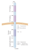
Targeting the epidermal growth factor receptor in epithelial ovarian cancer: current knowledge and future challenges
- PMID: 20037743
- PMCID: PMC2796463
- DOI: 10.1155/2010/568938
Targeting the epidermal growth factor receptor in epithelial ovarian cancer: current knowledge and future challenges
Abstract
The epidermal growth factor receptor is overexpressed in up to 60% of ovarian epithelial malignancies. EGFR regulates complex cellular events due to the large number of ligands, dimerization partners, and diverse signaling pathways engaged. In ovarian cancer, EGFR activation is associated with increased malignant tumor phenotype and poorer patient outcome. However, unlike some other EGFR-positive solid tumors, treatment of ovarian tumors with anti-EGFR agents has induced minimal response. While the amount of information regarding EGFR-mediated signaling is considerable, current data provides little insight for the lack of efficacy of anti-EGFR agents in ovarian cancer. More comprehensive, systematic, and well-defined approaches are needed to dissect the roles that EGFR plays in the complex signaling processes in ovarian cancer as well as to identify biomarkers that can accurately predict sensitivity toward EGFR-targeted therapeutic agents. This new knowledge could facilitate the development of rational combinatorial therapies to sensitize tumor cells toward EGFR-targeted therapies.
Figures


Similar articles
-
Challenges for the application of EGFR-targeting peptide GE11 in tumor diagnosis and treatment.J Control Release. 2022 Sep;349:592-605. doi: 10.1016/j.jconrel.2022.07.018. Epub 2022 Jul 22. J Control Release. 2022. PMID: 35872181 Review.
-
Implications of EGFR inhibition in ovarian cancer cell proliferation.Gynecol Oncol. 2008 Jun;109(3):411-7. doi: 10.1016/j.ygyno.2008.02.030. Gynecol Oncol. 2008. PMID: 18423824
-
Estrogen receptor-positive human epithelial ovarian carcinoma cells respond to the antitumor drug suramin with increased proliferation: possible insight into ER and epidermal growth factor signaling interactions in ovarian cancer.Gynecol Oncol. 2004 Sep;94(3):705-12. doi: 10.1016/j.ygyno.2004.05.059. Gynecol Oncol. 2004. PMID: 15350362
-
MET Gene Amplification and MET Receptor Activation Are Not Sufficient to Predict Efficacy of Combined MET and EGFR Inhibitors in EGFR TKI-Resistant NSCLC Cells.PLoS One. 2015 Nov 18;10(11):e0143333. doi: 10.1371/journal.pone.0143333. eCollection 2015. PLoS One. 2015. PMID: 26580964 Free PMC article.
-
Epidermal growth factor receptor dependence in human tumors: more than just expression?Oncologist. 2002;7 Suppl 4:31-9. doi: 10.1634/theoncologist.7-suppl_4-31. Oncologist. 2002. PMID: 12202786 Review.
Cited by
-
Combined effects of EGFR and hedgehog signaling blockade on inhibition of head and neck squamous cell carcinoma.Int J Clin Exp Pathol. 2017 Sep 1;10(9):9816-9828. eCollection 2017. Int J Clin Exp Pathol. 2017. PMID: 31966869 Free PMC article.
-
Aspirin Blocks EGF-stimulated Cell Viability in a COX-1 Dependent Manner in Ovarian Cancer Cells.J Cancer. 2013 Sep 27;4(8):671-8. doi: 10.7150/jca.7118. eCollection 2013. J Cancer. 2013. PMID: 24155779 Free PMC article.
-
In-silico docking analysis of bioactive compounds sourced from Punica granatum peel extract: Approaching a precision solution for ovarian cancer.J Ayurveda Integr Med. 2025 Jul-Aug;16(4):101125. doi: 10.1016/j.jaim.2025.101125. Epub 2025 Jul 2. J Ayurveda Integr Med. 2025. PMID: 40609197 Free PMC article.
-
Optimizing molecular-targeted therapies in ovarian cancer: the renewed surge of interest in ovarian cancer biomarkers and cell signaling pathways.J Oncol. 2012;2012:737981. doi: 10.1155/2012/737981. Epub 2012 Feb 6. J Oncol. 2012. PMID: 22481932 Free PMC article.
-
EGFR/HER-targeted therapeutics in ovarian cancer.Future Med Chem. 2012 Mar;4(4):447-69. doi: 10.4155/fmc.12.11. Future Med Chem. 2012. PMID: 22416774 Free PMC article. Review.
References
-
- Roh MH, Kindelberger D, Crum CP. Serous tubal intraepithelial carcinoma and the dominant ovarian mass: clues to serous tumor origin? American Journal of Surgical Pathology. 2009;33(3):376–383. - PubMed
-
- Feeley KM, Wells M. Precursor lesions of ovarian epithelial malignancy. Histopathology. 2001;38(2):87–95. - PubMed
-
- Dimova I, Zaharieva B, Raitcheva S, Dimitrov R, Doganov N, Toncheva D. Tissue microarray analysis of EGFR and erbB2 copy number changes in ovarian tumors. International Journal of Gynecological Cancer. 2006;16(1):145–151. - PubMed
-
- Lin C-K, Chao T-K, Yu C-P, Yu M-H, Jin J-S. The expression of six biomarkers in the four most common ovarian cancers: correlation with clinicopathological parameters. APMIS. 2009;117(3):162–175. - PubMed
-
- Brustmann H. Epidermal growth factor receptor expression in serous ovarian carcinoma: an immunohistochemical study with galectin-3 and cyclin D1 and outcome. International Journal of Gynecological Pathology. 2008;27(3):380–389. - PubMed
Grants and funding
LinkOut - more resources
Full Text Sources
Other Literature Sources
Research Materials
Miscellaneous

