

Catalysis and inhibition of Mycobacterium tuberculosis methionine aminopeptidase
- PMID: 20038112
- PMCID: PMC2820511
- DOI: 10.1021/jm901624n
Catalysis and inhibition of Mycobacterium tuberculosis methionine aminopeptidase
Abstract
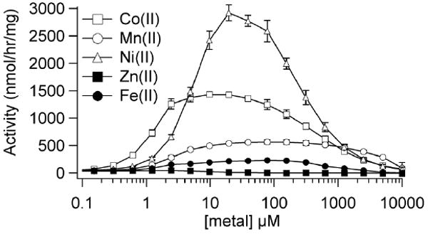
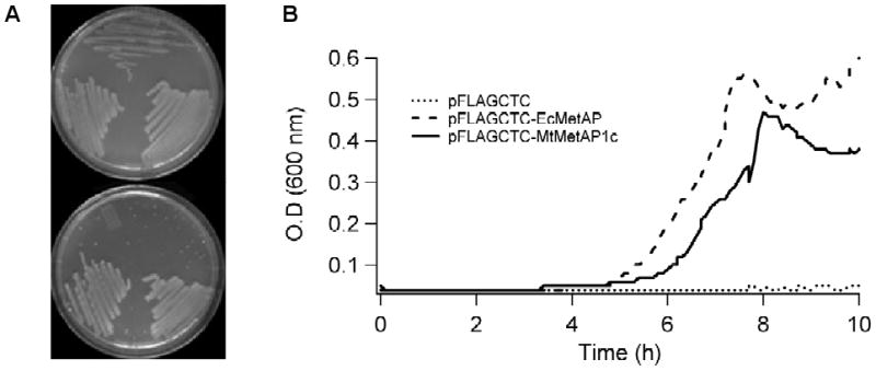
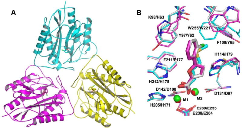
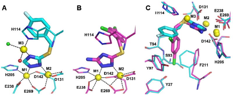
Methionine aminopeptidase (MetAP) carries out an important cotranslational N-terminal methionine excision of nascent proteins and represents a potential target to develop antibacterial and antitubercular drugs. We cloned one of the two MetAPs in Mycobacterium tuberculosis (MtMetAP1c from the mapB gene) and purified it to homogeneity as an apoenzyme. Its activity required a divalent metal ion, and Co(II), Ni(II), Mn(II), and Fe(II) were among activators of the enzyme. Co(II) and Fe(II) had the tightest binding, while Ni(II) was the most efficient cofactor for the catalysis. MtMetAP1c was also functional in E. coli cells because a plasmid-expressed MtMetAP1c complemented the essential function of MetAP in E. coli and supported the cell growth. A set of potent MtMetAP1c inhibitors were identified, and they showed high selectivity toward the Fe(II)-form, the Mn(II)-form, or the Co(II) and Ni(II) forms of the enzyme, respectively. These metalloform selective inhibitors were used to assign the metalloform of the cellular MtMetAP1c. The fact that only the Fe(II)-form selective inhibitors inhibited the cellular MtMetAP1c activity and inhibited the MtMetAP1c-complemented cell growth suggests that Fe(II) is the native metal used by MtMetAP1c in an E. coli cellular environment. Finally, X-ray structures of MtMetAP1c in complex with three metalloform-selective inhibitors were analyzed, which showed different binding modes and different interactions with metal ions and active site residues.
Figures
Similar articles
-
Advances in Bacterial Methionine Aminopeptidase Inhibition.Curr Top Med Chem. 2016;16(4):397-414. doi: 10.2174/1568026615666150813145410. Curr Top Med Chem. 2016. PMID: 26268344 Free PMC article. Review.
-
Amino-terminal extension present in the methionine aminopeptidase type 1c of Mycobacterium tuberculosis is indispensible for its activity.BMC Biochem. 2011 Jul 5;12:35. doi: 10.1186/1471-2091-12-35. BMC Biochem. 2011. PMID: 21729287 Free PMC article.
-
Expression and characterization of Mycobacterium tuberculosis methionine aminopeptidase type 1a.Bioorg Med Chem Lett. 2010 May 1;20(9):2776-9. doi: 10.1016/j.bmcl.2010.03.067. Epub 2010 Mar 19. Bioorg Med Chem Lett. 2010. PMID: 20363127 Free PMC article.
-
FE(II) is the native cofactor for Escherichia coli methionine aminopeptidase.J Biol Chem. 2008 Oct 3;283(40):26879-85. doi: 10.1074/jbc.M804345200. Epub 2008 Jul 31. J Biol Chem. 2008. PMID: 18669631 Free PMC article.
-
Structure and function of the methionine aminopeptidases.Biochim Biophys Acta. 2000 Mar 7;1477(1-2):157-67. doi: 10.1016/s0167-4838(99)00271-x. Biochim Biophys Acta. 2000. PMID: 10708856 Review.
Cited by
-
Advances in Bacterial Methionine Aminopeptidase Inhibition.Curr Top Med Chem. 2016;16(4):397-414. doi: 10.2174/1568026615666150813145410. Curr Top Med Chem. 2016. PMID: 26268344 Free PMC article. Review.
-
Analogs of N'-hydroxy-N-(4H,5H-naphtho[1,2-d]thiazol-2-yl)methanimidamide inhibit Mycobacterium tuberculosis methionine aminopeptidases.Bioorg Med Chem. 2012 Jul 15;20(14):4507-13. doi: 10.1016/j.bmc.2012.05.022. Epub 2012 May 17. Bioorg Med Chem. 2012. PMID: 22704656 Free PMC article.
-
Amino-terminal extension present in the methionine aminopeptidase type 1c of Mycobacterium tuberculosis is indispensible for its activity.BMC Biochem. 2011 Jul 5;12:35. doi: 10.1186/1471-2091-12-35. BMC Biochem. 2011. PMID: 21729287 Free PMC article.
-
Discovery of Inhibitors of Burkholderia pseudomallei Methionine Aminopeptidase with Antibacterial Activity.ACS Med Chem Lett. 2013 Jul 1;4(8):699-703. doi: 10.1021/ml400034m. ACS Med Chem Lett. 2013. PMID: 24376907 Free PMC article.
-
Anti-Tuberculosis Potential of OJT008 against Active and Multi-Drug-Resistant Mycobacterium Tuberculosis: In Silico and In Vitro Inhibition of Methionine Aminopeptidase.Int J Mol Sci. 2023 Dec 5;24(24):17142. doi: 10.3390/ijms242417142. Int J Mol Sci. 2023. PMID: 38138972 Free PMC article.
References
-
- Fauci AS. Multidrug-Resistant and Extensively Drug-Resistant Tuberculosis: The National Institute of Allergy and Infectious Diseases Research Agenda and Recommendations for Priority Research. J Infect Dis. 2008;197:1493–1498. - PubMed
-
- Vaughan MD, Sampson PB, Honek JF. Methionine in and out of proteins: targets for drug design. Curr Med Chem. 2002;9:385–409. - PubMed
-
- Addlagatta A, Quillin ML, Omotoso O, Liu JO, Matthews BW. Identification of an SH3-binding motif in a new class of methionine aminopeptidases from Mycobacterium tuberculosis suggests a mode of interaction with the ribosome. Biochemistry. 2005;44:7166–7174. - PubMed
-
- Wilcox DE. Binuclear Metallohydrolases. Chem Rev. 1996;96:2435–2458. - PubMed
Publication types
MeSH terms
Substances
Grants and funding
LinkOut - more resources
Full Text Sources
Molecular Biology Databases
