Comparative Study
. 2010 Feb 8;11(2):369-76.
doi: 10.1021/bm900983b.

Amelogenin nanoparticles in suspension: deviations from spherical shape and pH-dependent aggregation

Affiliations
Comparative Study

Amelogenin nanoparticles in suspension: deviations from spherical shape and pH-dependent aggregation

Barbara Aichmayer et al. Biomacromolecules. .

Abstract

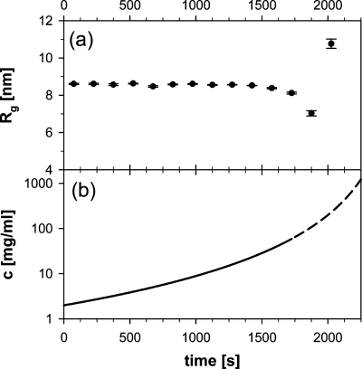
It is well-known that amelogenin self-assembles to form nanoparticles, usually referred to as amelogenin nanospheres, despite the fact that not much is known about their actual shape in solution. In the current paper, we combine SAXS and DLS to study the three-dimensional shape of the recombinant amelogenins rP172 and rM179. Our results show for the first time that amelogenins build oblate nanoparticles in suspension using experimental approaches that do not require the proteins to be in contact with a support material surface. The SAXS studies give evidence for the existence of isolated amelogenin nano-oblates with aspect ratios in the range of 0.45-0.5 at pH values higher than pH 7.2 and show an aggregation of these nano-oblates at lower pH values. The role of the observed oblate shape in the formation of chain-like structures at physiological conditions is discussed as a key factor in the biomineralization of dental enamel.


Figures

Figure 1
Figure 1
Small-angle X-ray scattering (intensity I vs modulus of the scattering vector Q) of rP172 (2 mg/mL) in 80 mM Tris-HCl at 4 °C and a measured pH value of 8.1. The experimental data, collected with a Nanostar laboratory instrument, correspond to the black dots with error bars and are not consistent with the scattering function for monodisperse spheres (dotted gray line, calculated for spheres of 10.5 nm in radius, 0%PD). The solid gray line shows a fit with the scattering function for monodisperse (0%PD) oblate ellipsoids, which describes the data very well. The fitted size parameters were 5.5 and 12.2 nm for the shorter (= rotational) and longer half axes, respectively.
Figure 2
Figure 2
Radius of gyration Rg vs time determined from SAXS measurements (Nanostar laboratory instrument) of a 2 mg/mL suspension of rP172 prepared in 80 mM Tris-HCl buffer adjusted to pH 7.2 at 37 °C. The temperature dependence of the buffer was used to change the pH value by means of heating and cooling the sample. The boxes with different gray scales correspond to different temperatures and measured pH values. At pH 7.2, Rg shows a remarkable increase. Larger error bars indicate deviations from the Guinier approximation, which is only valid for well-separated particles.
Figure 3
Figure 3
Radius of gyration Rg determined from SAXS measurements (Nanostar laboratory instrument) of rP172 (2 mg/mL) in 80 mM Tris-HCl buffers that were adjusted to different pH values. The temperature during all the measurements was 25 °C. The increase in Rg at pH values lower than pH 7.2 together with the increase in the size of the error bars results from a pH-induced aggregation of protein nanoparticles.
Figure 4
Figure 4
(a) Radius of gyration Rg determined from in situ synchrotron SAXS measurements (μ-Spot beamline, BESSY II, HZB) of rP172 at 20 °C and a measured pH value of 7.9 and (b) calculated concentration c of the protein suspension vs time. A 5 μL droplet without any sample container was levitated by using an ultrasound trap. Solvent evaporation led to an increase in the concentration, but Rg was not affected over a wide range of concentrations. At very high protein concentrations, shortly before drying, the evaporation rate presumably differs from the function used to calculate the concentration (see also Materials and Methods). Hence, these values (dashed part of the curve) should only be seen as rough estimates.
Figure 5
Figure 5
Revised core−shell model for the structure of amelogenin nanoparticles based on SAXS and DLS measurements of rP172 and rM179 at pH > 7.2. The SAXS measurements reflect the dimensions of the more electron-dense oblate core (R1 = R2, R3), whereas the hydrodynamic radius RH describes the overall size, including the loose shell, which affects the mobility of the particle as measured by means of DLS. The scheme is consistent with the radii given in Tables 1 and 2.

Similar articles

Cited by

References

    1. Fincham A. G.; Moradian-Oldak J.; Simmer J. P. J. Struct. Biol. 1999, 126 (3), 270–299. - PubMed
    1. Paine M. L.; White S. N.; Luo W.; Fong H.; Sarikaya M.; Snead M. L. Matrix Biol. 2001, 20 (5−6), 273–292. - PubMed
    1. Margolis H. C.; Beniash E.; Fowler C. E. J. Dent. Res. 2006, 85 (9), 775–793. - PubMed
    1. Zeichner-David M. Matrix Biol. 2001, 20 (5−6), 307–316. - PubMed
    1. Aoba T.; Moreno E. C. Calcif. Tissue Int. 1987, 41 (2), 86–94. - PubMed

Publication types