. 2009 Dec 29:10:166.
doi: 10.1186/1471-2474-10-166.

Articular cartilage mineralization in osteoarthritis of the hip

Affiliations

Articular cartilage mineralization in osteoarthritis of the hip

Martin Fuerst et al. BMC Musculoskelet Disord. .

Abstract

Background: The aim of this study was to examine the frequency of articular cartilage calcification in patients with end-stage hip OA. Further, its impact on the clinical situation and the OA severity are analyzed.

Methods: Eighty patients with OA of the hip who consecutively underwent total hip replacement were prospectively evaluated, and 10 controls were included. The patients' X-rays were analyzed for the presence of articular cartilage mineralization. A Harris Hip Score (HHS) was preoperatively calculated for every patient.Slab specimens from the femoral head of bone and cartilage and an additional square centimeter of articular cartilage from the main chondral defect were obtained from each patient for analysis of mineralization by digital contact radiography (DCR). Histological grading was also performed. In a subset of 20 patients, minerals were characterized with an electron microscope (FE-SEM).

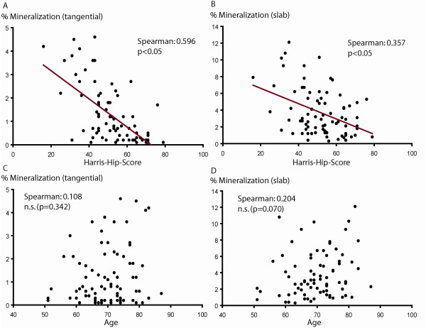
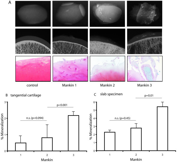
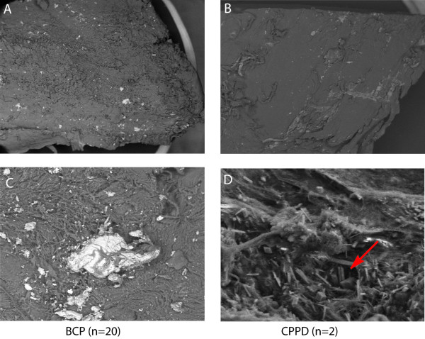
Results: Calcifications were seen in all OA cartilage and slab specimens using DCR, while preoperative X-rays revealed calcification in only 17.5%. None of the control cartilage specimens showed mineralization. There was a highly significant inverse correlation between articular cartilage calcification and preoperative HHS. Histological OA grade correlated positively with the amount of matrix calcification. FE-SEM analysis revealed basic calcium phosphate (BCP) as the predominant mineral; CPPD crystals were found in only two patients.

Conclusions: Articular cartilage calcification is a common event in osteoarthritis of the hip. The amount of calcification correlates with clinical symptoms and histological OA grade.


Figures

Figure 1
Figure 1
Cartilage mineralization and clinical relationships. Contact radiography (DCR) of tangential cut cartilage and slab specimens revealed mineralization in all articular cartilage specimens. The mean area of mineralization was 1.25% (± 0.14 SE, range 0.1-4.6%) for the tangential cut cartilage and 3.69% (± 0.31 SE, range 0.3-12.1) for the slab specimens. Scatter plots show a highly significant inverse correlation between HHS score and mean cartilage calcification for both the tangential cut articular cartilage and the slab specimens (A and B) (p < 0.01, p = 0.01). There was no correlation between patient age and the mean area of cartilage mineralization (C and D) (p = 0.342, p = 0.070).
Figure 2
Figure 2
Cartilage mineralization and correlation with the degree of histologic changes. Histological assessment was performed on tangential cut cartilage using a modified Mankin scoring system. Three representative histological sections from each Mankin grade and one from the control group are shown with their corresponding DCRs. Whereas the 10 controls show intact articular cartilage with no mineralization detected, an increase in matrix mineralization with further cartilage destruction was observed by DCR (A). A statistically significant positive correlation between OA grade and articular cartilage mineralization for both the slab specimens and the tangential cut articular cartilage was observed in specimens graded as Mankin II and III (I-II: p = 0.036; II-III: p = 0.001) (B, C).
Figure 3
Figure 3
Electron microscope analysis of cartilage mineralization. FE-SEM analysis in combination with EDX was performed on a subset of 20 consecutive specimens out of the total 80. All areas of mineralization identified by DCR were proven to be calcium phosphate aggregates using this technique. The FE-SEM studies detected one nearly amorphous (A, C) and one idiomorphic mineral (B, D) phase with different chemical compositions. The two mineral phases contained calcium-phosphate ratios similar to BCP (A, C) and CPPD crystals (B, D). BCP crystals were found in all patients, CPPD crystals additionally in two patients. Panel A show a cartilage specimen with apatite calcification at 15× magnification; panel C the same specimen at 500× magnification. Panel B (15×) shows a patient with CPPD crystals. In panel D (500× magnification), the typical rhomboid CPPD crystals (arrow) are visible.

Similar articles

Cited by

References

    1. Ryan LM, McCarthy DJ. In: Arthritis and allied conditions. Koopman WJ, editor. Baltimore: Wiliams & Wilkins; 1997. Calcium pyrophosphate crystal deposition disease, pseudogout and articular chondrocalcinosis; pp. 2103–5.
    1. Derfus BA, Kurian JB, Butler JJ, Daft LJ, Carrera GF, Ryan LM, Rosenthal AK. The high prevalence of pathologic calcium crystals in pre-operative knees. J Rheumatol. 2002;29(3):570–4. - PubMed
    1. Halverson PB, McCarty DJ. Patterns of radiographic abnormalities associated with basic calcium phosphate and calcium pyrophosphate dihydrate crystal deposition in the knee. Ann Rheum Dis. 1986;45(7):603–5. doi: 10.1136/ard.45.7.603. - DOI - PMC - PubMed
    1. Sokoloff L, Varma AA. Chondrocalcinosis in surgically resected joints. Arthritis Rheum. 1988;31(6):750–6. doi: 10.1002/art.1780310608. - DOI - PubMed
    1. Abreu M, Johnson K, Chung CB, De Lima JE Jr, Trudell D, Terkeltaub R, Pe S, Resnick D. Calcification in calcium pyrophosphate dihydrate (CPPD) cristalline deposits in the knee: anatomic, radiographic, MR imaging, and histologic study in cadavers. Skeletal Radiol. 2004;33(7):392–8. doi: 10.1007/s00256-004-0767-9. - DOI - PubMed

Publication types

MeSH terms

Substances

LinkOut - more resources