

Forebrain projections of arcuate neurokinin B neurons demonstrated by anterograde tract-tracing and monosodium glutamate lesions in the rat
- PMID: 20038444
- PMCID: PMC2823949
- DOI: 10.1016/j.neuroscience.2009.12.053
Forebrain projections of arcuate neurokinin B neurons demonstrated by anterograde tract-tracing and monosodium glutamate lesions in the rat
Abstract
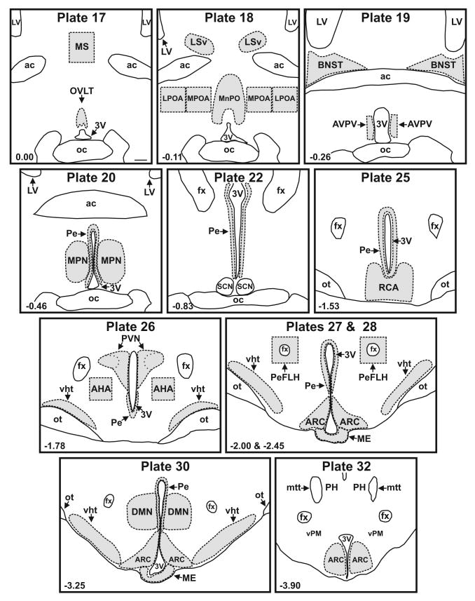
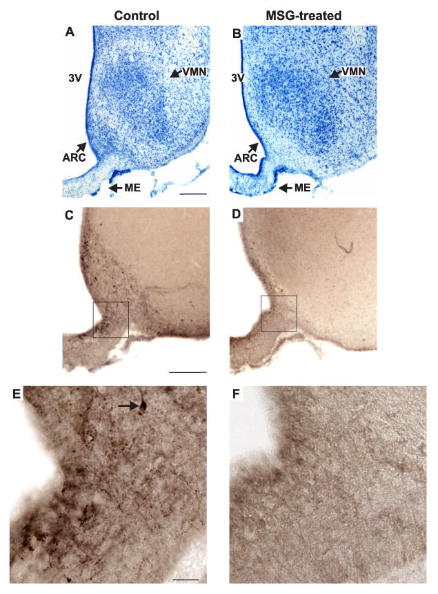
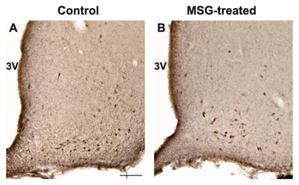
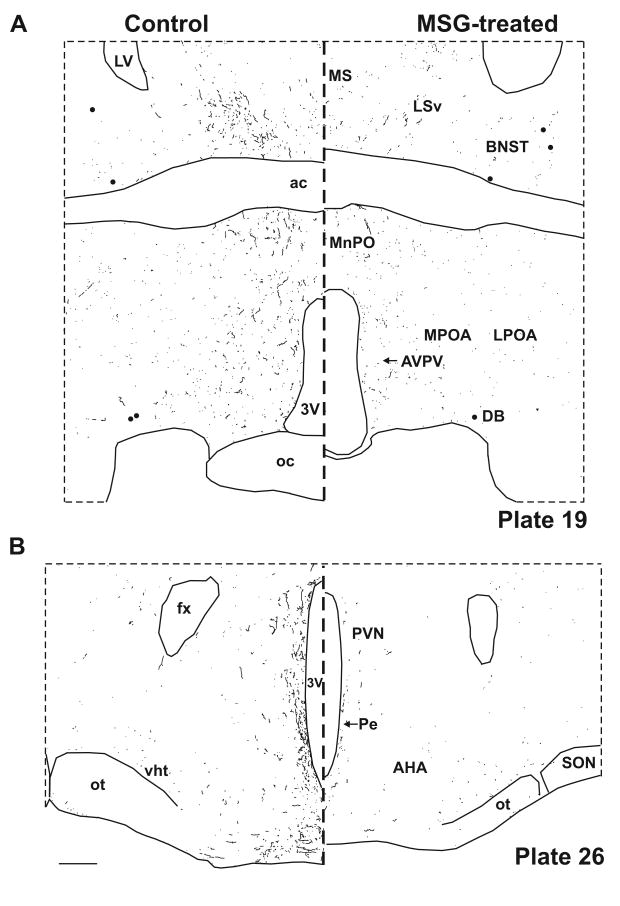
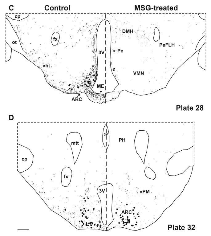
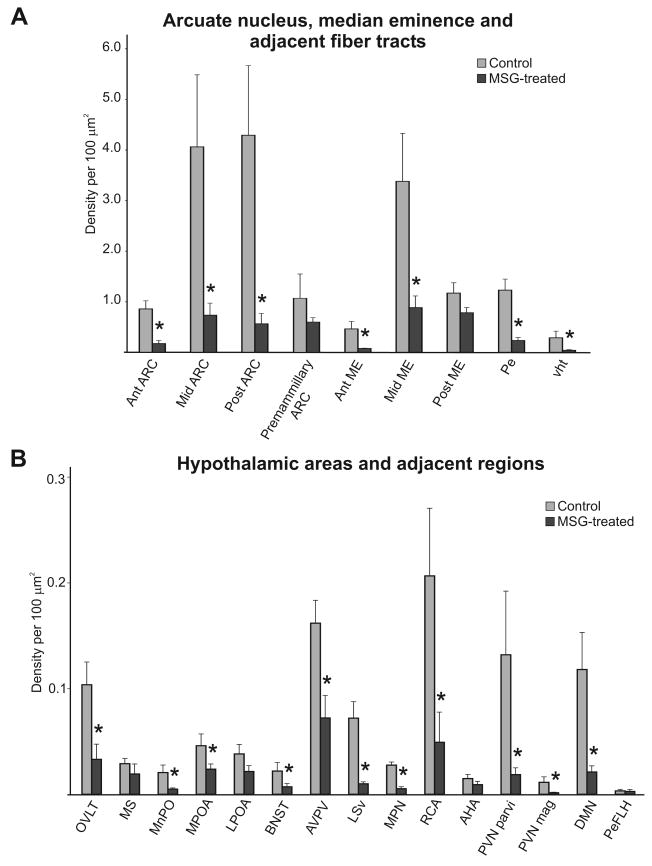
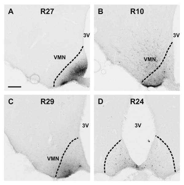
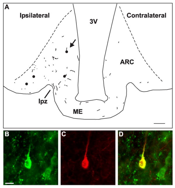
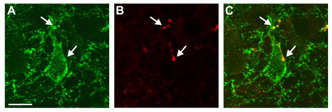
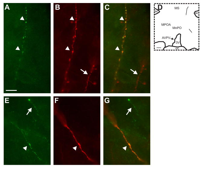
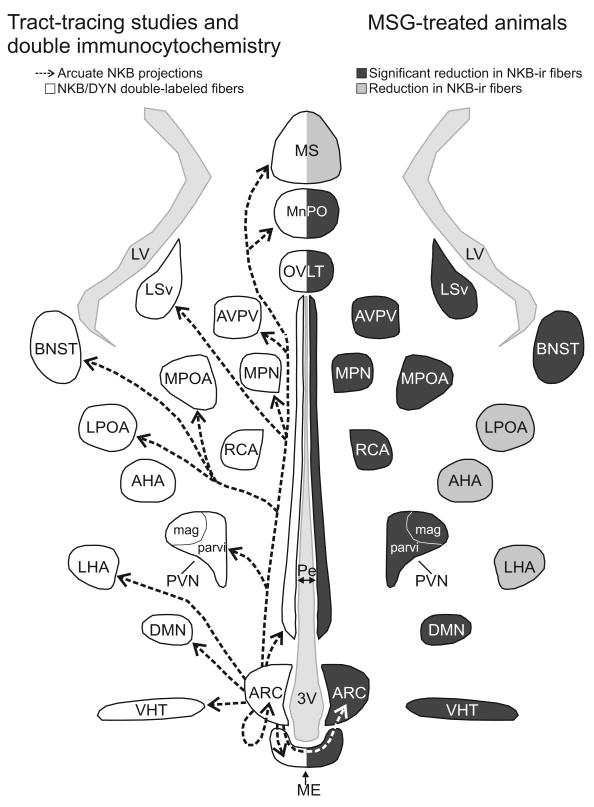
Neurokinin B (NKB) and kisspeptin receptor signaling are essential components of the reproductive axis. A population of neurons resides within the arcuate nucleus of the rat that expresses NKB, kisspeptin, dynorphin, NK3 receptors and estrogen receptor alpha (ERalpha). Here we investigate the projections of these neurons using NKB-immunocytochemistry as a marker. First, the loss of NKB-immunoreactive (ir) somata and fibers was characterized after ablation of the arcuate nucleus by neonatal injections of monosodium glutamate. Second, biotinylated dextran amine was injected into the arcuate nucleus and anterogradely labeled NKB-ir fibers were identified using dual-labeled immunofluorescence. Four major projection pathways are described: (1) local projections within the arcuate nucleus bilaterally, (2) projections to the median eminence including the lateral palisade zone, (3) projections to a periventricular pathway extending rostrally to multiple hypothalamic nuclei, the septal region and BNST and dorsally to the dorsomedial nucleus and (4) Projections to a ventral hypothalamic tract to the lateral hypothalamus and medial forebrain bundle. The diverse projections provide evidence that NKB/kisspeptin/dynorphin neurons could integrate the reproductive axis with multiple homeostatic, behavioral and neuroendocrine processes. Interestingly, anterograde tract-tracing revealed NKB-ir axons originating from arcuate neurons terminating on other NKB-ir somata within the arcuate nucleus. Combined with previous studies, these experiments reveal a bilateral interconnected network of sex-steroid responsive neurons in the arcuate nucleus of the rat that express NKB, kisspeptin, dynorphin, NK3 receptors and ERalpha and project to GnRH terminals in the median eminence. This circuitry provides a mechanism for bilateral synchronization of arcuate NKB/kisspeptin/dynorphin neurons to modulate the pulsatile secretion of GnRH.
Copyright (c) 2010 IBRO. Published by Elsevier Ltd. All rights reserved.
Figures
Similar articles
-
Coexpression of dynorphin and neurokinin B immunoreactivity in the rat hypothalamus: Morphologic evidence of interrelated function within the arcuate nucleus.J Comp Neurol. 2006 Oct 10;498(5):712-26. doi: 10.1002/cne.21086. J Comp Neurol. 2006. PMID: 16917850
-
Colocalisation of dynorphin a and neurokinin B immunoreactivity in the arcuate nucleus and median eminence of the sheep.J Neuroendocrinol. 2006 Jul;18(7):534-41. doi: 10.1111/j.1365-2826.2006.01445.x. J Neuroendocrinol. 2006. PMID: 16774502
-
Morphologic evidence that neurokinin B modulates gonadotropin-releasing hormone secretion via neurokinin 3 receptors in the rat median eminence.J Comp Neurol. 2005 Aug 29;489(3):372-86. doi: 10.1002/cne.20626. J Comp Neurol. 2005. PMID: 16025449
-
Neurokinin B and the hypothalamic regulation of reproduction.Brain Res. 2010 Dec 10;1364:116-28. doi: 10.1016/j.brainres.2010.08.059. Epub 2010 Aug 25. Brain Res. 2010. PMID: 20800582 Free PMC article. Review.
-
The Roles of Neurokinins and Endogenous Opioid Peptides in Control of Pulsatile LH Secretion.Vitam Horm. 2018;107:89-135. doi: 10.1016/bs.vh.2018.01.011. Epub 2018 Feb 13. Vitam Horm. 2018. PMID: 29544644 Review.
Cited by
-
Kisspeptin Regulation of Neuronal Activity throughout the Central Nervous System.Endocrinol Metab (Seoul). 2016 Jun;31(2):193-205. doi: 10.3803/EnM.2016.31.2.193. Epub 2016 May 27. Endocrinol Metab (Seoul). 2016. PMID: 27246282 Free PMC article. Review.
-
Arcuate kisspeptin/neurokinin B/dynorphin (KNDy) neurons mediate the estrogen suppression of gonadotropin secretion and body weight.Endocrinology. 2012 Jun;153(6):2800-12. doi: 10.1210/en.2012-1045. Epub 2012 Apr 16. Endocrinology. 2012. PMID: 22508514 Free PMC article.
-
KNDy (kisspeptin/neurokinin B/dynorphin) neurons are activated during both pulsatile and surge secretion of LH in the ewe.Endocrinology. 2012 Nov;153(11):5406-14. doi: 10.1210/en.2012-1357. Epub 2012 Sep 18. Endocrinology. 2012. PMID: 22989631 Free PMC article.
-
The Effects of Estrogens on Neural Circuits That Control Temperature.Endocrinology. 2021 Aug 1;162(8):bqab087. doi: 10.1210/endocr/bqab087. Endocrinology. 2021. PMID: 33939822 Free PMC article. Review.
-
Minireview: kisspeptin/neurokinin B/dynorphin (KNDy) cells of the arcuate nucleus: a central node in the control of gonadotropin-releasing hormone secretion.Endocrinology. 2010 Aug;151(8):3479-89. doi: 10.1210/en.2010-0022. Epub 2010 May 25. Endocrinology. 2010. PMID: 20501670 Free PMC article. Review.
References
-
- Abel TW, Voytko ML, Rance NE. The effects of hormone replacement therapy on hypothalamic neuropeptide gene expression in a primate model of menopause. J Clin Endocrinol Metab. 1999;84:2111–2118. - PubMed
-
- Adachi S, Yamada S, Takatsu Y, Matsui H, Kinoshita M, Takase K, Sugiura H, Ohtaki T, Matsumoto H, Uenoyama Y, Tsukamura H, Inoue K, Maeda K. Involvement of anteroventral periventricular metastin/kisspeptin neurons in estrogen positive feedback action on luteinizing hormone release in female rats. J Reprod Dev. 2007;53:367–378. - PubMed
-
- Amstalden M, Coolen LM, Hemmerle AM, Billings HJ, Connors JM, Goodman RL, Lehman MN. Neurokinin 3 receptor immunoreactivity in the septal region, preoptic area and hypothalamus of the female sheep: colocalization in neurokinin B Cells of the arcuate nucleus but not in gonadotrophin-releasing hormone neurones. J Neuroendocrinol. 2009 In press. - PMC - PubMed
-
- Badger TM, Millard WJ, Martin JB, Rosenblum PM, Levenson SE. Hypothalamic-pituitary function in adult rats treated neonatally with monosodium glutamate. Endocrinology. 1982;111:2031–2038. - PubMed
-
- Barry J, Hoffman GE, Wray S. LHRH-containing systems. In: Björklund A, Hökfelt T, editors. Handbook of Chemical Neuroanatomy Vol 4 GABA and neuropeptides in the CNS Part I. Amsterdam: Elsevier Science; 1985. pp. 166–215.
Publication types
MeSH terms
Substances
Grants and funding
LinkOut - more resources
Full Text Sources
Other Literature Sources
