. 2010 Jan;135(1):29-42.
doi: 10.1085/jgp.200910305.

Inactivation of the KcsA potassium channel explored with heterotetramers

Affiliations

Inactivation of the KcsA potassium channel explored with heterotetramers

Dvir Rotem et al. J Gen Physiol. 2010 Jan.

Abstract

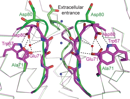
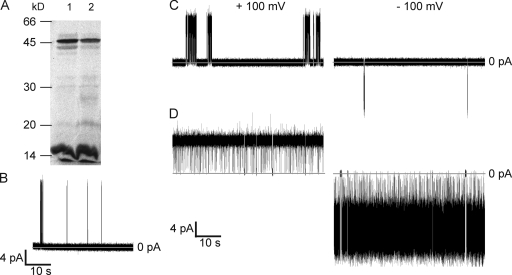
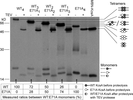
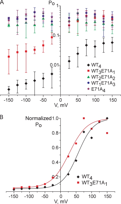
The tetrameric prokaryotic potassium channel KcsA is activated by protons acting on the intracellular aspect of the protein and inactivated through conformational changes in the selectivity filter. Inactivation is modulated by a network of interactions within each protomer between the pore helix and residues at the external entrance of the channel. Inactivation is suppressed by the E71A mutation, which perturbs the stability of this network. Here, cell-free protein synthesis followed by protein purification by sodium dodecyl sulfate-polyacrylamide gel electrophoresis was used to produce heterotetramers of KcsA that contain different combinations of wild-type and E71A subunits. Single-channel recordings from these heterotetramers reveal how the network of interactions in individual protomers affects ionic conduction and channel inactivation, suggesting that the latter is a cooperative process.


Figures

Figure 1.
Figure 1.
Superposition of the selectivity filter region of the WT KcsA structure (PDB entry 1K4C; magenta) and the flipped E71A KcsA structure (PDB entry 2ATK; green). There is a network of interactions between the side chains of Asp80, Glu71, and Trp67 in the WT channel. This network is broken in the mutated channel, and the side chains of Asp80 are exposed at the extracellular entrance of the channel. K+ ion binding sites inside the selectivity filter are presented as blue spheres and water molecules as red spheres. One KcsA protomer is omitted for clarity. Both of these structures are believed to represent conductive forms of the selectivity filter exhibiting four K+ ion binding sites with an overall occupancy of two K+ ions. The superposition was created with PyMOL (http://www.pymol.org).
Figure 2.
Figure 2.
Expression of functional KcsA channels by coupled IVTT. (A) Assembly of KcsA channels during IVTT. Products of IVTT were separated in a 12.5% SDS polyacrylamide gel supplemented with 10 mM KCl. Lane 1, WT KcsA; lane 2, E71A KcsA. The proteins were labeled with [35S]methionine and visualized by exposure of the dried gel to x-ray film. (B–D) Representative single-channel recordings of KcsA purified by SDS-PAGE. (B) WT KcsA in POPE/POPG (3:1) bilayers at +150 mV. (C) WT KcsA in DPhPC bilayers at ±100 mV. (D) E71A KcsA in DPhPC bilayers at ±100 mV. The measurements were made in 200 mM KCl, buffered with 10 mM HEPES, pH 7.0, in the cis chamber and 200 mM KCl, buffered with 10 mM succinic acid, pH 4.0, in the trans chamber. The traces were digitally filtered at 1 kHz for display.
Figure 3.
Figure 3.
The addition of a peptide extension to the N terminus of KcsA does not affect its activity. I-V curves of WT (squares) and E71A (triangles) KcsA without (filled symbols) or with the G8N extension at the N terminus (open symbols). Each data point represents the mean ± SD from at least three separate bilayers. All the measurements were done in 200 mM KCl, buffered with 10 mM HEPES, pH 7.0, in the cis chamber and 200 mM KCl, buffered with 10 mM succinic acid, pH 4.0, in the trans chamber.
Figure 4.
Figure 4.
Expression of heteromeric KcsA channels by coupled IVTT. The samples were separated in a 12.5% SDS polyacrylamide gel supplemented with 10 mM KCl. The proteins were labeled with [35S]methionine and visualized by exposure of the dried gel to x-ray film. Lanes 1–5: protein products from plasmid DNAs encoding WT-long and E71A-short in the ratios 4:0, 1:3, 2:2, 3:1, and 0:4. A schematic representation of the distribution of subunits in the heterotetramers is shown (right).
Figure 5.
Figure 5.
Electrophoretically purified KcsA heteromers. The five WT/E71A heteromers were purified by SDS-PAGE as described in Materials and methods and rerun in a 12.5% SDS polyacrylamide gel supplemented with 10 mM KCl, with or without treatment with TEV protease. The WT subunits had the long extension and the E71A subunits the short extension. Without treatment with the protease, the relative mobilities of the heteromers take on a staircase appearance because of the different total masses of the extensions. After proteolysis with TEV, all of the heteromers have the same mobility as a KcsA tetramer with an eight-Asn extension at the N terminus (right lane, M8N-KcsA; M, Met). The proteins were labeled with [35S]methionine and visualized by exposure of the dried gel to x-ray film. A schematic representation of the distribution of subunits in the heterotetramers is shown (right). The table at the bottom of the figure presents the ratios of WT KcsA and E71A KcsA subunits from spontaneously dissociated tetramers (not treated with TEV protease) on the same gel. The ratios were calculated with Quantity One software (Bio-Rad Laboratories).
Figure 6.
Figure 6.
Representative single-channel recordings of WT/E71A heteromers purified by SDS-PAGE. All measurements were done at ±100 mV in 200 mM KCl, buffered with 10 mM HEPES, pH 7.0, in the cis chamber and 200 mM KCl, buffered with 10 mM succinic acid, pH 4.0, in the trans chamber. The traces were digitally filtered at 1 kHz for display. All-points histograms of the traces are shown.
Figure 7.
Figure 7.
Currents through WT4 and E71A4 at pH 7.0 and 3.0 in the cis chamber. I-V curves of WT4 (squares) and E71A4 (triangles) at pH 7.0 (filled symbols) and 3.0 (open symbols) in the cis chamber. Each data point represents the mean ± SD from at least three separate bilayers. All the measurements were done in 200 mM KCl, buffered with 10 mM succinic acid, pH 4.0, in the trans chamber and with 200 mM KCl, buffered with 10 mM HEPES, pH 7.0, or with 10 mM succinic acid, pH 3.0, in the cis chamber.
Figure 8.
Figure 8.
(A) I-V relationships for homotetramers and heterotetramers of KcsA. Diamonds, WT4; squares, WT3E71A1; triangles, WT2E71A2; circles, WT1E71A3; crosses, E71A4. Each data point represents the mean ± SD from at least four separate bilayers. (B) Unitary conductance values of WT/E71A heteromers at −100 mV. Each data point represents the mean ± SD from at least 11 separate bilayers. The measurements were done in 200 mM KCl, buffered with 10 mM HEPES, pH 7.0, in the cis chamber and 200 mM KCl, buffered with 10 mM succinic acid, pH 4.0, in the trans chamber.
Figure 9.
Figure 9.
Voltage dependences of the open probabilities of WT/E71A heteromers. (A) Plots of open probability (Po) versus voltage. Diamonds, WT4; squares, WT3E71A1; triangles, WT2E71A2; circles, WT1E71A3; stars, E71A4. Each data point represents the mean ± SD from at least three separate bilayers. The measurements were done in 200 mM KCl, buffered with 10 mM HEPES, pH 7.0, in the cis chamber and 200 mM KCl, buffered with 10 mM succinic acid, pH 4.0, in the trans chamber. (B) Normalized values of the data shown in A for WT4 and WT3E71A1. Values were normalized to the maximum Po value, which was set at Po = 1. Diamonds, WT4; squares, WT3E71A1. The continuous curves are the best fits to Boltzmann functions for WT4 (black) and WT3E71A1 (red).
Figure 10.
Figure 10.
A model for inactivation of KcsA channels as an allosteric process. Upon inactivation, there is a concerted transition of all four selectivity filter sequences (TVGYG) from the active conformation (green symbols) to the inactive conformation (red symbols). The conformation of the selectivity filter (the “active site” by analogy with allosteric enzymes) is regulated by networks of interactions between the pore helix and the external entrance of the channel that are formed independently within each subunit of the tetramer (the “regulatory sites” by analogy with allosteric enzymes). The selectivity filter sequence of each WT KcsA protomer is stabilized in its inactive conformation (striped symbols). On the other hand, the regulatory sites of E71A protomers are defective and do not support the stabilization of the inactive conformation of the selectivity filter sequences (open symbols). Under highly depolarizing potentials (top panel), the network of interactions is weakened (Cordero-Morales et al., 2006a). Under these conditions, the concerted transition of all four selectivity filter sequences into inactive conformations happens only when all four are stabilized by their regulatory site. When the membrane potential is negative (bottom panel), the network of interactions in the regulatory site becomes stronger. As a result, the concerted transition of the four selectivity filter sequences into inactive conformations occurs even when one of the sequences is not stabilized in the inactive conformation by its regulatory site. Pale red symbols correspond to unfavorable states of the KcsA channel.

Similar articles

Cited by

References

    1. Baker K.A., Tzitzilonis C., Kwiatkowski W., Choe S., Riek R. 2007. Conformational dynamics of the KcsA potassium channel governs gating properties. Nat. Struct. Mol. Biol. 14:1089–1095 10.1038/nsmb1311 - DOI - PMC - PubMed
    1. Bayley H., Jayasinghe L. 2004. Functional engineered channels and pores (Review). Mol. Membr. Biol. 21:209–220 10.1080/09687680410001716853 - DOI - PubMed
    1. Blunck R., Cordero-Morales J.F., Cuello L.G., Perozo E., Bezanilla F. 2006. Detection of the opening of the bundle crossing in KcsA with fluorescence lifetime spectroscopy reveals the existence of two gates for ion conduction. J. Gen. Physiol. 128:569–581 10.1085/jgp.200609638 - DOI - PMC - PubMed
    1. Braha O., Walker B., Cheley S., Kasianowicz J.J., Song L., Gouaux J.E., Bayley H. 1997. Designed protein pores as components for biosensors. Chem. Biol. 4:497–505 10.1016/S1074-5521(97)90321-5 - DOI - PubMed
    1. Brelidze T.I., Niu X., Magleby K.L. 2003. A ring of eight conserved negatively charged amino acids doubles the conductance of BK channels and prevents inward rectification. Proc. Natl. Acad. Sci. USA. 100:9017–9022 10.1073/pnas.1532257100 - DOI - PMC - PubMed

Publication types

MeSH terms