. 2010 Mar;30(5):1130-44.
doi: 10.1128/MCB.00999-09. Epub 2009 Dec 28.

Evolutionarily conserved function of RRP36 in early cleavages of the pre-rRNA and production of the 40S ribosomal subunit

Affiliations

Evolutionarily conserved function of RRP36 in early cleavages of the pre-rRNA and production of the 40S ribosomal subunit

Marie Gérus et al. Mol Cell Biol. 2010 Mar.

Abstract

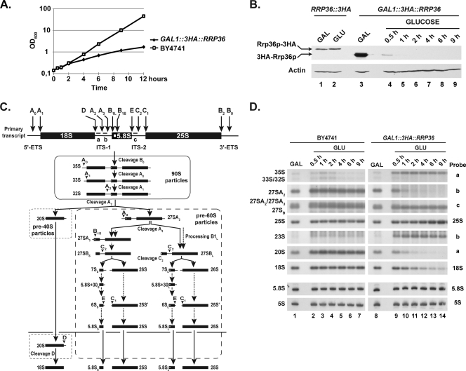
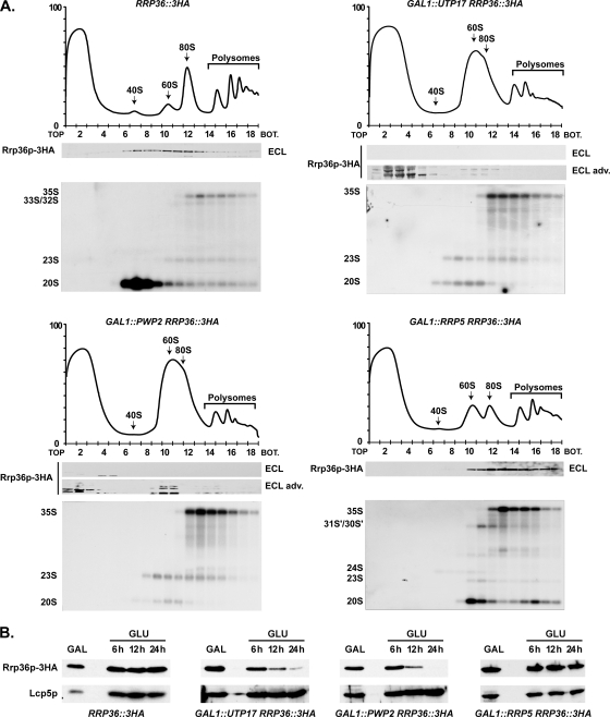
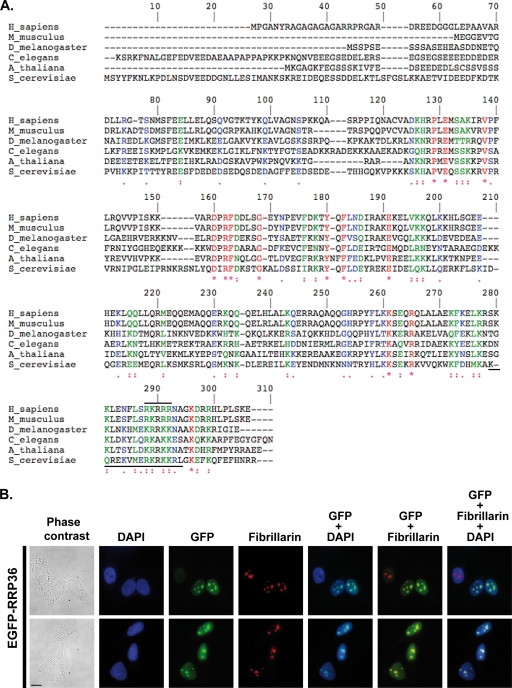
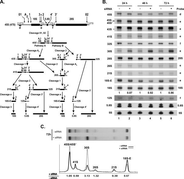
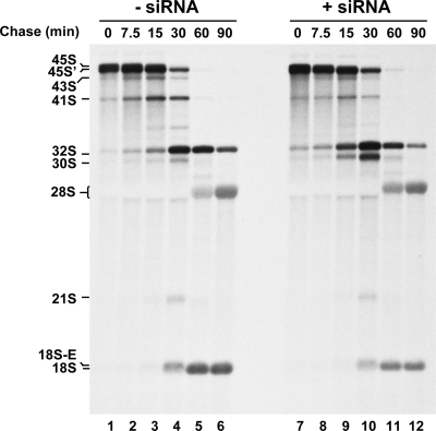
Ribosome biogenesis in eukaryotes is a major cellular activity mobilizing the products of over 200 transcriptionally coregulated genes referred to as the rRNA and ribosome biosynthesis regulon. We investigated the function of an essential, uncharacterized gene of this regulon, renamed RRP36. We show that the Rrp36p protein is nucleolar and interacts with 90S and pre-40S preribosomal particles. Its depletion affects early cleavages of the 35S pre-rRNA and results in a rapid decrease in mature 18S rRNA levels. Rrp36p is a novel component of the 90S preribosome, the assembly of which has been suggested to result from the stepwise incorporation of several modules, including the tUTP/UTP-A, PWP2/UTP-B, and UTP-C subcomplexes. We show that Rrp36p depletion does not impair the incorporation of these subcomplexes and the U3 small nucleolar RNP into preribosomes. In contrast, depletion of components of the UTP-A or UTP-B modules, but not Rrp5p, prevents Rrp36p recruitment and reduces its accumulation levels. In parallel, we studied the human orthologue of Rrp36p in HeLa cells, and we show that the function of this protein in early cleavages of the pre-rRNA has been conserved through evolution in eukaryotes.


Figures

FIG. 1.
FIG. 1.
Rrp36p is a nucleolar protein cosedimenting with preribosomes in yeast cells. (A) Subcellular localization of Rrp36p. The RRP36::3HA strain transformed with a plasmid expressing GFP-Nop1p was grown exponentially, and cell samples were treated for fluorescence microscopy. The two rightmost columns show merged images. (B) Sedimentation profile of Rrp36p-3HA on a sucrose gradient. A total extract prepared from RRP36::3HA cells growing exponentially was sedimented through a sucrose gradient, and 18 fractions were collected. The corresponding A254 profile is displayed with the characteristic annotated peaks. One-half of each fraction was precipitated by TCA, and Rrp36p-3HA was detected in these samples by Western blotting using anti-HA antibodies. RNAs contained in the other half of each fraction were purified, and specific molecules were detected by Northern blotting using the indicated probes (see Fig. 3C and Table S1 in the supplemental material for details).
FIG. 2.
FIG. 2.
Rrp36p interacts with 90S and pre-40S preribosomal particle components in yeast cells. (A) Immunoprecipitation (IP) experiments using IgG-conjugated Sepharose and total cellular extracts prepared from strain BMA64 (devoid of TAP-tagged protein; lanes 1 and 2) and from strains expressing TAP-Rrp36p (lanes 3 and 4) or Pwp2p-TAP (lanes 5 and 6). About 1/5 of the proteins retained in each purified sample (IP) and 1/50 of the proteins contained in each input extract (T) were analyzed by Western blotting using rabbit PAP to detect the TAP-tagged proteins (WB PAP). The RNAs extracted from each purified sample (IP) and from a fraction of each input extract corresponding to 1/50 of the amount used as starting material for the immunoprecipitation (T) were analyzed by primer extension (PE) and/or Northern blot analyses. Signal intensities were quantified from PhosphorImager data using MultiGauge software, and the percentages of input RNAs precipitated in each experiment are indicated below the IP lanes (bg, background level). (B) TAP of TAP-Rrp36p under native and stringent conditions. TAP-Rrp36p was affinity purified from GAL1::TAP::RRP36 cells under both native (20,400 × g; lane 2) and stringent (180,000 × g; lane 4) conditions (see Materials and Methods). As a control, the parental BMA64 WT strain, devoid of TAP-tagged protein, was handled similarly in each case. Approximately 1/10 of the final purified samples were separated by SDS-polyacrylamide gel electrophoresis and stained with PageBlue protein staining solution. The remaining samples were subjected to mass spectrometry analyses. CBP-tagged Rrp36p is indicated with a dot in lanes 2 and 4. Lane 1, molecular weight markers (MWM). (C) Proteins specifically associated with Rrp36p. The list includes all the nonribosomal proteins detected by mass spectrometry in the TAP-Rrp36p purifications but not in the mock purifications. Ribosomal proteins have been omitted from this list.
FIG. 3.
FIG. 3.
Rrp36p depletion affects early cleavages of the pre-rRNA and the production of the mature 18S rRNA in yeast cells. The GAL1::3HA::RRP36 or BY4741 (WT) strain was shifted from a galactose- to a glucose-based medium and maintained under conditions of exponential growth. (A) Growth curve of yeast cells undergoing Rrp36p depletion. Growth of the strains was followed by measuring the optical density of the cultures at 600 nm (OD600) at different times after the nutritional shift. (B) Depletion of 3HA-Rrp36p following the nutritional shift. Total proteins were extracted from the RRP36::3HA strain grown on galactose (GAL)- or glucose (GLU)-containing medium and from the GAL1::3HA::RRP36 strain before transfer to the glucose-based medium (GAL) and at different times after transfer to the glucose-based medium (GLUCOSE). The accumulation of 3HA-tagged Rrp36p proteins and actin (loading control) was assessed by Western blotting using anti-HA and actin-specific antibodies, respectively. Note that Rrp36p-3HA is slightly larger than 3HA-Rrp36p due to the insertion of a linker sequence, in addition to the 3HA tag, into the Rrp36p-3HA fusion protein. (C) Scheme of the pre-rRNA processing pathway in S. cerevisiae. Endonucleolytic cleavages and exonucleolytic trimmings are marked by solid and dotted arrows, respectively. The positions of the oligonucleotide probes used to detect the different pre-rRNAs analyzed in this study (a, b, and c) are shown on the initial precursor; their sequences are detailed in Table S1 in the supplemental material. (D) Early pre-rRNA processing defects in cells undergoing Rrp36p depletion. The BY4741 and GAL1::3HA::RRP36 strains were shifted from a galactose- to a glucose-based medium, and culture samples were collected before the nutritional shift (GAL) and at different times after the nutritional shift (GLU). Total RNAs were extracted from these cell samples, and the accumulation of the different (pre-)rRNAs was analyzed by Northern blotting using the indicated oligonucleotide probes (see Fig. 3C and Table S1 in the supplemental material).
FIG. 4.
FIG. 4.
Depletion of Rrp36p in yeast does not affect the incorporation of components of the UTP-A, UTP-B, or UTP-C modules within preribosomes. Strains expressing TAP-tagged versions of Utp15p, Utp17p, Pwp2p, or Utp22p that were otherwise WT (lower panels) or expressing 3HA-Rrp36p under the control of the GAL1 promoter (upper panels) were transferred from galactose- to glucose-based medium and grown for 6 h. Total extracts prepared from these cells were sedimented through sucrose gradients. Western blot experiments were carried out using rabbit PAP to detect TAP-tagged proteins in the different fractions. BOT, bottom.
FIG. 5.
FIG. 5.
Components of the UTP-A and UTP-B complexes, but not Rrp5p, are required for the incorporation of Rrp36p into preribosomes. (A) Sedimentation profile of Rrp36p-3HA in WT cells (upper left panel) or in cells lacking Utp17p (upper right panel), Pwp2p (lower left panel), or Rrp5p (lower right panel). The GAL1::UTP17, GAL1::PWP2, and GAL1::RRP5 strains expressing Rrp36p-3HA were shifted from galactose- to glucose-containing medium and grown for 16, 18, or 14 h, respectively, to deplete the corresponding protein according to the method described in reference . As a control, the RRP36::3HA strain grown in the presence of glucose was used. Total extracts prepared from these cell samples were sedimented through sucrose gradients. The proteins contained in one-half of each fraction were analyzed by Western blotting using anti-HA antibodies to detect Rrp36p-3HA (note that Rrp36p-3HA was detected using regular ECL in WT and Rrp5p-depleted cells and the more sensitive “ECL Advance” [ECL adv.] kit in Utp17p- and Pwp2p-depleted cells). Total RNAs were extracted from the other half of each fraction, and the pre-rRNAs were detected by Northern blotting using probe a (Fig. 3C). BOT, bottom. (B) Accumulation levels of Rrp36p-3HA in WT or Utp17p-, Pwp2p-, or Rrp5p-depleted cells. The WT, GAL1::UTP17, GAL1::PWP2, and GAL1::RRP5 strains expressing Rrp36p-3HA were transferred from a galactose- to a glucose-containing medium, and cultures were maintained under conditions of exponential growth. Total protein extracts prepared from cell samples harvested before the nutritional shift (GAL) and 6, 12, and 24 h after the nutritional shift (GLU) were analyzed by Western blotting. Rrp36p-3HA was detected using anti-HA antibodies and regular ECL. Lcp5p was detected using specific antibodies.
FIG. 6.
FIG. 6.
RRP36 accumulates in the nucleoli of HeLa cells. (A) Alignment of S. cerevisiae Rrp36p with homologous proteins in Arabidopsis thaliana (NP_172725.2), Caenorhabditis elegans (NP_503179.1), Drosophila melanogaster (NP_650367.1), Mus musculus (NP_659106.1), and Homo sapiens (NP_149103.1), identified using the NCBI BLAST program and aligned using ClustalW. Identical residues are indicated by red characters and stars, strongly similar residues by green characters and double dots, and weakly similar residues by blue characters and single dots. The horizontal solid black lines mark potential nuclear localization signals in the yeast and human sequences. (B) Nucleolar localization of RRP36 in HeLa cells. An immunofluorescence experiment was performed using antifibrillarin antibodies and fixed HeLa cells electroporated with plasmid pEGFP-RRP36. From left to right, images represent cells visualized by phase-contrast microscopy, DAPI staining (blue), EGFP-RRP36 signal (green), and antifibrillarin signal (red); the rightmost three columns represent merged images. Scale bar, 10 μm.
FIG. 7.
FIG. 7.
Depletion of RRP36 mRNA in HeLa cells impairs early cleavages of the pre-rRNA. (A) Scheme of the pre-rRNA processing pathway in HeLa cells. The positions of the oligonucleotide probes used to detect the pre-rRNAs analyzed in this study (d, e, and f) are shown on the initial precursor; their sequences are described in Table S1 in the supplemental material. (B) Pre-rRNA processing defects in siRNA-treated HeLa cells. Northern blot analysis of total RNAs extracted from HeLa cells treated (+) with siRNAs against RRP36 mRNA for 24, 48, or 72 h or left untreated (−). The different (pre-)rRNA species were detected using the specified oligonucleotide probes (Fig. 7A; also see Table S1 in the supplemental material). The second and third hybridization panels from the top correspond to different exposures of the same hybridized RNAs. The values shown below the 18S rRNA panel correspond to the 18S/28S ratios calculated from PhosphorImager quantifications of the signals corresponding to each species in four experiments, with the value for control cells arbitrarily set to 1 (−siRNA). (C) Quantification of the accumulation levels of precursors to 18S rRNA in siRNA-treated and control cells. The accumulation of precursors to 18S rRNA in siRNA-treated cells and control cells after 72 h of treatment (Fig. 7B [72 h, probe e]) was quantified from PhosphorImager data. The intensity profiles of the different maturation intermediates in siRNA-treated cells (gray line) and control cells (black line) are depicted. The value given below each species designation corresponds to the ratio of its accumulation levels in siRNA-treated cells (+siRNA) to those in control cells (−siRNA), after normalization to the amount of 28S rRNA.
FIG. 8.
FIG. 8.
Depletion of RRP36 mRNA in HeLa cells affects the production of the mature 18S rRNA. Neosynthesized RNAs from HeLa cells transfected with siRNAs targeting RRP36 mRNA (lanes 7 to 12) or mock transfected (lanes 1 to 6) were pulse-labeled with l-methyl 3H methionine 48 h posttransfection. Cells were subsequently rinsed to remove radioactive methionine, incubated in regular DMEM, and harvested at the indicated time points. The different (pre-)rRNA species are indicated on the left.

Similar articles

Cited by

References

    1. Andersen, J. S., C. E. Lyon, A. H. Fox, A. K. Leung, Y. W. Lam, H. Steen, M. Mann, and A. I. Lamond. 2002. Directed proteomic analysis of the human nucleolus. Curr. Biol. 12:1-11. - PubMed
    1. Baudin, A., O. Ozier-Kalogeropoulos, A. Denouel, F. Lacroute, and C. Cullin. 1993. A simple and efficient method for direct gene deletion in Saccharomyces cerevisiae. Nucleic Acids Res. 21:3329-3330. - PMC - PubMed
    1. Bernstein, K. A., J. E. Gallagher, B. M. Mitchell, S. Granneman, and S. J. Baserga. 2004. The small-subunit processome is a ribosome assembly intermediate. Eukaryot. Cell 3:1619-1626. - PMC - PubMed
    1. Billy, E., T. Wegierski, F. Nasr, and W. Filipowicz. 2000. Rcl1p, the yeast protein similar to the RNA 3′-phosphate cyclase, associates with U3 snoRNP and is required for 18S rRNA biogenesis. EMBO J. 19:2115-2126. - PMC - PubMed
    1. Bowman, L. H., B. Rabin, and D. Schlessinger. 1981. Multiple ribosomal RNA cleavage pathways in mammalian cells. Nucleic Acids Res. 9:4951-4966. - PMC - PubMed

Publication types

MeSH terms