. 2010 Feb 19;285(8):5347-60.
doi: 10.1074/jbc.M109.076976. Epub 2009 Dec 28.

Lysine 63-linked polyubiquitination of TAK1 at lysine 158 is required for tumor necrosis factor alpha- and interleukin-1beta-induced IKK/NF-kappaB and JNK/AP-1 activation

Affiliations

Lysine 63-linked polyubiquitination of TAK1 at lysine 158 is required for tumor necrosis factor alpha- and interleukin-1beta-induced IKK/NF-kappaB and JNK/AP-1 activation

Yihui Fan et al. J Biol Chem. .

Abstract

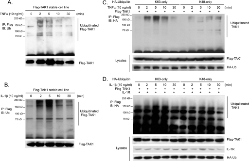
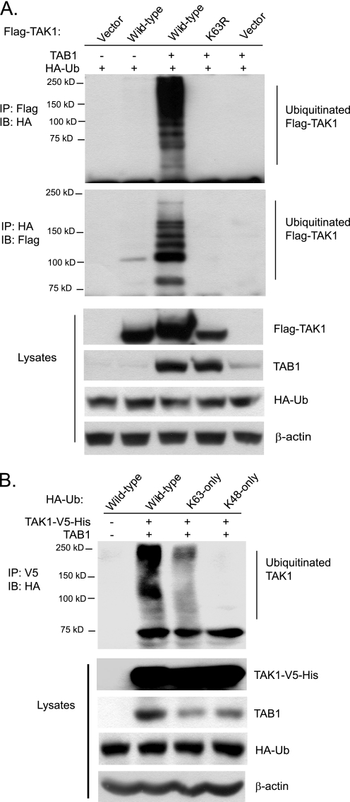
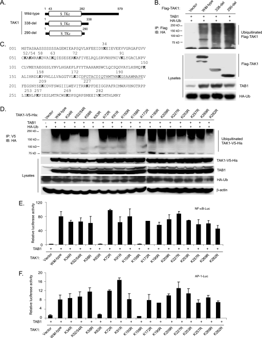
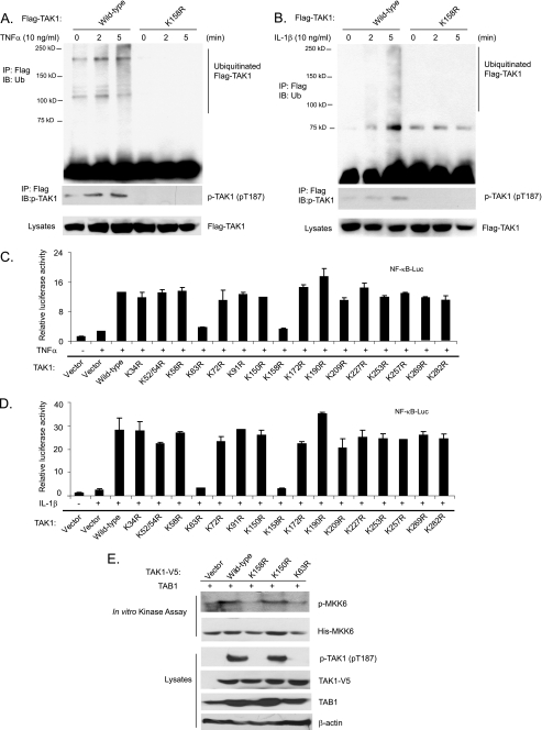
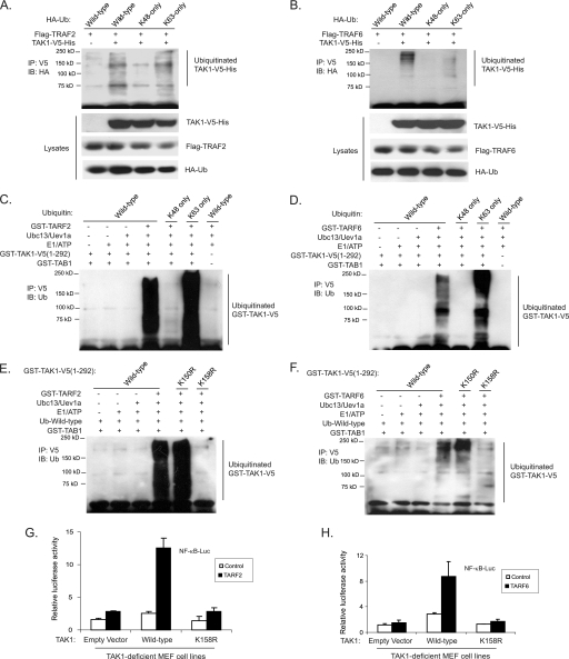
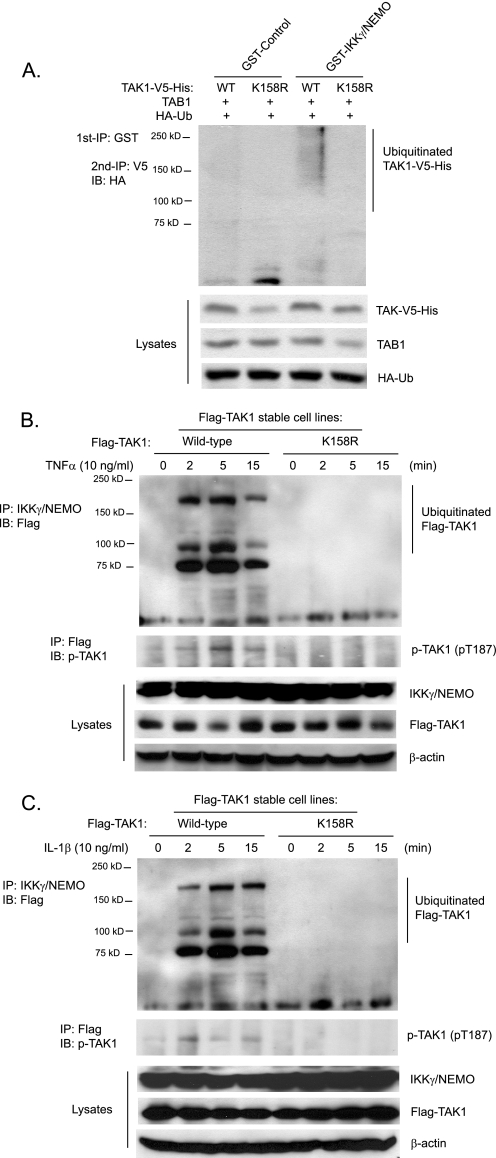
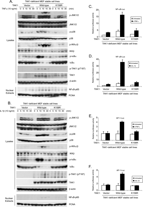
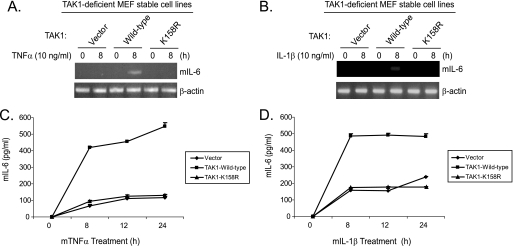
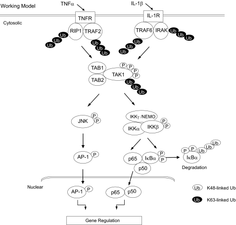
Transforming growth factor-beta-activated kinase 1 (TAK1) plays an essential role in the tumor necrosis factor alpha (TNFalpha)- and interleukin-1beta (IL-1beta)-induced IkappaB kinase (IKK)/nuclear factor-kappaB (NF-kappaB) and c-Jun N-terminal kinase (JNK)/activator protein 1 (AP-1) activation. Here we report that TNFalpha and IL-1beta induce Lys(63)-linked TAK1 polyubiquitination at the Lys(158) residue within the kinase domain. Tumor necrosis factor receptor-associated factors 2 and 6 (TRAF2 and -6) act as the ubiquitin E3 ligases to mediate Lys(63)-linked TAK1 polyubiquitination at the Lys(158) residue in vivo and in vitro. Lys(63)-linked TAK1 polyubiquitination at the Lys(158) residue is required for TAK1-mediated IKK complex recruitment. Reconstitution of TAK1-deficient mouse embryo fibroblast cells with TAK1 wild type or a TAK1 mutant containing a K158R mutation revealed the importance of this site in TNFalpha and IL-1beta-mediated IKK/NF-kappaB and JNK/AP-1 activation as well as IL-6 gene expression. Our findings demonstrate that Lys(63)-linked polyubiquitination of TAK1 at Lys(158) is essential for its own kinase activation and its ability to mediate its downstream signal transduction pathways in response to TNFalpha and IL-1beta stimulation.


Figures

FIGURE 1.
FIGURE 1.
TNFα and IL-1β induce Lys63-linked TAK1 polyubiquitination. A and B, TNFα and IL-1β induce TAK1 polyubiquitination. HeLa cells with stable expression of FLAG-TAK1 were either untreated or treated with TNFα (10 ng/ml) (A) and IL-1β (10 ng/ml) (B) for the times indicated and subsequently lysed. FLAG-TAK1 proteins in the cell lysates were immunoprecipitated (IP) with anti-FLAG antibodies and immunoblotted (IB) with anti-ubiquitin antibodies to detect the presence of ubiquitinated FLAG-TAK1. C, TNFα induces Lys63-linked TAK1 polyubiquitination. Expression vectors encoding FLAG-TAK1 were co-transfected into HEK-293T cells with expression vectors encoding HA-ubiquitin-Lys63 only and Lys48 only, respectively. Then cells were either untreated or treated with TNFα (10 ng/ml) for the time points indicated and subsequently lysed. FLAG-TAK1 proteins in the cell lysates were immunoprecipitated with anti-FLAG antibodies and immunoblotted with anti-HA antibodies to detect the presence of ubiquitinated FLAG-TAK1. D, IL-1β induces Lys63-linked TAK1 polyubiquitination. Expression vectors encoding FLAG-TAK1 and IL-1R were co-transfected into HEK-293T cells with expression vectors encoding HA-ubiquitin-Lys63-only and Lys48-only, respectively. Then cells were either untreated or treated with IL-1β (10 ng/ml) for the times indicated and subsequently lysed. FLAG-TAK1 proteins in the cell lysates were immunoprecipitated with anti-FLAG antibodies and immunoblotted with anti-HA antibodies to detect the presence of ubiquitinated FLAG-TAK1.
FIGURE 2.
FIGURE 2.
Co-overexpression of TAK1/TAB1 induces Lys63-linked TAK1 polyubiquitination. A, co-overexpression of TAK1/TAB1 induces TAK1 polyubiquitination. Expression vectors encoding FLAG-TAK1 and HA-ubiquitin were co-transfected into HEK-293T cells with control vector and expression vectors encoding TAB1, respectively. FLAG-TAK1 proteins in the transfected cells were immunoprecipitated (IP) with anti-FLAG antibodies (or anti-HA antibodies) and immunoblotted (IB) with anti-HA antibodies (or anti-FLAG antibodies) to detect the presence of ubiquitinated FLAG-TAK1. B, co-overexpression of TAK1/TAB1 induces Lys63-linked TAK1 polyubiquitination. Expression vectors encoding TAK1-V5-His and TAB1 were co-transfected into HEK-293T cells with control vector and expression vectors encoding HA-ubiquitin wild type, Lys63-only, and Lys48-only, respectively. TAK1-V5-His proteins in the transfected cells were immunoprecipitated with anti-V5 antibodies and immunoblotted with anti-HA antibodies to detect the presence of ubiquitinated TAK1-V5-His.
FIGURE 3.
FIGURE 3.
Co-overexpression of TAK1/TAB1 induces Lys63-linked TAK1 polyubiquitination at the Lys158 residue within the kinase domain. A, schematic representation of TAK1 wild type and deletion mutants with the kinase domain indicated. B, co-overexpression of the TAK1/TAB1-induced TAK1 polyubiquitination site is located within the kinase domain. Expression vectors encoding HA-ubiquitin and TAB1 were co-transfected into HEK-293T cells with control vector and expression vectors encoding FLAG-TAK1 wild type and two deletion mutants (338-del and 290-del), respectively. FLAG-TAK1 proteins in the transfected cells were immunoprecipitated (IP) with anti-FLAG antibodies and immunoblotted (IB) with anti-HA antibodies to detect the presence of ubiquitinated FLAG-TAK1. C, TAK1 primary sequence with the lysine residues within its N-terminal 290 amino acids indicated. The kinase activation loop is underlined. D, co-overexpression of TAK1/TAB1 induces TAK1 polyubiquitination at Lys158 within the kinase domain. Expression vectors encoding HA-ubiquitin and TAB1 were co-transfected into HEK-293T cells with control vector and expression vectors encoding TAK1-V5-His wild type and 16 lysine to arginine mutants, respectively. TAK1-V5-His proteins in the transfected cells were immunoprecipitated with anti-V5 antibodies and immunoblotted with anti-HA antibodies to detect the presence of ubiquitinated TAK1-V5-His. E and F, the effect of overexpression of TAK1 lysine to arginine mutants with TAB1 on TAK1/TAB1-induced NF-κB (E) and AP-1 (F) activation. TAB1 expression vectors, NF-κB luciferase reporter, and control Renilla luciferase reporter vectors were co-transfected into TAK1-deficient MEF cells with empty vector or expression vectors encoding TAK1 wild type and lysine to arginine mutants, respectively. The relative luciferase activity was measured 48 h later and normalized with the Renilla activity. Error bars, ±S.D. in triplicate experiments.
FIGURE 4.
FIGURE 4.
TNFα and IL-1β induce TAK1 polyubiquitination at the Lys158 residue. A and B, TNFα and IL-1β induce TAK1 polyubiquitination at Lys158. HeLa cells with stable expression of FLAG-TAK1 wild type and K158R mutant were either untreated or treated with TNFα (10 ng/ml) (A) and IL-1β (10 ng/ml) (B) for the time points indicated and subsequently lysed. FLAG-TAK1 proteins in the cell lysates were immunoprecipitated (IP) with anti-FLAG antibodies and immunoblotted (IB) with anti-ubiquitin antibodies to detect the presence of ubiquitinated FLAG-TAK1. C and D, the effect of overexpression of TAK1 lysine to arginine mutants on TNFα-induced (C) and IL-1β-induced (D) NF-κB activation. NF-κB luciferase reporter and control Renilla luciferase reporter vectors were co-transfected into TAK1-deficient MEF cells with empty vector or expression vectors encoding TAK1 wild type and lysine to arginine mutants for 48 h, respectively. Cells were then either untreated or treated with TNFα (1 ng/ml) and IL-1β (1 ng/ml) for 6 h. The relative luciferase activity was measured and normalized with the Renilla activity. The error bars indicate ±S.D. in triplicate experiments. E, TAK1 Lys158 is required for TAK1 activation. TAB1 were transfected into HEK-293T cells with TAK1-V5 wild type, K158R, K150R, and K63R, respectively. TAK1-V5 proteins were immunoprecipitated from cell extracts with anti-V5 antibodies for an in vitro kinase assay using recombinant His-MKK6 as a substrate.
FIGURE 5.
FIGURE 5.
TRAF2 and TRAF6 mediate Lys63-linked TAK1 polyubiquitination at the Lys158 residue in vivo and in vitro. A and B, overexpression of TRAF2 (A) and TRAF6 (B) induces Lys63-linked TAK1 polyubiquitination. Expression vectors encoding TAK1-V5-His and TRAF2 (A) or TRAF6 (B) were co-transfected into HEK-293T cells with control vector and expression vectors encoding HA-ubiquitin wild type, Lys63-only and Lys48-only, respectively. TAK1-V5-His proteins in the transfected cells were immunoprecipitated (IP) with anti-V5 antibodies and immunoblotted (IB) with anti-HA antibodies to detect the presence of ubiquitinated TAK1-V5-His. C and D, TRAF2 (C) and TRAF6 (D) mediate Lys63-linked TAK1 polyubiquitination in vitro. Recombinant GST-TAK1-V5-(1–292), GST-TAB1, E1, E2, (Ubc13/Uev1a), and GST-TRAF2 (C) or GST-TRAF6 (D) were co-incubated in the ubiquitination buffer with recombinant ubiquitin wild type, Lys48-only, and Lys63-only, respectively. After 2 h, GST-TAK1-V5-(1–292) proteins were immunoprecipitated with anti-V5 antibodies and immunoblotted with anti-ubiquitin antibodies to detect the presence of ubiquitinated TAK1-V5-(1–292). E and F, TRAF2 (E) and TRAF6 (F) mediate Lys63-linked TAK1 polyubiquitination at Lys158 in vitro. Recombinant ubiquitin wild type, GST-TAB1, E1, E2 (Ubc13/Uev1a), and GST-TRAF2 (E) or GST-TRAF6 (F) were co-incubated in the ubiquitination buffer with GST-TAK1-V5-(1–292) wild type, K158R, or K150R, respectively. After 2 h, GST-TAK1-V5-(1–292) wild type and mutant proteins were immunoprecipitated with anti-V5 antibodies and immunoblotted with anti-ubiquitin antibodies to detect the presence of ubiquitinated TAK1-V5-(1–292). G and H, TAK1 lysine 158 to arginine mutant inhibits TRAF2-induced (G) and TRAF6-induced (H) NF-κB activation. TRAF2 or TRAF6 expression vectors, NF-κB luciferase reporter, and control Renilla luciferase reporter vectors were co-transfected into TAK1-deficient MEF cells with empty vector or expression vectors encoding TAK1 wild type and lysine 158 to arginine (K158R) mutant. The relative luciferase activity was measured 48 h later and normalized with the Renilla activity. Error bars, ±S.D. in triplicate experiments.
FIGURE 6.
FIGURE 6.
IKKγ/NEMO binds to polyubiquitinated TAK1. A, recombinant IKKγ/NEMO pulls down polyubiquitinated TAK1. Expression vectors encoding TAB1 and HA-ubiquitin were co-transfected into HEK-293T cells with expression vectors encoding TAK1-V5-His wild type and K158R mutant, respectively. The cell lysates were co-incubated with recombinant GST control and GST-IKKγ/NEMO proteins bound to glutathione-agarose beads for 2 h, respectively. GST control and GST-IKKγ/NEMO proteins bound to glutathione-agarose beads were first precipitated with centrifugation and boiled. Then co-precipitated TAK1-V5-His proteins were immunoprecipitated (IP) with anti-V5 antibodies and immunoblotted (IB) with anti-HA antibodies to detect the presence of polyubiquitinated TAK1-V5-His proteins. B and C, TNFα (B) and IL-1β (C) induce association of IKKγ/NEMO with TAK1 wild type but not K158R mutant proteins. HeLa cells with stable expression of FLAG-TAK1 wild type and K158R mutant were either untreated or treated with TNFα (10 ng/ml) (B) and IL-1β (10 ng/ml) (C) for the times indicated and subsequently lysed. Endogenous IKKγ/NEMO proteins in the cell lysates were immunoprecipitated with anti-NEMO antibodies and immunoblotted with anti-FLAG antibodies to detect the co-immunoprecipitation of FLAG-TAK1.
FIGURE 7.
FIGURE 7.
Polyubiquitination of TAK1 at Lys158 residue is required for TNFα- and IL-1β-induced optimal IKK/NF-κB and JNK/AP-1 activation. A and B, expression of the TAK1 K158R mutant inhibits TNFα-induced (A) and IL-1β-induced (B) JNK, p38, IKK, and IκBα phosphorylation as well as IκBα degradation and NF-κB nuclear translocation. TAK1-deficient MEF cells were transduced with the retrovirus encoding the vector control, TAK1 wild type, or TAK1 K158R mutant and subsequently selected with puromycin (2 μg/ml) to establish the TAK1-deficient MEF cell lines with the stable expression of either TAK1 wild type or K158R mutant. TAK1-deficient, TAK1 wild type, and K158R mutant reconstituted MEF cells were untreated or treated with TNFα (10 ng/ml) (A) and IL-1β (10 ng/ml) (B) for the times indicated and subsequently harvested. Whole cell extracts and nuclear extracts were subjected to SDS-PAGE and immunoblotted (IB) with antibodies indicated. β-Actin was detected as a loading control for whole cell extracts, and PCNA was used as a loading control for nuclear extracts. C–F, expression of the TAK1 K158R mutant inhibits TNFα (C and E) and IL-1β (D and F)-induced NF-κB (C and D) and AP-1 (E and F) reporter activities. NF-κB (C and D) and AP-1 (E and F) reporter and 20 ng of Renilla-Luc plasmids were cotransfected into TAK1-deficient MEF control, TAK1 wild type, and K158R reconstituted cells for 48 h. Cells were then either untreated or treated with TNFα (1 ng/ml) and IL-1β (1 ng/ml) for 6 h. The relative luciferase activity was measured and normalized with the Renilla activity. Error bars, ±S.D. in triplicate experiments.
FIGURE 8.
FIGURE 8.
Polyubiquitination of TAK1 at Lys158 residue is required for TNFα and IL-1β-induced IL-6 gene expression. A and B, expression of the TAK1 K158R mutant inhibits IL-1-induced IL-6 gene transcription. TAK1-deficient, TAK1 wild type, and K158R mutant reconstituted MEF cells were untreated or treated with TNFα (10 ng/ml) (A) and IL-1β (10 ng/ml) (B) for 8 h and subsequently harvested for extraction of total RNA using TRIzol reagent. 1 μg of total RNA was used to synthesize first-strand cDNA using a reverse transcription kit according to the manufacturer's instructions. These synthesized cDNAs were used as templates for mouse IL-6 PCR amplification. The PCR products were resolved in 2% agarose gel. C and D, expression of the TAK1 K158R mutant inhibits IL-1-induced IL-6 production. TAK1-deficient, TAK1 wild type, and K158R mutant reconstituted MEF cells were untreated or treated with TNFα (2 ng/ml) (C) and IL-1β (2 ng/ml) (D) for the times indicated. The supernatants from the cell cultures were collected and subjected to the mouse IL-6 ELISA analysis according to the manufacturer's instruction.
FIGURE 9.
FIGURE 9.
A working model for the role of Lys63-linked TAK1 polyubiquitination in TNFα- and IL-1β-mediated IKK/NF-κB and JNK/AP-1 activation. TNFα and IL-1β induces TAK1 polyubiquitination at Lys158 residue and phosphorylation at the Thr178, Thr184, Thr187, and Ser192 residues for TAK1 activation. In turn, activated TAK1 mediates IKK/NF-κB and JNK/AP-1 activation as well as NF-κB- and AP-1-dependent IL-6 gene expression in the cells.

Similar articles

Cited by

References

    1. Chen G., Goeddel D. V. (2002) Science 296, 1634–1635 - PubMed
    1. Dunne A., O'Neill L. A. (2003) Sci. STKE 2003, re3. - PubMed
    1. Häcker H., Karin M. (2006) Sci. STKE 2006, re13. - PubMed
    1. Karin M., Gallagher E. (2005) IUBMB Life 57, 283–295 - PubMed
    1. Li S., Wang L., Dorf M. E. (2009) Mol. Cell 33, 30–42 - PMC - PubMed

Publication types

MeSH terms