. 2010 Mar 5;285(10):7686-96.
doi: 10.1074/jbc.M109.089623. Epub 2009 Dec 28.

Contribution of asparagine residues to the stabilization of a proteinaceous antigen-antibody complex, HyHEL-10-hen egg white lysozyme

Affiliations

Contribution of asparagine residues to the stabilization of a proteinaceous antigen-antibody complex, HyHEL-10-hen egg white lysozyme

Akiko Yokota et al. J Biol Chem. .

Abstract

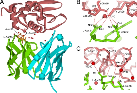
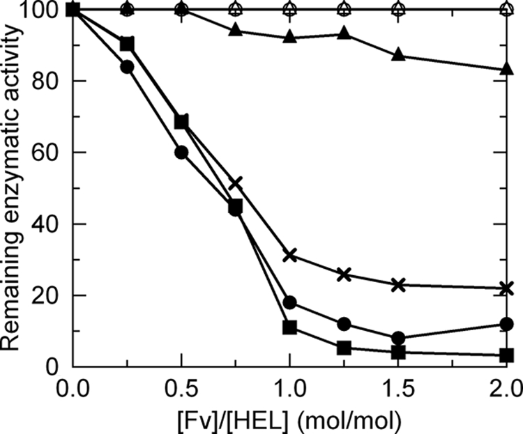
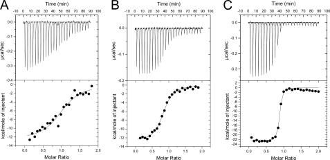
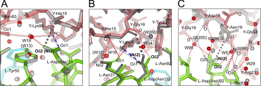
Many germ line antibodies have asparagine residues at specific sites to achieve specific antigen recognition. To study the role of asparagine residues in the stabilization of antigen-antibody complexes, we examined the interaction between hen egg white lysozyme (HEL) and the corresponding HyHEL-10 variable domain fragment (Fv). We introduced Ala and Asp substitutions into the Fv side chains of L-Asn-31, L-Asn-32, and L-Asn-92, which interact directly with residues in HEL via hydrogen bonding in the wild-type Fv-HEL complex, and we investigated the interactions between these mutant antibodies and HEL. Isothermal titration calorimetric analysis showed that all the mutations decreased the negative enthalpy change and decreased the association constants of the interaction. Structural analyses showed that the effects of the mutations on the structure of the complex could be compensated for by conformational changes and/or by gains in other interactions. Consequently, the contribution of two hydrogen bonds was minor, and their abolition by mutation resulted in only a slight decrease in the affinity of the antibody for its antigen. By comparison, the other two hydrogen bonds buried at the interfacial area had large enthalpic advantage, despite entropic loss that was perhaps due to stiffening of the interface by the bonds, and were crucial to the strength of the interaction. Deletion of these strong hydrogen bonds could not be compensated for by other structural changes. Our results suggest that asparagine can provide the two functional groups for strong hydrogen bond formation, and their contribution to the antigen-antibody interaction can be attributed to their limited flexibility and accessibility at the complex interface.


Figures

FIGURE 1.
FIGURE 1.
Interaction between HyHEL-10 Fv and HEL. A, overall structure of the wild-type HyHEL-10 Fv-HEL complex. The C-α schematic diagrams of VL, VH, and HEL are shown in green, cyan, and pink, respectively. The residues investigated in this study are shown in orange. Interfacial water molecules bridging Fv and HEL are represented by red balls. B and C, local structure around the target sites investigated in this study. Interfacial Asn residues at sites 31, 32 (B), and 92 (C) in the VL participate in the antigen-antibody interaction by the formation of direct hydrogen bonds with the antigen. The contacting residues in VL and HEL are shown by green and pink sticks, respectively. Direct hydrogen bonds and indirect hydrogen bonds (via interfacial water molecules) between the antigen and antibody are indicated by red dotted lines. The figures were generated with WebLab Viewer (Molecular Simulations Inc., San Diego).
FIGURE 2.
FIGURE 2.
Inhibition of lysozyme enzymatic activity by HyHEL-10 Fv. Experimental conditions are provided in the text. Symbols used are as follows: solid squares, wild type; open circles, LN31A; solid circles, LN31D; open triangle, LN32A; solid triangles, LN32D; solid crosses, LN92D.
FIGURE 3.
FIGURE 3.
Thermodynamic analyses of interactions between HyHEL-10 Fv mutants and HEL by isothermal titration calorimetry. Thermogram and titration curves for LN31A-HEL (A), LN32D-HEL (B), and LN92D-HEL (C) are shown. The base line obtained by titrating each mutant Fv solution (50 μm) with buffer was subtracted from the thermogram obtained by titrating the corresponding Fv solution with the HEL solution (5 μm).
FIGURE 4.
FIGURE 4.
Comparison of local structures at the mutation site between mutant Fv-HEL and wild-type Fv-HEL complexes. A, LN31D-HEL; B, LN32D-HEL; C, LN92D-HEL. C-α atoms of all polypeptide chains of each mutant complex are superimposed on those of the wild-type complex. Wild-type complex is shown in gray. Residues of VL, VH, and HEL in the mutant Fv-HEL complexes are shown in green, cyan, and pink, respectively. The positions marked W correspond to the water molecules (parentheses indicate wild-type water molecules) shown as red balls. Hydrogen bonds in the mutant Fv-HEL complexes and wild-type complex are depicted as red dotted lines and gray dotted lines, respectively. Salt bridges in the mutant complexes are depicted as blue broken lines. The hydrogen bonding (observed in the wild-type Fv-HEL complex) that is abolished in each mutation is represented as a gray thick dashed line.
FIGURE 5.
FIGURE 5.
Comparison of local structures at sites other than the mutation site between mutant Fv-HEL and wild-type Fv-HEL complexes. Local structures around l-Asn-92 in the LN32D-HEL complex (A) and around l-Asn-32 in the LN92D-HEL complex (B) are shown. Hydrogen bonds conserved in mutant and wild-type Fv-HEL complexes are omitted to facilitate visualization. Refer to Fig. 4 for details.

Similar articles

Cited by

References

    1. Böhm H. J., Schneider G. (eds) (2003) Protein-Ligand Interactions: From Molecular Recognition to Drug Design (Methods and Principles in Medicinal Chemistry) Vol. 19, 1st Ed., pp. 21–51, Wiley-VCH, Weinheim, Germany
    1. Kabat E. A., Wu T. T., Bilofsky H. (1976) Proc. Natl. Acad. Sci. U.S.A. 73, 4471–4473 - PMC - PubMed
    1. Kabat E. A., Wu T. T, Bilofsky H. (1977) J. Biol. Chem. 252, 6609–6616 - PubMed
    1. Padlan E. A. (1990) Proteins Struct. Funct. Genet. 7, 112–124 - PubMed
    1. Mian I. S., Bradwell A. R., Olson A. J. (1991) J. Mol. Biol. 217, 133–151 - PubMed

Publication types

LinkOut - more resources