. 2010 Feb 26;285(9):6477-88.
doi: 10.1074/jbc.M109.064907. Epub 2009 Dec 28.

Constitutively active inflammasome in human melanoma cells mediating autoinflammation via caspase-1 processing and secretion of interleukin-1beta

Affiliations

Constitutively active inflammasome in human melanoma cells mediating autoinflammation via caspase-1 processing and secretion of interleukin-1beta

Miyako Okamoto et al. J Biol Chem. .

Abstract

Interleukin-1beta (IL-1beta) is a pleiotropic cytokine promoting inflammation, angiogenesis, and tissue remodeling as well as regulation of immune responses. Although IL-1beta contributes to growth and metastatic spread in experimental and human cancers, the molecular mechanisms regulating the conversion of the inactive IL-1beta precursor to a secreted and active cytokine remains unclear. Here we demonstrate that NALP3 inflammasome is constitutively assembled and activated with cleavage of caspase-1 in human melanoma cells. Late stage human melanoma cells spontaneously secrete active IL-1beta via constitutive activation of the NALP3 inflammasome and IL-1 receptor signaling, exhibiting a feature of autoinflammatory diseases. Unlike human blood monocytes, these melanoma cells require no exogenous stimulation. In contrast, NALP3 functionality in intermediate stage melanoma cells requires activation of the IL-1 receptor to secrete active IL-1beta; cells from an early stage of melanoma require stimulation of the IL-1 receptor plus the co-stimulant muramyl dipeptide. The spontaneous secretion of IL-1beta from melanoma cells was reduced by inhibition of caspase-1 or the use of small interfering RNA directed against ASC. Supernatants from melanoma cell cultures enhanced macrophage chemotaxis and promoted in vitro angiogenesis, both prevented by pretreating melanoma cells with inhibitors of caspases-1 and -5 or IL-1 receptor blockade. These findings implicate IL-1-mediated autoinflammation as contributing to the development and progression of human melanoma and suggest that inhibiting the inflammasome pathway or reducing IL-1 activity can be a therapeutic option for melanoma patients.


Figures

FIGURE 1.
FIGURE 1.
Late stage melanoma but not early stage melanoma constitutively synthesizes and secretes IL-1β. A, mean (± S.E., n = 3 experiments) 24-h levels of intracellular IL-1β (lysates) and secreted IL-1β (supernatants) in various cell lines as described under “Experimental Procedures.” The result of one representative experiment is shown. The error bars are from one experiment done thrice. B, mean (± S.E., n = 3 experiments) 24-h levels of intracellular (upper) and secreted (lower) IL-1β from cell lines as indicated above each graph. The dose-response concentrations of IL-1α stimulation are shown under the horizontal axis. LPS was used at 1.0 μg/ml. *, p < 0.05; **, p < 0.01; ***, p < 0.001 compared with unstimulated (constitutive) levels indicated as base line. The level of IL-1β (2077 pg/ml) secreted from LPS-stimulated THP-1 cells is shown above the bar.
FIGURE 2.
FIGURE 2.
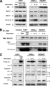
Inflammasome components are expressed, assembled, and activated in late stage human melanoma cell lines. A, Western blot (WB) analysis of inflammasome components (ASC, NALP1, and NALP3) and IL-1β in THP-1, 1205Lu, and HS294T cells. THP-1 cells were primed with PMA and stimulated for 24 h with IL-1α (10 ng/ml) or LPS (1 μg/ml). Melanoma cells were unstimulated or stimulated for 24 h with IL-1α (10 ng/ml), and the lysates were prepared. The data shown are representative of three separate experiments. B, THP-1, 1205Lu, and HS294T cells were incubated for 24 h in the absence or presence of LPS (1 μg/ml) or IL-1α (10 ng/ml). The cell lysates were immunoprecipitated (IP) with anti-ASC antibody (see “Experimental Procedures”) and then subjected to SDS-PAGE and probed with anti-NALP3 antibody. Western blotting with anti-ASC confirmed the presence of ASC in the immunoprecipitates. C, Western blot analysis of THP-1, 1205Lu, and HS294T cells. Unstimulated constitutive levels are shown in the left lane, whereas the right lane is after 24 h in the presence of LPS (1 μg/ml) or IL-1α (10 ng/ml). The data are one of three representative experiments.
FIGURE 3.
FIGURE 3.
The role of inflammasome in the secretion of IL-1β in human melanoma cells. A, upper panels, 1205Lu and HS294T cells were transfected with siRNA (2 nm) against ASC or scrambled RNA indicated as control. 24 h following transfection, the medium was replaced, and the cells were incubated for additional 24 h. The lysates were subjected to Western blotting with anti-ASC antibody. As shown, staining for β-actin was used to control loading. Lower panels, mean (± S.E., n = 4) 24-h secretion of constitutive IL-1β levels was measured in supernatants of the transfectants. The error bars are from one experiment done in quadruplicates, and the experiments were replicated thrice. ***, p < 0.001 compared with the scrambled control. B, 1205Lu and HS294T cells were treated with Z-VAD (2 μm), Z-YVAD (2 μm), Z-WHED (2 μm), or Me2SO control for 30 min, followed by stimulation with IL-1α (10 ng/ml). Cultures without either stimulant are indicated as base line. The data are shown as the means ± S.E. (n = 4). The error bars are from one experiment done in quadruplicate, and the experiments were replicated in triplicate. *, p < 0.05; **, p < 0.01; ***, p < 0.001 compared with the control (no caspase inhibitors).
FIGURE 4.
FIGURE 4.
Secreted products of melanoma cells induce production and secretion of IL-6, IL-8, and MCP-1 in human melanoma cells and enhance macrophage chemotaxis and angiogenesis in vitro. A, mean (± S.E., n = 3) 24-h levels of intracellular and secreted IL-1β, IL-6, IL-8, and MCP-1 from unstimulated 1205Lu cells in the absence (−) or presence (+) of IL-1Ra (10 μg/ml). The error bars are from one experiment done in triplicate, and the experiments were replicated thrice. *, p < 0.05; **, p < 0.01; ***, p < 0.001 compared with cells incubated without IL-1Ra. B, 1205Lu MCM (see “Experimental Procedures”) was added to THP-1 cells. After 24 h, the levels of intracellular IL-1β and IL-1α were measured in the lysates. The effect of IL-1Ra (10 μg/ml) present during the generation of the MCM is shown as MCM/IL-1Ra, and the effect of IL-1Ra added after the generation of MCM is shown as MCM+IL-1Ra. The data are shown as the means ± S.E. (n = 3). The error bars are from one experiment done in triplicate, and the experiments were replicated thrice. *, p < 0.05; **, p < 0.01; ***, p < 0.001 compared with MCM without IL-1Ra. C, THP-1 migration to 1205Lu MCM. The effects of Z-VAD (2 μm), Z-YVAD (2 μm), and IL-1Ra (10 μg/ml) present during the generation of the MCM are shown. The data are presented as the means ± S.E. migration (n = 3) (see “Experimental Procedures”). The error bars are from one experiment done in triplicate, and the experiments were replicated thrice. *, p < 0.05; **, p < 0.01; ***, p < 0.001 compared with MCM without inhibitors. D, left panel, mean (± S.E., n = 3) IL-8 level in 1205Lu MCM. The effects of Z-VAD (2 μm), Z-YVAD (2 μm), and IL-1Ra (10 μg/ml) present during the generation of the MCM are shown. The error bars are from one experiment done in triplicate, and the experiments were replicated thrice. *, p < 0.05; **, p < 0.01; ***, p < 0.001 compared with MCM without inhibitors. Center panel, HUVECs were cultured on Matrigel for 17 h with medium only (control), 1205Lu MCM (MCM), and 1205Lu MCM generated in the presence of Z-VAD (2 μm) (MCM/Z-VAD) or IL-1Ra (10 μg/ml) (MCM/IL-1Ra). Tube formation was visualized under a microscope. Bar, 1 mm. Each experiment was performed in sextuplicate, and the experiments were replicated thrice. Representative pictures are shown. Right panel, tube formation by HUVECs incubated with 1205Lu MCM. The effects of Z-VAD (2 μm) and IL-1Ra (10 μg/ml) present during the generation of the MCM are shown. The data are presented as the means ± S.E. tube formation (n = 6). The error bars are from one experiment done in sextuplicate, and the experiments were replicated thrice. *, p < 0.05; **, p < 0.01; ***, p < 0.001 compared with MCM without inhibitors.
FIGURE 5.
FIGURE 5.
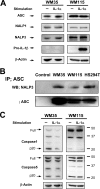
Inflammasome components are expressed, assembled, and activated in early and intermediate stage human melanoma cell lines. A, Western blot (WB) analysis of inflammasome components (ASC, NALP1, and NALP3) and IL-1β in WM35 and WM115 cells. The cells were unstimulated or stimulated for 24 h with IL-1α (10 ng/ml), and the lysates were prepared. The data shown are representative of three separate experiments. B, cell lysates of WM35, WM115, and HS294T cells were immunoprecipitated (IP) with anti-ASC antibody (see “Experimental Procedures”), then subjected to SDS-PAGE, and probed with anti-NALP3 antibody. Western blotting with anti-ASC confirmed the presence of ASC in the immunoprecipitates. C, Western blot analysis of WM35 and WM115 cells. The unstimulated constitutive levels are shown in the left lane, whereas the right lane is after 24 h in the presence of IL-1α (10 ng/ml). The data are one of three representative experiments.
FIGURE 6.
FIGURE 6.
Clinical metastatic melanoma specimens show constitutive secretion of IL-1β and activation of caspases. A, immunohistochemistry of representative clinical melanoma sections. Sample 1 is from primary melanoma, whereas samples 6 and 7 are from metastatic melanoma. Bar, 40 μm. B, summary of the staining. C, mean (± S.E., n = 3 experiments) 24-h levels of intracellular IL-1β (lysates) and secreted IL-1β (supernatants) in tumor cells from primary melanoma (sample 1) or metastatic melanoma (samples 2, 4, 6, and 7). The result of one representative experiment is shown. The error bars are from one experiment done in triplicate. D, Western blot analysis of caspases-1 and -5 from human metastatic melanoma tumor cells (tumors 2 and 6). Melanoma cells were unstimulated, and the lysates were prepared. The data shown are representative of three separate experiments.
FIGURE 7.
FIGURE 7.
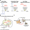
Diagrams representing the role of IL-1β and inflammasome in human melanoma. A, the inflammasome is constitutively activated in human melanoma cells (autoactive Signal 2). Late stage melanoma cells secrete IL-1β without exogenous stimuli via autoactive Signal 1 (activation of IL-1R signaling) and autoactive Signal 2. In contrast, intermediate stage human melanoma cells require Signal 1, and early stage human melanoma cells require further stimulation with Signal 1 and co-stimulant for IL-1β secretion. B, IL-1β released from human melanoma cells is biologically active inducing production and secretion of not only IL-1β itself but also other cytokines and chemokines such as IL-6, IL-8, and MCP-1 from human melanoma cells. Together, IL-1β-secreting human melanoma cells recruit stromal cells and induce angiogenesis, leading to tumor progression. C, IL-1β and its downstream mediators released from late stage human melanoma cells induce IL-1 from stromal cells. IL-1 secreted from stromal cells augments autoinflammatory loop of late stage human melanoma cells. IL-1 secreted from stromal cells can also induce IL-1 secretion from intermediate stage human melanoma cells, triggering an autoinflammatory loop.

References

    1. Ostrand-Rosenberg S., Sinha P. (2009) J. Immunol. 182, 4499–4506 - PMC - PubMed
    1. Balkwill F., Mantovani A. (2001) Lancet 357, 539–545 - PubMed
    1. Balkwill F., Coussens L. M. (2004) Nature 431, 405–406 - PubMed
    1. Dinarello C. A. (2009) Annu Rev. Immunol. 27, 519–550 - PubMed
    1. Paugh B. S., Bryan L., Paugh S. W., Wilczynska K. M., Alvarez S. M., Singh S. K., Kapitonov D., Rokita H., Wright S., Griswold-Prenner I., Milstien S., Spiegel S., Kordula T. (2009) J. Biol. Chem. 284, 3408–3417 - PMC - PubMed

Publication types