

Cardiac myosin is a substrate for zipper-interacting protein kinase (ZIPK)
- PMID: 20038585
- PMCID: PMC2820737
- DOI: 10.1074/jbc.C109.076489
Cardiac myosin is a substrate for zipper-interacting protein kinase (ZIPK)
Abstract
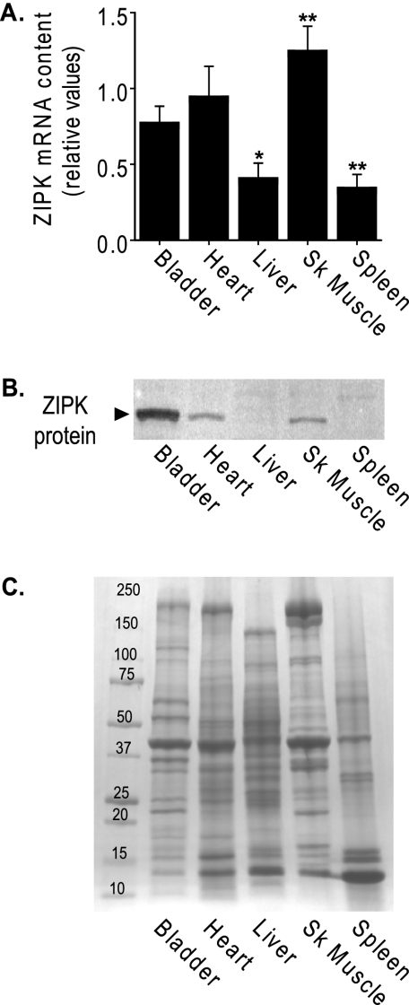
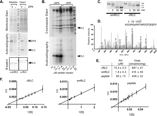
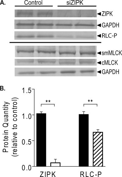
Zipper-interacting protein kinase (ZIPK) is a member of the death-associated protein kinase family associated with apoptosis in nonmuscle cells where it phosphorylates myosin regulatory light chain (RLC) to promote membrane blebbing. ZIPK mRNA and protein are abundant in heart tissue and isolated ventricular neonatal rat cardiac myocytes. An unbiased substrate search performed with purified ZIPK on heart homogenates led to the discovery of a prominent 20-kDa protein substrate identified as RLC of ventricular myosin. Biochemical analyses showed ZIPK phosphorylated cardiac RLC at Ser-15 with a V(max) value 2-fold greater than the value for smooth/nonmuscle RLC; cardiac RLC is a favorable biochemical substrate. Knockdown of ZIPK in cardiac myocytes by small interfering RNA significantly decreased the extent of RLC Ser-15 phosphorylation. Thus, ZIPK may act as a cardiac RLC kinase and thereby affect contractility.
Figures
Similar articles
-
Functional and Molecular Characterisation of Heart Failure Progression in Mice and the Role of Myosin Regulatory Light Chains in the Recovery of Cardiac Muscle Function.Int J Mol Sci. 2021 Dec 22;23(1):88. doi: 10.3390/ijms23010088. Int J Mol Sci. 2021. PMID: 35008512 Free PMC article.
-
Ca2+-independent contraction of longitudinal ileal smooth muscle is potentiated by a zipper-interacting protein kinase pseudosubstrate peptide.Am J Physiol Gastrointest Liver Physiol. 2009 Aug;297(2):G361-70. doi: 10.1152/ajpgi.00112.2009. Epub 2009 Jun 18. Am J Physiol Gastrointest Liver Physiol. 2009. PMID: 19541925
-
Chemical genetics of zipper-interacting protein kinase reveal myosin light chain as a bona fide substrate in permeabilized arterial smooth muscle.J Biol Chem. 2011 Oct 21;286(42):36978-91. doi: 10.1074/jbc.M111.257949. Epub 2011 Aug 31. J Biol Chem. 2011. PMID: 21880706 Free PMC article.
-
The regulation of smooth muscle contractility by zipper-interacting protein kinase.Can J Physiol Pharmacol. 2007 Jan;85(1):79-87. doi: 10.1139/y06-103. Can J Physiol Pharmacol. 2007. PMID: 17487247 Review.
-
Signaling to myosin regulatory light chain in sarcomeres.J Biol Chem. 2011 Mar 25;286(12):9941-7. doi: 10.1074/jbc.R110.198697. Epub 2011 Jan 21. J Biol Chem. 2011. PMID: 21257758 Free PMC article. Review.
Cited by
-
Defining the Sarcomeric Proteoform Landscape in Ischemic Cardiomyopathy by Top-Down Proteomics.J Proteome Res. 2023 Mar 3;22(3):931-941. doi: 10.1021/acs.jproteome.2c00729. Epub 2023 Feb 17. J Proteome Res. 2023. PMID: 36800490 Free PMC article.
-
The significance of regulatory light chain phosphorylation in cardiac physiology.Arch Biochem Biophys. 2011 Jun 15;510(2):129-34. doi: 10.1016/j.abb.2011.02.013. Epub 2011 Feb 21. Arch Biochem Biophys. 2011. PMID: 21345328 Free PMC article. Review.
-
A post-MI power struggle: adaptations in cardiac power occur at the sarcomere level alongside MyBP-C and RLC phosphorylation.Am J Physiol Heart Circ Physiol. 2016 Aug 1;311(2):H465-75. doi: 10.1152/ajpheart.00899.2015. Epub 2016 May 27. Am J Physiol Heart Circ Physiol. 2016. PMID: 27233767 Free PMC article.
-
Cardiac myosin light chain is phosphorylated by Ca2+/calmodulin-dependent and -independent kinase activities.Proc Natl Acad Sci U S A. 2016 Jul 5;113(27):E3824-33. doi: 10.1073/pnas.1600633113. Epub 2016 Jun 20. Proc Natl Acad Sci U S A. 2016. PMID: 27325775 Free PMC article.
-
Functional and Molecular Characterisation of Heart Failure Progression in Mice and the Role of Myosin Regulatory Light Chains in the Recovery of Cardiac Muscle Function.Int J Mol Sci. 2021 Dec 22;23(1):88. doi: 10.3390/ijms23010088. Int J Mol Sci. 2021. PMID: 35008512 Free PMC article.
References
-
- Kobayashi T., Solaro R. J. (2005) Annu. Rev. Physiol. 67, 39–67 - PubMed
-
- Olsson M. C., Patel J. R., Fitzsimons D. P., Walker J. W., Moss R. L. (2004) Am. J. Physiol. Heart Circ. Physiol. 287, H2712–2718 - PubMed
-
- Sweeney H. L., Stull J. T. (1986) Am. J. Physiol. 250, C657–660 - PubMed
-
- Silver P. J., Buja L. M., Stull J. T. (1986) J. Mol. Cell. Cardiol. 18, 31–37 - PubMed
Publication types
MeSH terms
Substances
Grants and funding
LinkOut - more resources
Full Text Sources
Molecular Biology Databases
