. 2010 Mar;192(5):1201-11.
doi: 10.1128/JB.01495-09. Epub 2009 Dec 28.

Flavobacterium johnsoniae gldN and gldO are partially redundant genes required for gliding motility and surface localization of SprB

Affiliations

Flavobacterium johnsoniae gldN and gldO are partially redundant genes required for gliding motility and surface localization of SprB

Ryan G Rhodes et al. J Bacteriol. 2010 Mar.

Abstract

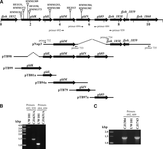
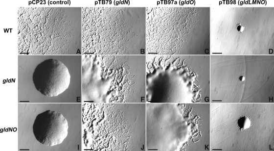
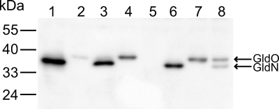
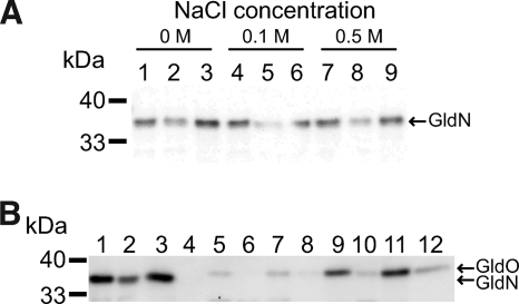
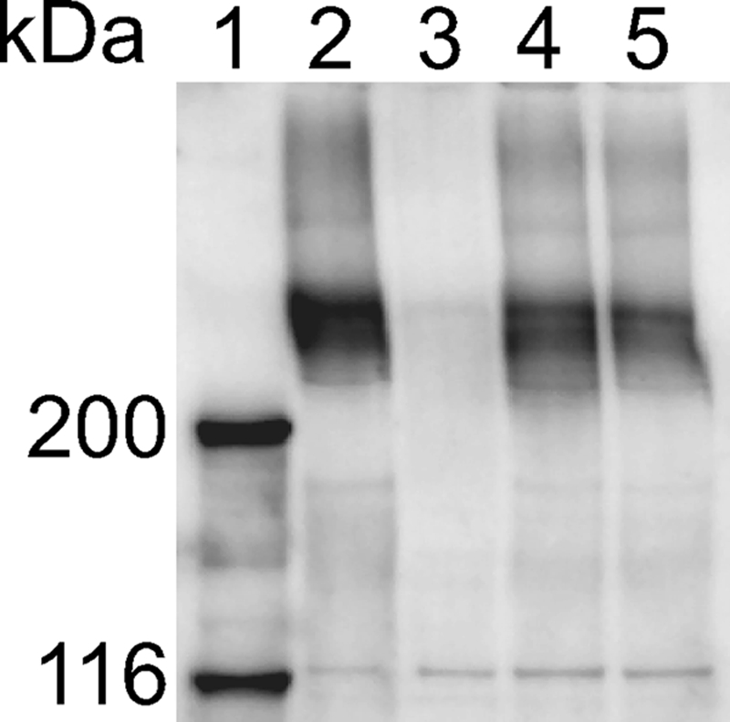
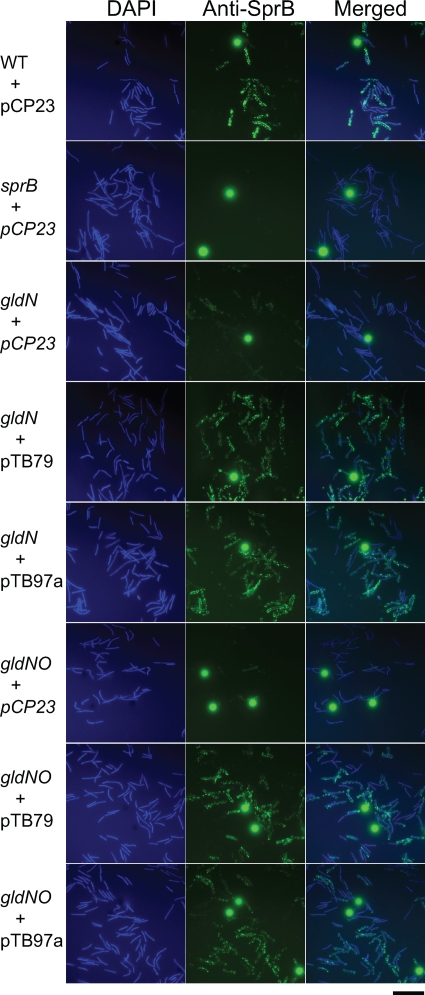
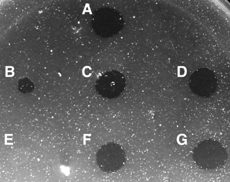
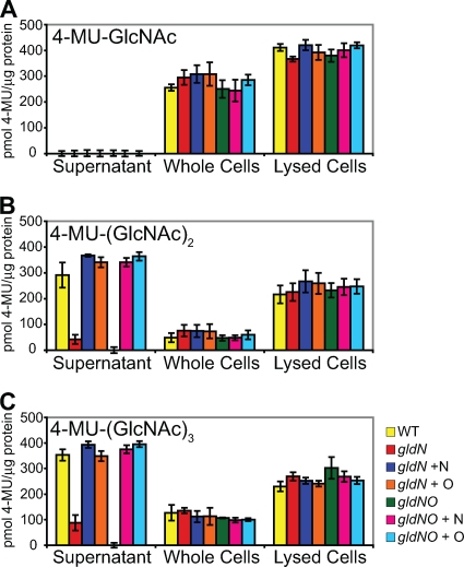
Cells of the gliding bacterium Flavobacterium johnsoniae move rapidly over surfaces. Mutations in gldN cause a partial defect in gliding. A novel bacteriophage selection strategy was used to aid construction of a strain with a deletion spanning gldN and the closely related gene gldO in an otherwise wild-type F. johnsoniae UW101 background. Bacteriophage transduction was used to move a gldN mutation into F. johnsoniae UW101 to allow phenotypic comparison with the gldNO deletion mutant. Cells of the gldN mutant formed nonspreading colonies on agar but retained some ability to glide in wet mounts. In contrast, cells of the gldNO deletion mutant were completely nonmotile, indicating that cells require GldN, or the GldN-like protein GldO, to glide. Recent results suggest that Porphyromonas gingivalis PorN, which is similar in sequence to GldN, has a role in protein secretion across the outer membrane. Cells of the F. johnsoniae gldNO deletion mutant were defective in localization of the motility protein SprB to the cell surface, suggesting that GldN may be involved in secretion of components of the motility machinery. Cells of the gldNO deletion mutant were also deficient in chitin utilization and were resistant to infection by bacteriophages, phenotypes that may also be related to defects in protein secretion.


Figures

FIG. 1.
FIG. 1.
Map of the gldNO region and characterization of gldN and gldNO mutations. (A) Map of the gldNO region. Numbers below the map refer to kilobase pairs of sequence. The sites of Himar insertions are indicated by triangles, and primers used for plasmid construction or for verification of strains are listed. The regions of DNA carried by the plasmid used to make the gldNO deletion (pNap3) and the regions of DNA carried by the complementation plasmids pTB79, pTB81a, pTB94, pTB97a, pTB98, and pTB99 are indicated beneath the map. (B) Verification of loss of gldN and gldO in the deletion mutant CJ1631A. PCR using primers 692 and 939, which flank the deletion, amplified a 3.4-kbp product from wild-type cells (UW101) and amplified a 1.5-kbp product from cells of the deletion mutant CJ1631A. PCR using primers 684 and 694, which lie within the deleted region, resulted in a 1.1-kbp product from wild-type cells and no product from cells of the deletion mutant. (C) Verification of transduction of HimarEm2 from F. johnsoniae CJ1304 (derived from F. johnsoniae MM101) into F. johnsoniae UW101. PCR using primer 692, which anneals within gldM, and primer 609, which anneals near the end of HimarEm2, amplified a 1.4-kbp product from the original gldN mutant CJ1304 and from the transductant CJ1743 but not from wild-type F. johnsoniae UW101.
FIG. 2.
FIG. 2.
Photomicrographs of F. johnsoniae colonies. Colonies were incubated at 25°C on PY2 agar medium for 48 h, and photomicrographs were taken with a Photometrics CoolSNAPcf2 camera mounted on an Olympus IMT-2 phase-contrast microscope. (A) Wild-type (WT) F. johnsoniae UW101 with control vector pCP23. (B) Wild-type F. johnsoniae UW101 with pTB79, which carries gldN. (C) Wild-type F. johnsoniae UW101 with pTB97a, which carries gldO. (D) Wild-type F. johnsoniae UW101 with pTB98, which carries gldL, gldM, gldN, and gldO. (E) gldN mutant CJ1743 with pCP23. (F) gldN mutant CJ1743 with pTB79. (G) gldN mutant CJ1743 with pTB97a. (H) gldN mutant CJ1743 with pTB98. (I) gldNO deletion mutant CJ1631A with pCP23. (J) gldNO deletion mutant CJ1631A with pTB79. (K) gldNO deletion mutant CJ1631A with pTB97a. (L) gldNO deletion mutant CJ1631A with pTB98. Bars indicate 0.5 mm.
FIG. 3.
FIG. 3.
Immunodetection of GldN and GldO. Whole-cell extracts were examined for GldN and GldO by Western blot analysis. Lane 1, wild-type F. johnsoniae with control vector pCP23. Lane 2, gldN mutant CJ1743 with pCP23. Lane 3, gldN mutant CJ1743 with pTB79, which carries gldN. Lane 4, gldN mutant CJ1743 with pTB97a, which carries gldO. Lane 5, gldNO deletion mutant CJ1631A with pCP23. Lane 6, gldNO deletion mutant CJ1631A with pTB79, which carries gldN. Lane 7, gldNO deletion mutant CJ1631A with pTB97a, which carries gldO. Lane 8, gldNO deletion mutant CJ1631A with pTB98, which carries gldL, gldM, gldN, and gldO.
FIG. 4.
FIG. 4.
Localization of GldN and GldO. (A) Fractionation of GldN between soluble and particulate fractions. Cells of wild-type F. johnsoniae UW101 were disrupted and separated into soluble and membrane fractions. Equal amounts of each fraction based on the starting material were separated by SDS-PAGE, and GldN was detected by Western blot analysis. NaCl was added to some extracts to determine whether the increased ionic strength would alter the localization of GldN. Lanes 1, 4, and 7, whole cells. Lanes 2, 5, and 8, soluble fraction. Lanes 3, 6, and 9, particulate fraction. Lanes 1, 2, and 3, no salt added. Lanes 4, 5, and 6, 100 mM NaCl. Lanes 7, 8, and 9, 500 mM NaCl. (B) Fractionation of GldN and GldO. Wild-type and mutant cells were disrupted and separated into soluble and membrane fractions. Membranes were fractionated further by differential solubilization in Sarkosyl, and proteins were detected by Western blot analysis. Equal amounts of each fraction based on the starting material were loaded in each lane. Lanes 1 to 4, wild-type F. johnsoniae with control vector pCP23. Lanes 5 to 8, gldN mutant CJ1743 with pCP23. Lanes 9 to 12, gldNO deletion mutant CJ1631A with pTB97a, which carries gldO. Lanes 1, 5, and 9, whole-cell extracts. Lanes 2, 6, 10, soluble (cytoplasmic and periplasmic) fractions. Lanes 3, 7, and 11, Sarkosyl-soluble (cytoplasmic membrane) fractions. Lanes 4, 8, and 12, Sarkosyl insoluble (outer membrane) fractions.
FIG. 5.
FIG. 5.
Western blot analysis of SprB in cells of wild-type and mutant F. johnsoniae strains. Cells were disrupted using a French pressure cell, and samples were boiled in SDS-PAGE loading buffer. Proteins (25 μg per lane) were separated by electrophoresis, and SprB was detected using anti-SprB antibody. Lane 1, molecular weight markers. Lane 2, wild-type F. johnsoniae carrying control plasmid pCP23. Lane 3, sprB mutant FJ156 carrying pCP23. Lane 4, gldN mutant CJ1743 carrying pCP23. Lane 5, gldNO deletion mutant CJ1631A carrying pCP23.
FIG. 6.
FIG. 6.
Detection of surface-localized SprB protein by immunofluorescence microscopy. Cells of wild-type and mutant F. johnsoniae were exposed to DAPI and to anti-SprB antibodies followed by secondary antibodies conjugated to Alexa-488. The fluorescent spheres are InSpeck relative intensity control fluorescence beads. WT, wild-type F. johnsoniae UW101. sprB refers to the HimarEm2 sprB mutant FJ156. gldN refers to the HimarEm2 gldN mutant CJ1743. gldNO refers to the gldNO deletion mutant CJ1631A. pCP23 is a control vector, pTB79 carries gldN, and pTB97a carries gldO. The bar indicates 10 μm.
FIG. 7.
FIG. 7.
Mutations in gldN and gldO result in defects in chitin utilization. Approximately 106 cells of F. johnsoniae were spotted on MYA-chitin medium and incubated at 25°C for 4 days. (A) Wild-type F. johnsoniae UW101 with control vector pCP23. (B) gldN mutant CJ1743 with pCP23. (C) gldN mutant CJ1743 with pTB79, which carries gldN. (D) gldN mutant CJ1743 with pTB97a, which carries gldO. (E) gldNO deletion mutant CJ1631A with pCP23. (F) gldNO deletion mutant CJ1631A with pTB79. (G) gldNO deletion mutant CJ1631A with pTB97a.
FIG. 8.
FIG. 8.
Chitinase and β-N-acetyl-glucosaminidase activities. Chitinase and β-N-acetyl-glucosaminidase activities of intact cells, cell lysates, and culture supernatants of F. johnsoniae strains were determined using the synthetic substrates 4-MU-GlcNAc, 4-MU-(GlcNAc)2, and 4-MU-(GlcNAc)3. Equal amounts of each sample, based on the protein content of the cell suspension, were incubated with 10 nmol of synthetic substrate for 4 h at 37°C, and the amount of 4-MU released was determined by measuring fluorescence emission at 460 nm following excitation at 360 nm. Yellow, wild-type F. johnsoniae UW101 carrying control vector pCP23. Red, gldN mutant CJ1743 with pCP23. Dark blue, gldN mutant CJ1743 with pTB79, which carries gldN. Orange, gldN mutant CJ1743 with pTB97a, which carries gldO. Green, gldNO deletion mutant CJ1631A with pCP23. Pink, gldNO deletion mutant CJ1631A with pTB79. Light blue, gldNO deletion mutant CJ1631A with pTB97a. Error bars indicate standard errors.
FIG. 9.
FIG. 9.
Mutations in gldN and gldO result in resistance to bacteriophages. Bacteriophages (5 μl of lysates containing approximately 109 phage/ml) were spotted onto lawns of cells in CYE overlay agar. The plates were incubated at 25°C for 24 h to observe lysis. Bacteriophages were spotted in the following order from left to right, as indicated also by the numbers in panel A: top row, φCj1, φCj13, and φCj23; middle row, φCj28, φCj29, and φCj42; bottom row, φCj48 and φCj54. (A) Wild-type F. johnsoniae UW101 with control vector pCP23. (B) gldN mutant CJ1743 with pCP23. (C) gldN mutant CJ1743 with pTB79, which carries gldN. (D) gldN mutant CJ1743 with pTB97a, which carries gldO. (E) gldNO deletion mutant CJ1631A with pCP23. (F) gldNO deletion mutant CJ1631A with pTB79. (G) gldNO deletion mutant CJ1631A with pTB97a.

Similar articles

Cited by

References

    1. Agarwal, S., D. W. Hunnicutt, and M. J. McBride. 1997. Cloning and characterization of the Flavobacterium johnsoniae (Cytophaga johnsonae) gliding motility gene, gldA. Proc. Natl. Acad. Sci. U. S. A. 94:12139-12144. - PMC - PubMed
    1. Braun, T. F., M. K. Khubbar, D. A. Saffarini, and M. J. McBride. 2005. Flavobacterium johnsoniae gliding motility genes identified by mariner mutagenesis. J. Bacteriol. 187:6943-6952. - PMC - PubMed
    1. Braun, T. F., and M. J. McBride. 2005. Flavobacterium johnsoniae GldJ is a lipoprotein that is required for gliding motility. J. Bacteriol. 187:2628-2637. - PMC - PubMed
    1. Chang, L. Y. E., J. L. Pate, and R. J. Betzig. 1984. Isolation and characterization of nonspreading mutants of the gliding bacterium Cytophaga johnsonae. J. Bacteriol. 159:26-35. - PMC - PubMed
    1. Duchaud, E., M. Boussaha, V. Loux, J. F. Bernardet, C. Michel, B. Kerouault, S. Mondot, P. Nicolas, R. Bossy, C. Caron, P. Bessières, J. F. Gibrat, S. Claverol, F. Dumetz, M. L. Hénaff, and A. Benmansour. 2007. Complete genome sequence of the fish pathogen Flavobacterium psychrophilum. Nat. Biotechnol. 25:763-769. - PubMed

Publication types