. 2010 Jan 18;207(1):173-87.
doi: 10.1084/jem.20091873. Epub 2009 Dec 28.

Decoration of T-independent antigen with ligands for CD22 and Siglec-G can suppress immunity and induce B cell tolerance in vivo

Affiliations

Decoration of T-independent antigen with ligands for CD22 and Siglec-G can suppress immunity and induce B cell tolerance in vivo

Bao Hoa Duong et al. J Exp Med. .

Erratum in

  • J Exp Med. 2010 Feb 15;207(2):445. Paulson, James [corrected to Paulson, James C]

Abstract

Autoreactive B lymphocytes first encountering self-antigens in peripheral tissues are normally regulated by induction of anergy or apoptosis. According to the "two-signal" model, antigen recognition alone should render B cells tolerant unless T cell help or inflammatory signals such as lipopolysaccharide are provided. However, no such signals seem necessary for responses to T-independent type 2 (TI-2) antigens, which are multimeric antigens lacking T cell epitopes and Toll-like receptor ligands. How then do mature B cells avoid making a TI-2-like response to multimeric self-antigens? We present evidence that TI-2 antigens decorated with ligands of inhibitory sialic acid-binding Ig-like lectins (siglecs) are poorly immunogenic and can induce tolerance to subsequent challenge with immunogenic antigen. Two siglecs, CD22 and Siglec-G, contributed to tolerance induction, preventing plasma cell differentiation or survival. Although mutations in CD22 and its signaling machinery have been associated with dysregulated B cell development and autoantibody production, previous analyses failed to identify a tolerance defect in antigen-specific mutant B cells. Our results support a role for siglecs in B cell self-/nonself-discrimination, namely suppressing responses to self-associated antigens while permitting rapid "missing self"-responses to unsialylated multimeric antigens. The results suggest use of siglec ligand antigen constructs as an approach for inducing tolerance.


Figures
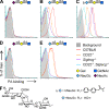
Figure 1.
Figure 1.
Binding analysis of various sialylated PA–glycan conjugates to wild-type compared with siglec mutant B cells. Results are representative of three independent experiments. (A–E) B cells of the indicated genotypes were stained with biotinylated PA conjugated with the following glycans: (A) NeuAcα2-6Galβ1-4GlcNAc, (B) NeuGc, (C) bNeuGc, (D) NeuAcα2-3Galβ1-4GlcNAc, and (E), NeuGcα2-3Galβ1-4GlcNAc. Binding was revealed using fluorescent streptavidin. Background staining by streptavidin alone is shown in gray. (F) Chemical structures of the NeuGc-containing sialosides.
Figure 2.
Figure 2.
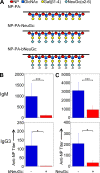
Design of sialylated and unsialylated TI-2–like conjugates and comparison of initial antibody responses elicited in C57BL/6 mice. (A) Structure of immunogenic and tolerogenic conjugates. Shown schematically are PA conjugates carrying both NP and the nonsiglec-binding carbohydrate Galβ1-4GlcNAc (NP–PA), compared with the conjugates NP–PA–NeuGc or NP–PA–bNeuGc. (B and C) Mice were immunized 7 d previously with 20 µg of the indicated conjugates in PBS. Unless otherwise stated, the antigen dose given in all experiments of this study was 20 µg in PBS. Results shown are representative of at least three independent experiments. All statistical tests given used the two-tailed Student’s t test. Means + SD are shown. *, P < 0.05; ***, P < 0.005. (B) Antibody response to NP–PA–bNeuGc (+) versus NP–PA (−; n = 4/group). (C) Antibody response to NP–PA–NeuGc (+) versus NP–PA (−; n = 7/group).
Figure 3.
Figure 3.
Serum anti-NP responses to unsialylated (−; NP–PA) or natively sialylated (+; NP–PA–NeuGc) compounds. (A–C) Responses of (A) wild-type, (B) CD22−/−, and (C) Siglecg−/− mice. Anti-NP titers were assessed on days 7 and 14 as indicated. (top) IgM anti-NP titers; (bottom) IgG3 anti-NP titers. Each point represents the response of an individual mouse of the indicated genotype. Results shown are representative of at least two independent experiments. Horizontal bars represent means. *, P < 0.05; and ***, P < 0.005 using the two-tailed Student’s t test. n.s., not significant.
Figure 4.
Figure 4.
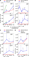
Analysis of tolerance induction by the sialylated antigens NP–PA–NeuGc and NP–PA–bNeuGc, and responses in siglec-deficient mice. (A–D) Functional assay of in vivo tolerance induction. Results shown are representative of at least two independent experiments. (A) Mice (n = 4/group) were challenged with NP65–PA–NeuGc on day 0 and rechallenged with NP200–PA on day 17. IgM and IgG3 antibody levels were assessed at the indicated time points. Control mice received either NP65–PA on day 0 and were rechallenged with NP200–PA as in the experimental group, or received just NP200–PA without pretreatment (green lines). #, a significant reduction in NP65–PA–NeuGc–pretreated mice at 7 d after challenge with NP200–PA (P = 8.84 × 10⁻⁵) compared to nonpretreated animals at 7 d after NP200–PA challenge (green line). (B) Mice (n = 4/group) were challenged with NP–PA–bNeuGc on day 0 and rechallenged with NP–PA on days 14 and 31. Control mice received NP-Ficoll on day 0 and were rechallenged with NP–PA as in the experimental group. (C and D) Tolerance induction in siglec-deficient mice. Serum IgM and IgG3 anti-NP responses of mice that were primed on day 0 with either NP–PA–bNeuGc or NP–PA and boosted on days 17 or 18 with NP–PA. (C) Responses in CD22−/− mice (n = 8 NP–PA; n = 9 NP–PA–bNeuGc). Secondary challenge was done on day 17. Because of the known weak TI-2 responses in CD22−/− mice, all conjugate injections were performed with a double dose of antigen (40 µg/mouse). (D) Responses in Siglecg−/− mice (n = 8 NP–PA; n = 9 NP–PA–bNeuGc). Secondary challenge was done on day 18. Shown are means + SD. *, P < 0.05; **, P < 0.01; and ***, P < 0.005 using the two-tailed Student’s t test.
Figure 5.
Figure 5.
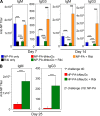
Effect of Ribi coadministration on tolerance induction and response to sialylated and unsialylated antigens. (A) Mice received the indicated conjugates in the presence or absence of Ribi; serum IgM and IgG3 anti-NP levels were measured on days 7 and 14. (B) Mice were treated with the tolerogenic compound NP–PA–bNeuGc with or without Ribi on day 0, and were rechallenged on day 18 with NP–PA. Shown are serum anti-NP titers monitored on day 25. Results are representative of three experiments. For all experimental groups, eight mice were analyzed. Shown are means + SD. All statistical tests given used the two-tailed Student’s t test. *, P < 0.05; ***, P < 0.005. n.s., not significant.
Figure 6.
Figure 6.
Flow cytometry and serum antibody analysis of in vivo responses of NP-specific B cells to sialylated and unsialylated conjugates. Rag1−/− mice that received 107 isolated splenic QM transgenic B cells were challenged with 40 µg of the indicated conjugates 2 h later. From the time of reconstitution they were labeled with BrdU as indicated in Materials and methods. Similar results were obtained in two independent experiments similar in design but using CFSE-prelabeled B cells rather than BrdU to measure cell division (e.g., Fig. S4). (A) At day 7 after reconstitution, spleen cells were analyzed for BrdU uptake and B cell marker expression. A lymphocyte gate was used for analysis. Plots shown were representative of mice receiving NP–PA–bNeuGc (n = 5), NP–PA (n = 5), PA–bNeuGc (n = 3), and PBS (n = 3). Percentages are shown. (B) Quantitation of percentages of IgM+ plasma cells as defined by high levels of cytoplasmic IgM. (C) Percentages of B220+ cells scoring positive for BrdU uptake. (D) Serum anti-NP IgM and IgG3 antibody titers of the indicated recipients obtained at day 7 after reconstitution/challenge. Shown are means + SD. *, P < 0.05; and ***, P < 0.005 using the two-tailed Student’s t test. n.s., not significant.
Figure 7.
Figure 7.
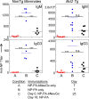
In vivo analysis of the effect of B cell–enforced Bcl2 expression on tolerance induction with NP–PA–bNeuGc. Bcl2 Tg or nontransgenic littermates were challenged with NP–PA–bNeuGc on day 0, followed by rechallenge with NP–PA on day 17. Shown are IgM and IgG3 antibody titers of sera obtained at day 7 after primary or secondary challenge. Transgenic and nontransgenic littermates were immunized and sera were taken in three independent, noncontemporaneous immunization experiments; these sera were analyzed together by ELISA. Note that one Bcl2 Tg mouse had an extremely high titer that is given on a different scale than the other data points. No statistically significant differences were seen between Bcl2 Tg and nontransgenic littermate responses to NP–PA only (Fig. S5). ***, P < 0.005 using the two-tailed Student’s t test. n.s., not significant.
Figure 8.
Figure 8.
In vitro analysis of conjugate binding and response by NP-specific B cells. (A) Flow cytometry analysis of conjugate binding to isolated NP-specific B cells from QM transgenic mice. PA ligands used in this assay carried biotin tags along with the indicated carbohydrate and hapten moieties. (B) Ca2+ mobilization response monitored by Fluo-4 dye fluorescence. (C) Analysis of proliferation induced by the indicated concentrations of antigens by 3[H]thymidine uptake. Cells were harvested at 39 h of culture after a 15-h labeling period. Shown are means + SD. *, P < 0.05; and ***, P < 0.005 using the two-tailed Student’s t test. (D–G) Western blot analysis of protein phosphorylation induced by the indicated conjugates. The arrow indicates the expected molecular weight of CD22 on the pTyr blot. The number of independent replicates of the experiments shown in the indicated sections were as follows: A, one; B, two; C, three; D, three; and E–G, one.

References

    1. Adams E., Basten A., Goodnow C.C. 1990. Intrinsic B-cell hyporesponsiveness accounts for self-tolerance in lysozyme/anti-lysozyme double-transgenic mice. Proc. Natl. Acad. Sci. USA. 87:5687–5691 doi:10.1073/pnas.87.15.5687 - DOI - PMC - PubMed
    1. Alugupalli K.R., Leong J.M., Woodland R.T., Muramatsu M., Honjo T., Gerstein R.M. 2004. B1b lymphocytes confer T cell-independent long-lasting immunity. Immunity. 21:379–390 doi:10.1016/j.immuni.2004.06.019 - DOI - PubMed
    1. Bachmann M.F., Rohrer U.H., Kündig T.M., Bürki K., Hengartner H., Zinkernagel R.M. 1993. The influence of antigen organization on B cell responsiveness. Science. 262:1448–1451 doi:10.1126/science.8248784 - DOI - PubMed
    1. Blixt O., Collins B.E., van den Nieuwenhof I.M., Crocker P.R., Paulson J.C. 2003. Sialoside specificity of the siglec family assessed using novel multivalent probes: identification of potent inhibitors of myelin-associated glycoprotein. J. Biol. Chem. 278:31007–31019 doi:10.1074/jbc.M304331200 - DOI - PubMed
    1. Bovin N.V., Korchagina E.Yu., Zemlyanukhina T.V., Byramova N.E., Galanina O.E., Zemlyakov A.E., Ivanov A.E., Zubov V.P., Mochalova L.V. 1993. Synthesis of polymeric neoglycoconjugates based on N-substituted polyacrylamides. Glycoconj. J. 10:142–151 doi:10.1007/BF00737711 - DOI - PubMed

Publication types

MeSH terms