. 2010 Jan 18;207(1):39-49.
doi: 10.1084/jem.20091933. Epub 2009 Dec 28.

The antiviral factor APOBEC3G improves CTL recognition of cultured HIV-infected T cells

Affiliations

The antiviral factor APOBEC3G improves CTL recognition of cultured HIV-infected T cells

Nicoletta Casartelli et al. J Exp Med. .

Abstract

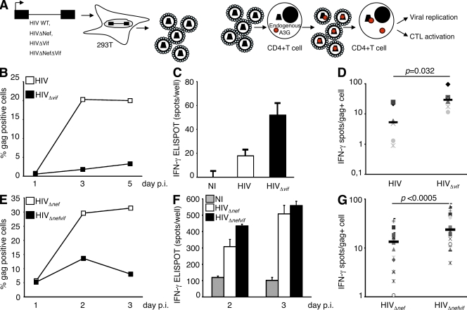
The cytidine deaminase APOBEC3G (A3G) enzyme exerts an intrinsic anti-human immunodeficiency virus (HIV) defense by introducing lethal G-to-A hypermutations in the viral genome. The HIV-1 viral infectivity factor (Vif) protein triggers degradation of A3G and counteracts this antiviral effect. The impact of A3G on the adaptive cellular immune response has not been characterized. We examined whether A3G-edited defective viruses, which are known to express truncated or misfolded viral proteins, activate HIV-1-specific (HS) CD8+ cytotoxic T lymphocytes (CTLs). To this end, we compared the immunogenicity of cells infected with wild-type or Vif-deleted viruses in the presence or absence of the cytidine deaminase. The inhibitory effect of A3G on HIV replication was associated with a strong activation of cocultivated HS-CTLs. CTL activation was particularly marked with Vif-deleted HIV and with viruses harboring A3G. Enzymatically inactive A3G mutants failed to enhance CTL activation. We also engineered proviruses bearing premature stop codons in their genome as scars of A3G editing. These viruses were not infectious but potently activated HS-CTLs. Therefore, the pool of defective viruses generated by A3G represents an underestimated source of viral antigens. Our results reveal a novel function for A3G, acting not only as an intrinsic antiviral factor but also as an inducer of the adaptive immune system.


Figures

Figure 1.
Figure 1.
Vif-deficient HIV-1 is a potent activator of HS-CTLs. (A) Viruses were produced by transfection of 293T cells (that do not express endogenous A3G). Viruses were then used to infect PHA-activated primary CD4+ T cells. Endogenous A3G (red) is incorporated into the progeny virions and exerts its editing activity starting from the second cycle of replication. (B) Activated T cells were incubated with HIVNL or HIVNLΔvif (100 ng/ml p24) and the kinetic of viral infection analyzed by intracellular Gagp24 FACS staining. (C) At day 5 after infection, infected cells were collected and used to stimulate HS-CTL clone EM40-F21 (2,500 clone/well) in an IFN-γ Elispot. Background IFN-γ production induced by uninfected cells is shown. Background IFN-γ productions by target cells alone have been subtracted and are at least three times lower than with HS-CTLs. Activation levels reached using SL9 peptide–loaded cells as positive control were ∼600 IFN-γ+ spots/well (not depicted). Data are mean (±SD) of triplicates. (D) Data from six experiments performed as in B and C, using primary CD4+ cells from three donors, are presented as IFN-γ+ spots to percentage of Gag+ cells (as measured by FACS) on a logarithmic scale. CD4+ T cells infected with Vif-deficient HIV-1 are more efficient than WT HIV-infected cells in activating HS-CTL (P = 0.032, Wilcoxon rank-sum test). Each symbol corresponds to an independent experiment. Horizontal bars indicate the mean IFN-γ/Gag+ cell ratio based on six experiments. (E) Activated primary CD4+ T cells were incubated with VSV-pseudotyped HIVNLΔnef or HIVNLΔnefvif and kinetic of viral infection monitored (as in A). (F) Infected cells were collected and used to stimulate EM40-F21 (as in C). Data are the mean (±SD) of triplicates. (G) Similar results were obtained with cells from eight donors infected at various inocula. Data are presented as IFN-γ+ spots to percentage of Gag+ cells on a logarithmic scale. CD4+ T cells infected with Vif-deficient HIVΔnef are more efficient than HIVΔnef-infected cells in activating HS-CTLs (P < 0.0005, Wilcoxon rank-sum test). Horizontal bars indicate the mean IFN-γ/Gag+ cell ratio. NI, noninfected cells; p.i., post infection.
Figure 2.
Figure 2.
Vif-deficient HIV-1 stimulates chemokine secretion of HS-CTLs. (A) Activated primary CD4+ T cells were incubated with VSV-pseudotyped HIVNLΔnef or HIVNLΔnefvif in the presence or absence of Nevirapine (NVP). At day 4 after infection, viral replication was monitored by FACS. The percentage of infected (Gag+) cells is indicated. (B) Infected cells were then used to stimulate HS-CTL clone EM40-F21 in an IFN-γ Elispot (2,500 clones/well). Activation levels reached, using SL9 peptide–loaded cells as positive control, were ∼600 IFN-γ+ spots/well (not depicted). Data are the mean (±SD) of triplicates and are representative of at least three independent experiments. (C) IFN-γ Elispot data are presented as IFN-γ+ spots to percentage of Gag+ cells. (D and G) 2.5 × 10⁵ HIVNLΔnef- and HIVNLΔnefvif-infected cells were co-cultured for 24 h with 5 × 10³ EM40-F21 (50/1 ratio). Culture supernatants were collected and the release of the indicated chemokine quantified by Luminex. Background secretions in the absence of HS-CTLs have been subtracted. HIV infection didn’t induce an increase of the background lymphokine productions by target CD4+ T cell alone (not depicted). (E and H) Chemokine quantifications presented as concentration to percentage of Gag+ cells. (F and I) Activated T cells were loaded with SL9 peptide at the indicated concentrations, co-cultured for 24 h with EM40-F21 (50/1 ratio), and chemokine release was measured by Luminex. Luminex data are the mean (±SD) of duplicates and are representative of three independent experiments.
Figure 3.
Figure 3.
A3G-mediated viral restriction enhances HS-CTL activation. (A) Viruses were produced upon cotransfection in 293T cells of HIV genome and plasmids encoding for A3G. CEM-A2+ cells were then infected. CEM-A2+ cells do not express A3G (blue); hence, exogenous A3G exerts its editing activity exclusively during the first cycle of replication. (B) CEM-A2+ cells were incubated with HIVSF2Δnef or HIVSF2Δnef + A3G (100 ng/ml p24) and the kinetic of viral infection analyzed by p24 FACS staining. (C) 48 h after infection, infected cells were collected and used to stimulate HS-CTL clone EM40-F21 in an IFN-γ Elispot (10,000 clones/well). Background IFN-γ production induced by uninfected cells is shown. Background IFN-γ productions by CEM-A2+ cells alone were close to zero. Activation levels with SL9 peptide–loaded cells were around 600 IFN-γ+ spots/well (not depicted). Data are the mean (±SD) of triplicates. (D) Data from nine independent experiments performed as in B and C using two HIV isolates (NLΔnef and SF2Δnef) are presented as IFN-γ+ spots to percentage of Gag+ cells on a logarithmic scale. CEM-A2+ cells infected with HIVΔnef + A3G are more efficient than HIVΔnef-infected cells in activating HS-CTL (P < 0.0005, Wilcoxon rank-sum test). Each symbol corresponds to an independent experiment. Horizontal bars indicate the mean IFN-γ/gag+ cell ratio based on the nine independent experiments. (E) To reach similar numbers of Gag+ cells, CEM-A2+ cells were infected with 5–10× more HIVSF2Δnef + A3G than HIVSF2Δnef (doses ranging from 20 to 100 ng p24/ml). The percentage of Gag+ cells achieved at 48 h after infection is indicated for six independent experiments. Using these experimental conditions, there is no difference in terms of gag+ cells between HIVSF2Δnef or HIVSF2Δnef + A3G-infected cells (P = 0.29, Wilcoxon rank-sum test). Horizontal bars indicate the mean percentage of Gag+ cells based on the six independent experiments. (F) Infected cells were then used in Elispot assays to stimulate EM40-F21 (10,000 clones/well). Results of six independent experiments are summarized. In the presence of A3G, there was a stronger CTL activation (P = 0.015, Wilcoxon rank-sum test). Each symbol in E and F corresponds to an independent experiment. Horizontal bars indicate the mean IFN-γ Elispot response based on the six independent experiments. (G) T1-B7 cells were incubated with HIVSF2 or HIVSF2 + A3G (100 ng/ml p24). 48 h after infection, cells were submitted to an IFN-γ Elispot assay to activate HIV Nef-specific CTLs restricted by HLA-B*0702 (104 CTL/well). HIV Nef-specific CTL lines were generated in three different HLA-B*0702 transgenic mice and used in two independent experiments. One representative experiment with one CTL line is shown. (H) As in F, HIV-infected cells were used to activate a control CTL line specific for CMV. Data are the mean (±SD) of triplicates.
Figure 4.
Figure 4.
The editing activity of A3G is required to enhance HS-CTL activation. (A) CEM-A2+ cells were infected with HIVNLΔnef produced without A3G or in the presence of WT and C-terminal domain mutants (H257R and C288S) of A3G (triangles, p24 doses: 20 and 10 ng/ml). 48 h after infection, viral infection was analyzed by Gagp24 staining (data are representative of four independent experiments) and infected cells were used to stimulate HS-CTL clone EM40-F21 in an IFN-γ Elispot (10,000 clones/well) (B). Background IFN-γ production induced by uninfected cells has been subtracted. Background IFN-γ productions by CEM-A2+ cells alone were close to zero. Activation levels reached using SL9 peptide–loaded cells as positive control were ∼600 IFN-γ+ spots/well (not depicted). (C) Data are presented as a ratio of IFN-γ Elispot to percentage of Gag+ cells. Impairing the editing activity of A3G abrogates A3G-mediated enhancement of CTL recognition. Data are the mean (±SD) of triplicates and are representative of four independent experiments.
Figure 5.
Figure 5.
Defective viruses with premature STOP codons are efficiently recognized by HS-CTLs. (A) Schematic representation of the strategy used to construct defective HIV (HIVNLΔnefP2 and HIVNLΔnefP3) mimicking A3G-mediated editing. (Top) Nucleotide and amino acid sequences of a fraction of gag gene (gagP2) targeted for mutagenesis. The G-to-A transition introduced by mutagenesis is underlined and the resulting amino acid change (W212 to STOP) is in bold. Nucleotide numbering is according to HIVLAI sequence. (Bottom) Schematic representation of Gag full length and truncated Gag proteins (GagP2 and GagP3) generated by mutagenesis. The SL9 peptide amino acid sequence within p17 is indicated. HeLa-A2+ cells were transfected with HIV, HIVNLΔnefP2, and HIVNLΔnefP3 proviruses. (B and C) Gag expression profiles were analyzed 2 d after transfection using anti-p17 antibody and Western blotting (B) or anti-p24 antibody and FACS (C). Data are representative of three independent experiments. For Western blotting, transfected cells were pelleted, lyzed, loaded on SDS-PAGE, and transferred to nitrocellulose. Membranes were then incubated with antibodies to p17 and actine. (D) HeLa-A2+ cells were transfected with the indicated doses of provirus. At 36 h after transfection, cells were used to activate HS-CTL clone EM40-F21 in an IFN-γ Elispot assay (10,000 clones/well). Data are the mean (±SD) of triplicates and are representative of three independent experiments. (E) Hela-A2 cells transfected with 1 µg of indicated HIV DNA were mock or Epoxomicin treated (1 µM) for 6 h prior to co-culture with HS-CTLs. Epoxomicin, a proteasome inhibitor, diminishes CTL activation. Background IFN-γ production induced by untransfected cells is shown. Background IFN-γ productions by target cells alone have been subtracted and are at least three times lower than with HS-CTLs. Activation levels reached using SL9 peptide–loaded cells as positive control were ∼400 IFN-γ+ spots/well (not depicted). Data are mean (±SD) of triplicates and are representative of two independent experiments.

Similar articles

Cited by

References

    1. An P., Bleiber G., Duggal P., Nelson G., May M., Mangeat B., Alobwede I., Trono D., Vlahov D., Donfield S., et al. 2004. APOBEC3G genetic variants and their influence on the progression to AIDS. J. Virol. 78:11070–11076 10.1128/JVI.78.20.11070-11076.2004 - DOI - PMC - PubMed
    1. Appay V., Douek D.C., Price D.A. 2008. CD8+ T cell efficacy in vaccination and disease. Nat. Med. 14:623–628 10.1038/nm.f.1774 - DOI - PubMed
    1. Armitage A.E., Katzourakis A., de Oliveira T., Welch J.J., Belshaw R., Bishop K.N., Kramer B., McMichael A.J., Rambaut A., Iversen A.K. 2008. Conserved footprints of APOBEC3G on Hypermutated human immunodeficiency virus type 1 and human endogenous retrovirus HERV-K(HML2) sequences. J. Virol. 82:8743–8761 10.1128/JVI.00584-08 - DOI - PMC - PubMed
    1. Cardinaud S., Moris A., Février M., Rohrlich P.S., Weiss L., Langlade-Demoyen P., Lemonnier F.A., Schwartz O., Habel A. 2004. Identification of cryptic MHC I–restricted epitopes encoded by HIV-1 alternative reading frames. J. Exp. Med. 199:1053–1063 10.1084/jem.20031869 - DOI - PMC - PubMed
    1. Chiu Y.L., Soros V.B., Kreisberg J.F., Stopak K., Yonemoto W., Greene W.C. 2005. Cellular APOBEC3G restricts HIV-1 infection in resting CD4+ T cells. Nature. 435:108–114 10.1038/nature03493 - DOI - PubMed

Publication types

MeSH terms