. 2010 Jan 18;207(1):189-206.
doi: 10.1084/jem.20091964. Epub 2009 Dec 28.

CD207+ CD103+ dermal dendritic cells cross-present keratinocyte-derived antigens irrespective of the presence of Langerhans cells

Affiliations

CD207+ CD103+ dermal dendritic cells cross-present keratinocyte-derived antigens irrespective of the presence of Langerhans cells

Sandrine Henri et al. J Exp Med. .

Erratum in

  • J Exp Med. 2010 Feb 15;207(2):447

Abstract

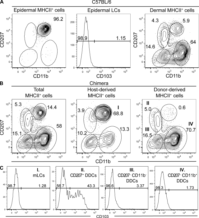
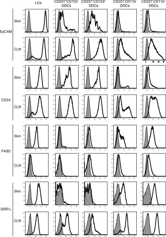
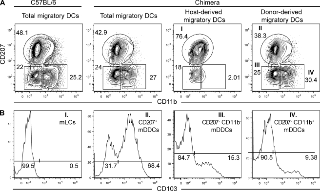
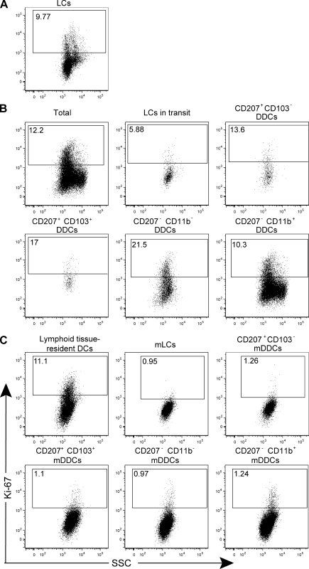
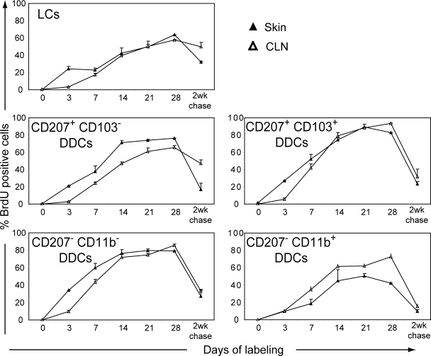
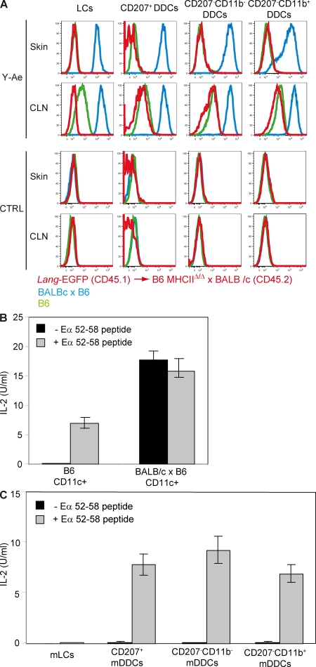
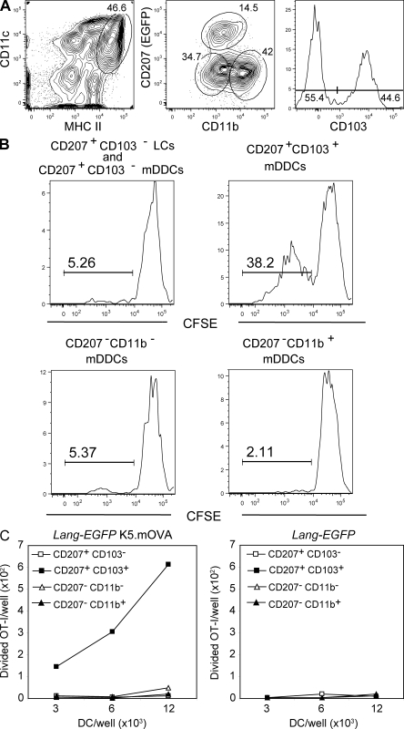
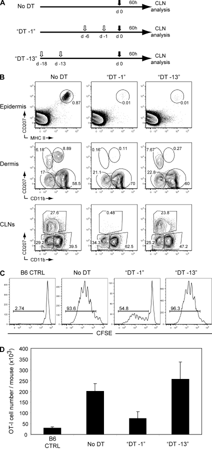
Recent studies have challenged the view that Langerhans cells (LCs) constitute the exclusive antigen-presenting cells of the skin and suggest that the dermal dendritic cell (DDC) network is exceedingly complex. Using knockin mice to track and ablate DCs expressing langerin (CD207), we discovered that the dermis contains five distinct DC subsets and identified their migratory counterparts in draining lymph nodes. Based on this refined classification, we demonstrated that the quantitatively minor CD207+ CD103+ DDC subset is endowed with the unique capability of cross-presenting antigens expressed by keratinocytes irrespective of the presence of LCs. We further showed that Y-Ae, an antibody that is widely used to monitor the formation of complexes involving I-Ab molecules and a peptide derived from the I-E alpha chain, recognizes mature skin DCs that express I-Ab molecules in the absence of I-E alpha. Knowledge of this extra reactivity is important because it could be, and already has been, mistakenly interpreted to support the view that antigen transfer can occur between LCs and DDCs. Collectively, these data revisit the transfer of antigen that occurs between keratinocytes and the five distinguishable skin DC subsets and stress the high degree of functional specialization that exists among them.


Figures

Figure 1.
Figure 1.
The skin contains five distinguishable DC subsets. Flow cytometry analysis of epidermal and dermal cell suspensions from C57BL/6 (B6) and B6 (CD45.1)→B6 (CD45.2) BM chimeras. After gating out autofluorescent cells using the AmCyan channel (den Haan et al., 2000; Wilson et al., 2003), MHCII+ cells were analyzed for expression of langerin (CD207) versus CD11b, and CD103 expression was determined on each of the specified DC subsets. (A) In B6 mice, the epidermis contains CD207high CD11bint LCs that are CD103 and the dermis contains four DC subsets that can be distinguished on the basis of CD11b and CD207 expression. (B) DCs found in the dermis of B6 (CD45.1)→B6 (CD45.2) BM chimeras were segregated into host-derived and donor-derived cells using CD45.1 staining (Fig. S1). Host-derived CD45.1 CD207high CD11bint cells correspond to mLCs that are in transit to CLNs. Donor-derived CD45.1+ DDCs segregate into CD207+ CD11blow, CD207 CD11b+, and CD207 CD11b subsets. (C) Analysis of the expression of CD103 on the four DC subsets present in the dermis showed that it is only expressed on a fraction of the CD207+ CD11blow subset and thus allows us to define five skin DC subsets. The percentages of cells found in each of the specified gates are indicated. Data shown are representative of at least 12 chimeric mice corresponding to six independent experiments.
Figure 2.
Figure 2.
Expression of EpCAM, CD24, F4/80, and SIRPα on the five skin DC subsets before and after their migration to CLNs. DC subsets were prepared as specified in Figs. 1 and 3 from dermis and CLNs from B6 (CD45.1)→B6 (CD45.2) BM chimeras and analyzed for the expression of EpCAM, CD24, F4/80, and SIRPα. Isotype control staining is shown by the shaded histograms. In steady-state skin, LCs and CD207 CD11b+ DDCs were homogeneously F4/80+. CD207 CD11b DDCs showed a heterogeneous pattern of F4/80 expression, whereas both subsets of CD207+ DDCs were F4/80. Upon migration to the CLNs, F4/80 expression was down-regulated on mLCs and CD207 mDDCs. If we except mLCs that showed a slight decrease in Sirpα levels, Sirpα expression was up-regulated or induced on all the remaining skin-derived migratory DC subsets. Data shown are representative of at least six chimeric mice corresponding to three independent experiments.
Figure 3.
Figure 3.
Draining CLNs contain five skin-derived DC subsets. Flow cytometry analysis of the skin-derived migratory DCs (MHCIIhigh CD11cinter-to-high) present in CLNs from C57BL/6 (B6) mice and B6 (CD45.1)→B6 (CD45.2) BM chimeras. MHCIIhigh CD11cinter-to-high DCs were analyzed for the expression of CD207 versus CD11b and CD103 expression determined on each of the specified DC subsets. (A) A single CD207+ CD11b−/low DC cluster is present in the CLNs of B6 mice. Unlike DDCs, LCs are radio resistant, and this permits the use of B6 (CD45.1)→B6 (CD45.2) chimeras to split the single CD207+ CD11b−/low DC cluster into host-derived mLCs and into CD103+ and CD103 donor-derived mDDCs. Two additional mDDC subsets with a CD207+ CD11b and CD207 CD11b+ phenotype can be identified in both C57BL/6 mice and B6 (CD45.1)→B6 (CD45.2) chimeras. (B) Expression of CD103 among the DC subsets found in the dermis of B6 (CD45.1)→B6 (CD45.2) BM chimeras. The percentages of cells found in each of the specified gates are indicated. Data shown are representative of at least 12 chimeric mice corresponding to six independent experiments.
Figure 4.
Figure 4.
The five skin DC subsets proliferate before their migration to LNs. (A–C) The five skin DC subsets (A and B) and their migratory counterparts found in the CLNs (C) were identified using B6 (CD45.1)→B6 (CD45.2) chimeras, as specified in Figs. 1 and 3, and analyzed for Ki-67 expression. Lymphoid tissue–resident DCs were defined on the basis of their CD11chigh MHCIIinter phenotype (Fig. S3). Positioning of the Ki-67+ gate is based on staining with isotype control antibody (Fig. S4). Data shown are representative of three independent experiments.
Figure 5.
Figure 5.
Comparison of the BrdU-labeling kinetics of the five distinct skin DC subsets before and after their migration to CLNs. BrdU was administered continuously for 28 d to a group of two B6 (CD45.1)→B6 (CD45.2) chimeric mice. After 28 d of continuous BrdU administration, a group of B6 (CD45.1)→B6 (CD45.2) chimeras was kept without BrdU and analyzed 2 wk later (2-wk chase). DC subsets were defined as described in Figs. 1 and 3, and the percentage of BrdU+ cells was determined. Data shown are representative of three independent experiments. Positioning of the positive gate is based on staining with isotype control antibody (not depicted). Error bars correspond to the SEM.
Figure 6.
Figure 6.
DDCs do not present Eα52-68 LC-derived peptides. (A) Flow cytometry analysis of LCs, CD207+, CD207 CD11b, and CD207 CD11b+ DDCs isolated from Langerin-EGFP→B6 MHCIIΔ/Δ x BALB/c chimeras (red) and from B6 (green) and B6 x BALB/c (blue) mice before (skin) and after (CLN) their migration to CLN. Cells were separately stained with Y-Ae and isotype control staining (CTRL). (B) CD11c+ DCs isolated from B6 and B6 x BALB/c mice were cultured together with H30 T cells in the presence or absence of 3 µg/ml Eα52-68 peptide. The content of IL-2 present in supernatant was determined after 24 h of culture. (C) LCs, CD207+, CD207 CD11b, and CD207 CD11b+ DDCs isolated from the CLNs of Langerin-EGFP→B6 MHCIIΔ/Δ x BALB/c chimeras were cultured together with H30 T cells that are specific for Eα52-68–I-Ab complexes and in the presence or absence of 3 µg/ml Eα52-68 peptide. The content of IL-2 present in supernatant was determined after 24 h of culture. Data shown are representative of three independent experiments. Error bars correspond to SEM.
Figure 7.
Figure 7.
CD207+ CD103+ mDDCs cross-present a keratinocyte-derived self antigen. (A) Gating strategy for isolation of CD207+ CD103+, CD207 CD11b, and CD207 CD11b+ mDDCs from Lang-EGFP K5.mOVA and Lang-EGFP CLNs. Skin-derived DCs were identified on the basis of their MHCIIhigh CD11cinter-to-high phenotype. Expression of CD207 (EGFP) and CD11b was assessed among MHCIIhigh CD11cinter-to-high DCs, leading to the isolation of CD207 CD11b−/low and CD207 CD11b+ mDDCs. CD207+ cells were categorized on the basis of CD103 expression. CD207+ CD103+ cells corresponded to CD207+ CD103+ mDDCs, whereas CD207+ CD103 cells corresponded to a mix of mLCs and CD207+ CD103 mDDCs. (B) DC subsets, sorted according to the scheme described in A, were cultured with CFSE-labeled OT-I transgenic T cells. Histograms corresponding to CFSE dilution are shown after gating on cells that stain positive with H-2Kb-OVA257-264 tetramers. (C) The absolute numbers of divided OT-I transgenic T cells, in response to DC subsets sorted from Lang-EGFP K5.mOVA or Lang-EGFP CLNs, are presented. Data are representative of three independent experiments.
Figure 8.
Figure 8.
CD207+ CD103+ mDDCs cross-present a keratinocyte-derived self antigen regardless of the presence of mLCs. (A) Experimental scheme for the depletion of LCs alone or of both LCs and CD207+ mDDCs in Lang-DTREGFP x B6 K5.mOVA mice. 1 µg DT (open arrows) was administered i.p. 18 and 13 (DT–13) or 6 and 1 (DT–1) d before adoptive transfer of 5 × 10⁶ CFSE-labeled OT-I CD8+ T cells (filled arrows). (B) Kinetics of reappearance of the various skin (epidermis and dermis) or skin-derived (CLN) DC subsets after DT ablation. DCs from DT–13 and DT–1 Lang-DTREGFP x B6 K5.mOVA mice were gated as MHCII+ (epidermis and dermis) and MHCIIhigh CD11cinter-to-high (CLNs) cells and then analyzed using CD207-MHCII (epidermis) and CD207)-CD11b (dermis and CLN) dot plots. Untreated Lang-DTREGFP x B6 K5.mOVA (no DT) mice are also shown. Windows correspond to LCs (epidermis) and to mLCs, CD207+ DDCs, CD207 CD11b, and CD207 CD11b+ (dermis). In the CLNs, windows correspond to CD207 CD11b, CD207 CD11b+, and mLCs + CD207+ DDCs. Numbers indicate the percentage of cells in the specified windows. (C and D) 60 h after adoptive transfer in the specified mice, the OT-I CD8+ T cells present in pooled skin-draining LNs were analyzed by flow cytometry for the percentage (C) and yield (D) of proliferating OT-I CD8+ T cells. Numbers in C indicate percent proliferated cells ± SEM. Data shown are representative of three independent experiments.

Similar articles

Cited by

References

    1. Allan R.S., Smith C.M., Belz G.T., van Lint A.L., Wakim L.M., Heath W.R., Carbone F.R. 2003. Epidermal viral immunity induced by CD8alpha+ dendritic cells but not by Langerhans cells. Science. 301:1925–1928 doi:10.1126/science.1087576 - DOI - PubMed
    1. Allenspach E.J., Lemos M.P., Porrett P.M., Turka L.A., Laufer T.M. 2008. Migratory and lymphoid-resident dendritic cells cooperate to efficiently prime naive CD4 T cells. Immunity. 29:795–806 doi:10.1016/j.immuni.2008.08.013 - DOI - PMC - PubMed
    1. Azukizawa H., Kosaka H., Sano S., Heath W.R., Takahashi I., Gao X.H., Sumikawa Y., Okabe M., Yoshikawa K., Itami S. 2003. Induction of T-cell-mediated skin disease specific for antigen transgenically expressed in keratinocytes. Eur. J. Immunol. 33:1879–1888 doi:10.1002/eji.200323630 - DOI - PubMed
    1. Bedoui S., Whitney P.G., Waithman J., Eidsmo L., Wakim L., Caminschi I., Allan R.S., Wojtasiak M., Shortman K., Carbone F.R., et al. 2009. Cross-presentation of viral and self antigens by skin-derived CD103+ dendritic cells. Nat. Immunol. 10:488–495 doi:10.1038/ni.1724 - DOI - PubMed
    1. Bennett C.L., Noordegraaf M., Martina C.A., Clausen B.E. 2007. Langerhans cells are required for efficient presentation of topically applied hapten to T cells. J. Immunol. 179:6830–6835 - PubMed

Publication types

MeSH terms