

Generation of PLZF+ CD4+ T cells via MHC class II-dependent thymocyte-thymocyte interaction is a physiological process in humans
- PMID: 20038602
- PMCID: PMC2812550
- DOI: 10.1084/jem.20091519
Generation of PLZF+ CD4+ T cells via MHC class II-dependent thymocyte-thymocyte interaction is a physiological process in humans
Abstract
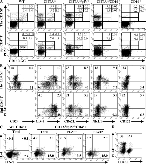
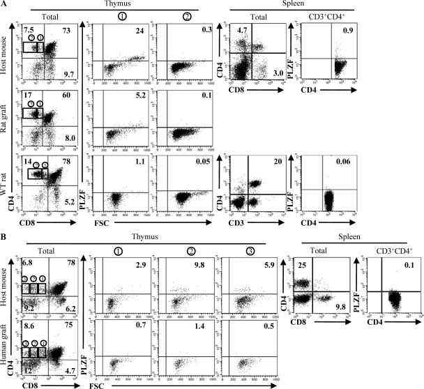
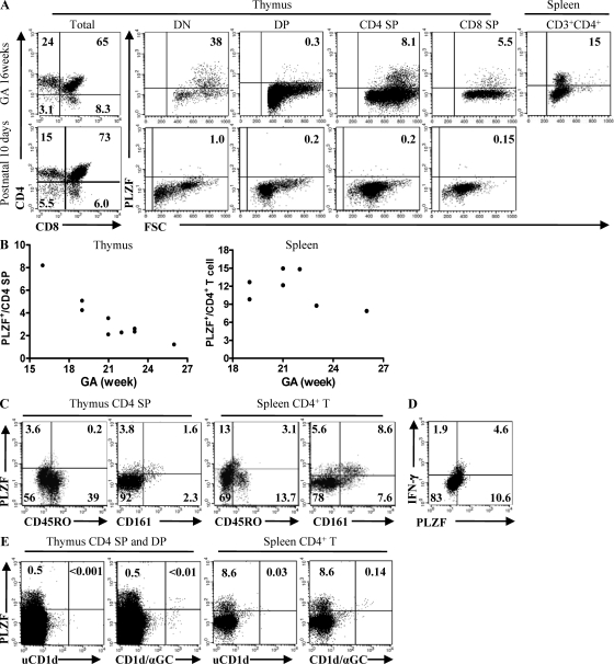
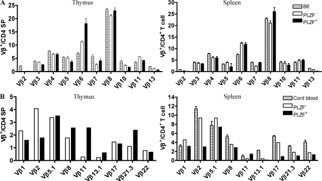
Human thymocytes, unlike mouse thymocytes, express major histocompatibility complex (MHC) class II molecules on their surface, especially during the fetal and perinatal stages. Based on this observation, we previously identified a novel developmental pathway for the generation of CD4+ T cells via interactions between MHC class II-expressing thymocytes (thymocyte-thymocyte [T-T] interactions) with a transgenic mouse system. However, the developmental dissection of this T-T interaction in humans has not been possible because of the lack of known cellular molecules specific for T-T CD4+ T cells. We show that promyelocytic leukemia zinc finger protein (PLZF) is a useful marker for the identification of T-T CD4+ T cells. With this analysis, we determined that a substantial number of fetal thymocytes and splenocytes express PLZF and acquire innate characteristics during their development in humans. Although these characteristics are quite similar to invariant NKT (iNKT) cells, they clearly differ from iNKT cells in that they have a diverse T cell receptor repertoire and are restricted by MHC class II molecules. These findings define a novel human CD4+ T cell subset that develops via an MHC class II-dependent T-T interaction.
Figures
Similar articles
-
MHC class II-restricted interaction between thymocytes plays an essential role in the production of innate CD8+ T cells.J Immunol. 2011 May 15;186(10):5749-57. doi: 10.4049/jimmunol.1002825. Epub 2011 Apr 8. J Immunol. 2011. PMID: 21478404 Free PMC article.
-
CD4(+) T cells from MHC II-dependent thymocyte-thymocyte interaction provide efficient help for B cells.Immunol Cell Biol. 2011 Nov;89(8):897-903. doi: 10.1038/icb.2011.8. Epub 2011 Mar 1. Immunol Cell Biol. 2011. PMID: 21358747 Free PMC article.
-
Innate PLZF+CD4+ αβ T cells develop and expand in the absence of Itk.J Immunol. 2014 Jul 15;193(2):673-87. doi: 10.4049/jimmunol.1302058. Epub 2014 Jun 13. J Immunol. 2014. PMID: 24928994 Free PMC article.
-
MHC class II-dependent T-T interactions create a diverse, functional and immunoregulatory reaction circle.Immunol Cell Biol. 2009 Jan;87(1):65-71. doi: 10.1038/icb.2008.85. Epub 2008 Nov 25. Immunol Cell Biol. 2009. PMID: 19030015 Review.
-
Development of PLZF-expressing innate T cells.Curr Opin Immunol. 2011 Apr;23(2):220-7. doi: 10.1016/j.coi.2010.12.016. Epub 2011 Jan 21. Curr Opin Immunol. 2011. PMID: 21257299 Free PMC article. Review.
Cited by
-
Leishmania major infection in humanized mice induces systemic infection and provokes a nonprotective human immune response.PLoS Negl Trop Dis. 2012;6(7):e1741. doi: 10.1371/journal.pntd.0001741. Epub 2012 Jul 24. PLoS Negl Trop Dis. 2012. PMID: 22848771 Free PMC article.
-
Natural Th1 cells: escape from neglect.Oncotarget. 2015 Sep 8;6(26):21795-6. doi: 10.18632/oncotarget.5489. Oncotarget. 2015. PMID: 26392409 Free PMC article. No abstract available.
-
Alternative memory in the CD8 T cell lineage.Trends Immunol. 2011 Feb;32(2):50-6. doi: 10.1016/j.it.2010.12.004. Epub 2011 Feb 1. Trends Immunol. 2011. PMID: 21288770 Free PMC article. Review.
-
PLZF(+) Innate T Cells Support the TGF-β-Dependent Generation of Activated/Memory-Like Regulatory T Cells.Mol Cells. 2016 Jun 30;39(6):468-76. doi: 10.14348/molcells.2016.0004. Epub 2016 Apr 20. Mol Cells. 2016. PMID: 27101876 Free PMC article.
-
Innate memory T cells.Adv Immunol. 2015;126:173-213. doi: 10.1016/bs.ai.2014.12.001. Epub 2015 Feb 7. Adv Immunol. 2015. PMID: 25727290 Free PMC article. Review.
References
Publication types
MeSH terms
Substances
LinkOut - more resources
Full Text Sources
Other Literature Sources
Molecular Biology Databases
Research Materials
