. 2010 Mar;184(3):707-16.
doi: 10.1534/genetics.109.113480. Epub 2009 Dec 28.

The Rim101p/PacC pathway and alkaline pH regulate pattern formation in yeast colonies

Affiliations

The Rim101p/PacC pathway and alkaline pH regulate pattern formation in yeast colonies

Sarah Piccirillo et al. Genetics. 2010 Mar.

Abstract

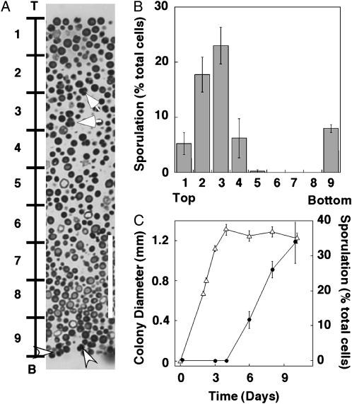
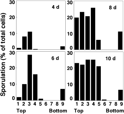
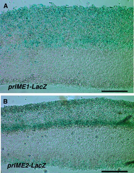
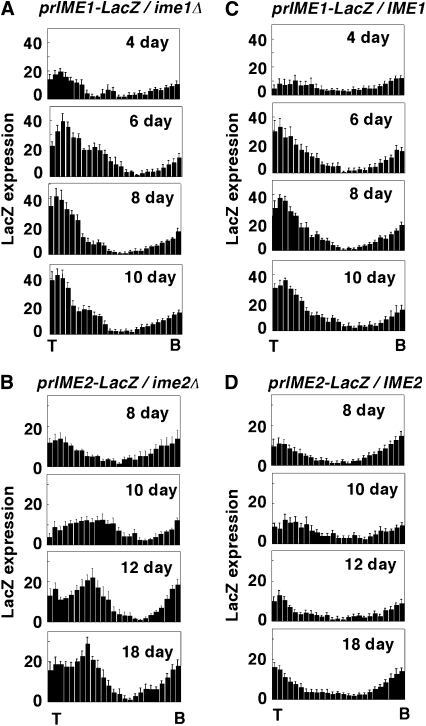
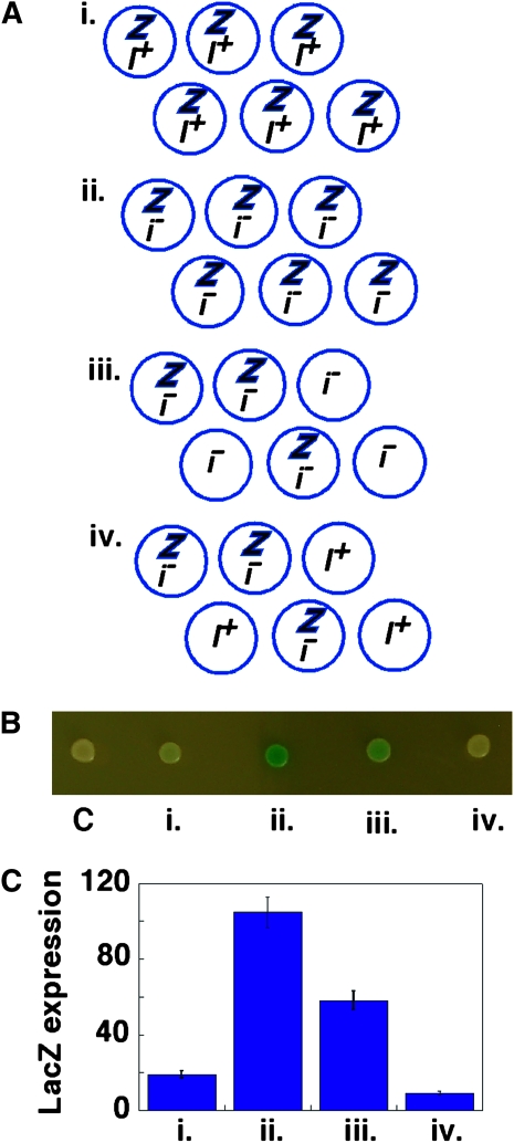
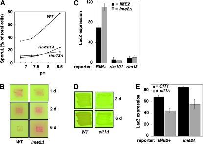
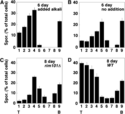
Multicellular organisms utilize cell-to-cell signals to build patterns of cell types within embryos, but the ability of fungi to form organized communities has been largely unexplored. Here we report that colonies of the yeast Saccharomyces cerevisiae formed sharply divided layers of sporulating and nonsporulating cells. Sporulation initiated in the colony's interior, and this region expanded upward as the colony matured. Two key activators of sporulation, IME1 and IME2, were initially transcribed in overlapping regions of the colony, and this overlap corresponded to the initial sporulation region. The development of colony sporulation patterns depended on cell-to-cell signals, as demonstrated by chimeric colonies, which contain a mixture of two strains. One such signal is alkaline pH, mediated through the Rim101p/PacC pathway. Meiotic-arrest mutants that increased alkali production stimulated expression of an early meiotic gene in neighboring cells, whereas a mutant that decreased alkali production (cit1Delta) decreased this expression. Addition of alkali to colonies accelerated the expansion of the interior region of sporulation, whereas inactivation of the Rim101p pathway inhibited this expansion. Thus, the Rim101 pathway mediates colony patterning by responding to cell-to-cell pH signals. Cell-to-cell signals coupled with nutrient gradients may allow efficient spore formation and spore dispersal in natural environments.


Figures

F<sc>igure</sc> 1.—
Figure 1.—
Patterns of sporulation in wild-type colonies. Approximately 300 cells/plate were inoculated on YNA medium and grown for 8 days at 30°. Isolated colonies were sectioned and then examined by light microscopy. (A) Cross section of embedded colony. Bar, 0.4 mm. (B) Higher magnification of approximately the region indicated by a box in A. The region of sporulation is indicated by a bracket marked “S”, and the unsporulated region indicated by a bracket marked “U”. The arrow indicates representative tetrad asci (four spores) and the arrowhead indicates representative dyad asci (two spores). Bar, 25 μm.
F<sc>igure</sc> 2.—
Figure 2.—
Sporulation initiates in central region of colonies. (A) A longitudinal section through the center of the colony, with the top (“T”) and bottom (“B”) of the colony indicated. To the left of the colony are 10 equally spaced marks that subdivide the colony into nine regions. Arrows indicate representative tetrad asci (four spores) and arrowheads indicate representative dyad asci (two spores). Bar, 50 μm. (B) Sporulation distribution in 6-day colonies. Bars represent percentage of sporulation in each of the nine regions of each image, as shown in A, from the top of the colony (left) to the bottom (right) (n = 4). (C) Timing of growth and sporulation in colonies. Colony diameter (open triangles) and percentage of sporulation (solid circles) are shown in colonies (SH1020) grown for various times on YNA medium (n = 5).
F<sc>igure</sc> 3.—
Figure 3.—
Time course of spore distribution in wild-type colonies. Wild-type colonies were sectioned after 4, 6, 8, or 10 days on YNA medium, and the spore distribution was analyzed as in Figure 2. A single colony in the height range of 0.8–1.0 mm was analyzed at each of the indicated times.
F<sc>igure</sc> 4.—
Figure 4.—
Expression of prIME1-LacZ and prIME2-LacZ in colonies. Colonies were sectioned after 12 days incubation on YNA medium containing X-gal. (A) prIME1-LacZ/ime1Δ strain (SH3830); (B) prIME2-LacZ/ime2Δ strain (SH3825). Bar, 0.1 mm.
F<sc>igure</sc> 5.—
Figure 5.—
Time course of prIME1-LacZ and prIME2-LacZ expression patterns and requirement of Ime1p or Ime2p for these patterns. Colonies were incubated for the indicated times on YNA medium containing X-gal and sectioned, and the distribution of LacZ expression was quantified as described in materials and methods (n = 3). Each bar in the distribution from left to right represents the average LacZ expression level in one of 25 bins from the top of the colony (“T”) to the bottom (“B”). (A) prIME1-LacZ expression levels in a prIME1-LacZ/ime1Δ strain; (B) prIME2-LacZ expression levels in a prIME2-LacZ/ime2Δ strain; (C) prIME1-LacZ expression levels in prIME1-LacZ/IME1 strain; (D) prIME2-LacZ expression levels in a prIME2-LacZ/IME2 strain.
F<sc>igure</sc> 6.—
Figure 6.—
Cell-to-cell signaling regulates sporulation efficiency in colonies. (A) Diagram represents chimeric colony experiments, in which prIME2-LacZ expression was compared in four colonies containing the following genotypes: (i) prIME2-LacZ/IME2 (SH3824) alone, (ii) prIME2-LacZ/ime2Δ (SH3825) alone, (iii) equal numbers of prIME2-LacZ/ime2Δ and ime2Δ/ime2Δ (SH3883), and (iv) equal numbers of prIME2-LacZ/ime2Δ and IME2/IME2 (SH3881). Z, prIME2-LacZ/ime2Δ; i, ime2Δ/ime2Δ; I+, IME2/IME2. (B) Colonies diagrammed in A grown for 6 days in YNA-2 medium. As a control (c), a LacZ colony (SH3881) was also spotted. (C) Quantification of pr-IME2-LacZ expression in colonies as described above (n = 5).
F<sc>igure</sc> 7.—
Figure 7.—
Alkaline pH, mediated through the Rim101p/PacC pathway, regulates colony sporulation efficiency. (A) Effect of pH on sporulation levels of wild type (SH3881), rim101Δ (SH4375), and rim13Δ (SH4415). Colonies were grown for 6 days on YNA-2 medium buffered to the indicated pH, and the percentage of cells in colonies that formed spores was assayed by microscopy after resuspending the complete colony (n = 3). (B) Production of alkali in wild type (WT) and ime2Δ mutant grown for 1, 2, or 6 days on YNA-2 media containing the pH indicator, phenol red. Red color in and around the patches of cells indicates an increased secretion of alkali from this strain. (C) RIM101 and RIM13 required to respond to neighboring cells. As indicated, chimeric colonies contained a prIME2-LacZ/ime2Δ reporter strain that is RIM+ (SH3825), rim101Δ (SH4378), or rim13Δ (SH4417) and signal strains that are either IME2+(SH3881) or ime2Δ (SH3883). LacZ expression was quantified after 12 days of growth to detect low rimΔ expression. Black bars indicate chimeras containing IME2+ signal strain, and gray bars indicate chimeras containing ime2Δ signal strain (n = 3). (D) CIT1 is required for alkali production. CIT1 (SH3881) and cit1Δ (SH4436) strains were patched to phenol red indicator medium containing both glucose and acetate (SPS2-0.3) to allow growth of the cit1Δ strain and photographed after 2 or 6 days of incubation. (E) CIT1 is required to stimulate sporulation in neighboring cells. As indicated, chimeric colonies contained prIME2-LacZ/IME2 reporter cells (SH3824) plus either the CIT1 or the cit1Δ strain described in D as signal cells. Black bars indicate chimeras containing CIT1+ signal strain, and gray bars indicate cit1Δ signal strain (n = 3). To ensure approximately equal numbers of cit1Δ and reporter cells in the mature chimeras, all chimeras were grown on medium containing both glucose and acetate (SPS2-0.3). Even in this medium, growth of the cit1Δ mutant is less than that of the wild type, so generation of mature chimeras containing approximately equal numbers of wild-type and cit1Δ strains required inoculating colonies with a 1:6 ratio of IME2 reporter to cit1Δ strain and a 1:10 ratio of ime2Δ reporter to cit1Δ strain; genotyping of 6-day chimeric colonies (see materials and methods) revealed that all contained from 40 to 50% reporter cells.
F<sc>igure</sc> 8.—
Figure 8.—
Effect of pH and Rim101p on sporulation patterns. (A and B) Addition of alkaline buffer expands sporulation region in colonies. Wild-type colonies (SH3881) were grown on YNA medium for 3 days under standard conditions before 1.0 m sodium phosphate buffer (pH 8.0) was added (A) or not (B) to a well at the center of the plate. Incubation was continued for 3 additional days, and then colonies were sectioned and analyzed for the distribution of sporulated cells in the colony as in Figure 2B (n = 3). (C and D) Rim101p is required for expansion of sporulation region in colonies. Either rim101Δ colonies (C) or wild-type colonies (D) were incubated for 8 days before colonies were sectioned and analyzed for the distribution of sporulated cells in the colony as in Figure 2B (n = 3).

Similar articles

Cited by

References

    1. Baillie, G. S., and L. J. Douglas, 1999. Role of dimorphism in the development of Candida albicans biofilms. J. Med. Microbiol. 48 671–679. - PubMed
    1. Colomina, N., E. Gari, C. Gallego, E. Herrero and M. Aldea, 1999. G1 cyclins block the Ime1 pathway to make mitosis and meiosis incompatible in budding yeast. EMBO J. 18 320–329. - PMC - PubMed
    1. Davis, D. A., 2009. How human pathogenic fungi sense and adapt to pH: the link to virulence. Curr. Opin. Microbiol. 12 365–370. - PubMed
    1. Diezmann, S., and F. S. Dietrich, 2009. Saccharomyces cerevisiae: population divergence and resistance to oxidative stress in clinical, domesticated and wild isolates. PLoS One 4 e5317. - PMC - PubMed
    1. Guttmann-Raviv, N., S. Martin and Y. Kassir, 2002. Ime2, a meiosis-specific kinase in yeast, is required for destabilization of its transcriptional activator, Ime1. Mol. Cell. Biol. 22 2047–2056. - PMC - PubMed

Publication types

MeSH terms

Substances