. 2009 Dec 28;187(7):1037-54.
doi: 10.1083/jcb.200904158.

Cellular IAPs inhibit a cryptic CD95-induced cell death by limiting RIP1 kinase recruitment

Affiliations

Cellular IAPs inhibit a cryptic CD95-induced cell death by limiting RIP1 kinase recruitment

Peter Geserick et al. J Cell Biol. .

Abstract

A role for cellular inhibitors of apoptosis (IAPs [cIAPs]) in preventing CD95 death has been suspected but not previously explained mechanistically. In this study, we find that the loss of cIAPs leads to a dramatic sensitization to CD95 ligand (CD95L) killing. Surprisingly, this form of cell death can only be blocked by a combination of RIP1 (receptor-interacting protein 1) kinase and caspase inhibitors. Consistently, we detect a large increase in RIP1 levels in the CD95 death-inducing signaling complex (DISC) and in a secondary cytoplasmic complex (complex II) in the presence of IAP antagonists and loss of RIP1-protected cells from CD95L/IAP antagonist-induced death. Cells resistant to CD95L/IAP antagonist treatment could be sensitized by short hairpin RNA-mediated knockdown of cellular FLICE-inhibitory protein (cFLIP). However, only cFLIP(L) and not cFLIP(S) interfered with RIP1 recruitment to the DISC and complex II and protected cells from death. These results demonstrate a fundamental role for RIP1 in CD95 signaling and provide support for a physiological role of caspase-independent death receptor-mediated cell death.


Figures

Figure 1.
Figure 1.
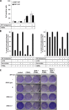
The IAP antagonist sensitizes SCC and HaCaT to DL-mediated apoptosis independent of autocrine TNF secretion. (A) HaCaT, MET1, or A5RT3 cells were either pretreated with 100 nM of the IAP antagonist (Ant) alone or in combination with 10 µg/ml TNF-R2-Fc for 30 min and then stimulated with TRAIL or CD95L. The viability of cells was analyzed by crystal violet assay after 18–24 h as indicated in Materials and methods. Mean and SEM of four independent experiments are shown. (B) For clonogenic assays, HaCaT cells were prestimulated with 100 nM of the IAP antagonist for 30 min followed by co-stimulation with 2.5 U/ml CD95L for 24 h. Colony formation was assayed as indicated in Materials and methods. One representative experiment of a total of three independent experiments is shown. (C–E) HaCaT cells were either prestimulated with 100 nM of the IAP antagonist for 30 min alone or stimulated/co-stimulated with 10 U/ml CD95L. (C) Cells were stained with annexin V–Cy5 and PI after 4 h and analyzed by FACS. (D) Cells were incubated for 8 h and subsequently analyzed for hypodiploidy by FACS analysis (see Materials and methods). (E) Cells were treated with 100 nM of the IAP antagonist, 2.5 U/ml CD95L, or the combination of both in the presence or absence of 10 µg/ml TNF-R2-Fc for the indicated time points. Western blot analysis was performed for the expression of cIAP1 and -2, caspase-8 and -3, PARP-1, FADD, and RIP1. β-Tubulin served as an internal loading control. One of two representative experiments is shown. The asterisk marks an unspecific band. MM, molecular mass.
Figure 2.
Figure 2.
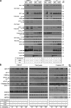
cIAPs specifically block CD95L-induced cell death of MEFs and human SCCs. (A) Transformed single knockout or DKO MEFs and respective control wild-type (WT) MEFs were treated with 10 ng/ml CD95L or 500 ng/ml TRAIL for 24 h in the presence or absence of 500 nM of the IAP antagonist (left). Cells were stained with PI and analyzed by flow cytometry. Four independent cIAP1 and -2 DKO MEFs were infected with inducible mouse cIAP1 or Flag-cIAP2 and induced with 10 nM 4-HT for 24 h. Cells were then treated with CD95L and TRAIL for 24 h in the presence or absence of the IAP antagonist, stained with PI, and analyzed by flow cytometry. The mean + SEM is shown throughout. (B) DKO 1.5 MEFs, infected with inducible mouse cIAP1 and Flag-cIAP2, were induced for 24 h with 4-HT. cIAP1 or Flag-cIAP2 expression was analyzed by Western blotting. β-Actin served as a loading control. (C) DKO 2.7 and DKO 3.1 MEFs were infected with inducible mouse XIAP and induced with the indicated concentrations of 4-HT for 24 h. Induction of XIAP was verified by Western blot analysis. Cells were treated with 20 ng/ml CD95L for 24 h in the presence or absence of 500 nM of the IAP antagonist, stained with PI, and analyzed by FACS. The mean + SEM of a total of six independent experiments is shown. (D) HaCaT cells were transduced with control vector or shRNA against cIAP1 or -2 or both. Western blot analysis shows expression of cIAP1 and -2. β-Actin served as a loading control. Subsequently, cells were analyzed for sensitivity to 2.5 U/ml CD95L in the presence or absence of 100 nM of the IAP antagonist 24 h later by crystal violet assay. (E) HaCaT cells were transduced with lentiviral control vector or inducible cIAP2 as previously described (Diessenbacher et al., 2008). Cells were induced with 4-HT as indicated and then treated with 2.5 U/ml CD95L for 24 h in the presence or absence of 100 nM of the IAP antagonist. Viability was examined by crystal violet assay 24 h later. (D and E) Mean + SEM of three independent experiments is shown. MM, molecular mass.
Figure 3.
Figure 3.
DR-mediated cell death in the presence of the IAP antagonist uses caspases and caspase-independent signaling pathways. (A) HaCaT cells were pre- or co-stimulated with 10 µM zVAD-fmk for 1 h and 100 nM of the IAP antagonist for 30 min. Subsequently, cells were stimulated with the indicated concentration of TRAIL or CD95L. The viability of cells was analyzed by crystal violet assay 18–24 h later as indicated in Materials and methods. Mean + SEM for three (TRAIL) or six (CD95L) independent experiments is shown. (B) HaCaT cells were either pretreated with 10 µM zVAD-fmk for 1 h or 100 nM of the IAP antagonist for 30 min. Cells were subsequently stimulated with 5 U/ml CD95L for 4 or 24 h. 5 µg/ml Hoechst 33342 and 5 pM SYTOX green were added for 15 min at 37°C, immediately followed by transmission (left) or fluorescence (right) microscopy. One of two independent experiments is representatively shown. (C) Stable knockdown of RIP1 in HaCaT cells was performed as indicated in Materials and methods and controlled by Western blot analysis for RIP1. Reprobing of the membrane with β-tubulin antibodies served as a control for protein loading. MM, molecular mass. (D) Transduced HaCaT cells as shown in C were prestimulated for 30 min with 100 nM of the IAP antagonist or diluent alone and subsequently stimulated with the indicated concentrations of TRAIL or CD95L for 18–24 h followed by crystal violet assay. Mean ± SEM of three (TRAIL) or four (CD95L) independent experiments is shown. (E) Transduced HaCaT cells, as described in C, were preincubated with 100 nM of the IAP antagonist (Ant) for 30 min and then stimulated with 0.5 U/ml CD95L. After 24 h, colony formation was assayed as indicated in Materials and methods. One of four representative independent experiments is shown. Bar, 10 µm.
Figure 4.
Figure 4.
RIP1 is an important regulator of DL-mediated cell death in the absence of cIAPs. (A) RIP1 knockout (KO) or wild type (WT) MEFs were stimulated with 10 ng/ml CD95L or 10 ng/ml CD95L and 500 nM of the IAP antagonist for 24 h and then assayed for cell viability using PI and flow cytometry. The mean + SEM of a minimum of three independent experiments is shown. (B) The combination of caspase inhibitor zVAD-fmk and RIP1K inhibitor Necrostatin-1 completely protects HaCaT cells from DL-mediated cell death in the presence of the IAP antagonist. HaCaT cells were separately prestimulated with 10 µM zVAD-fmk for 1 h, 50 µM Necrostatin-1 for 1 h, and 100 nM of the IAP antagonist for 30 min, followed by stimulation with 50 ng/ml TRAIL or 2.5 U/ml CD95L for 18–24 h and subsequent crystal violet assay. Mean + SEM of three (TRAIL) or six (CD95L) independent experiments is shown. (C) Transformed cIAP1 and -2 DKO 2.3 and 2.7 MEFs were treated with 10 ng/ml CD95L for 24 h in the presence or absence of 10 µM QVD and/or 50 µM Necrostatin-1 (Necro). Respective control wild-type MEFs were treated as the DKO MEFs but in the presence or absence of the IAP antagonist (Ant). Subsequently, cells were harvested, and an aliquot was replated in 6-well plates and cultured for another 4 d followed by crystal violet staining of colonies.
Figure 5.
Figure 5.
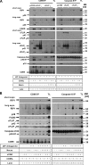
Induction of ligand-induced receptor-bound CD95 complex (DISC) or intracellular caspase-8–containing complex (complex II) in the presence or absence of the IAP antagonist. (A) The CD95 DISC was precipitated from MET1 or A5RT3 cells preincubated with 10 µM zVAD-fmk and 100 nM of the IAP antagonist for 1 h and subsequently treated with CD95L-Fc for 2 h. CD95L DISC (left) was precipitated as detailed in Materials and methods. Precipitation of receptor complexes after lysis (−) served as internal specificity control when compared with ligand affinity precipitates (IP; +). Equal amounts of DISC (CD95L IP) or complex II (caspase-8 IP) were subsequently analyzed by Western blotting for the indicated molecules. Equal amounts of total cellular lysates (TL) were loaded on the same gels to allow comparison of signal strength between CD95L-IP, complex II, and total cellular lysates. (B) Kinetics of DISC (left) or complex II (right) in the presence or absence of the IAP antagonist. The CD95 DISC was precipitated from parental HaCaT cells either prestimulated with 100 nM of the IAP antagonist and 10 µM zVAD-fmk for 1 h alone or the combination of both and subsequently stimulated with 250 U/ml CD95L for the indicated times. CD95L DISC (left) or complex II (right) was precipitated as detailed in Materials and methods and specified for A. MM, molecular mass.
Figure 6.
Figure 6.
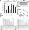
cFLIP is an important regulator of DL-mediated cell death in the absence of cIAPs. (A and B) HaCaT cells were transduced with cFLIPL (A) or cFLIPS (B) or control vector. Total cellular lysates were analyzed for cFLIP and caspase-8. β-Tubulin served as an internal control for protein loading. The mean + SEM of six independent experiments is shown. (C) Cells were prestimulated with 10 µM zVAD-fmk for 1 h, 50 µM Necrostatin-1 for 1 h, and 100 nM of the IAP antagonist for 30 min or diluent alone. Subsequently, cells were stimulated with 2.5 U/ml CD95L. The viability of cells was analyzed by crystal violet assay after 18–24 h. MM, molecular mass. (D) Transduced HaCaT cells were prestimulated with 100 nM of the IAP antagonist (Ant) for 30 min followed by co-stimulation with 2.5 U/ml CD95L. 24 h later, colony formation assay was performed as described in Materials and methods. One representative experiment of a total of three independent experiments is shown.
Figure 7.
Figure 7.
cFLIPL but not cFLIPS blocks the formation of complex II. (A) DISC or complex II formation in the presence or absence of the IAP antagonist. To allow for caspase activity, these experiments were performed without zVAD-fmk. HaCaT cells were stimulated with CD95L-Fc for 2 h. Subsequently, the CD95L DISC (left) was precipitated using ligand affinity precipitation as detailed in Materials and methods. Precipitation of receptor complexes after lysis (−) served as an internal specificity control when compared with ligand affinity precipitates (IP; +). Equal amounts of DISC (CD95L IP) or complex II (caspase-8 IP) were subsequently analyzed by Western blotting for the indicated molecules. Equal amounts of total cellular lysates (TL) were loaded on the same gels to allow comparison of signal strength between IP and total cellular lysates. (B) DISC or complex II formation in parental HaCaT. Cells were either pre- or co-stimulated with 10 µM zVAD-fmk, 50 µM Necrostatin-1, and 100 nM of the IAP antagonist for 1 h and subsequently stimulated with 250 U/ml CD95L for 2 h. The CD95L DISC (left) or complex II (right) was precipitated as detailed in Materials and methods and specified for A. Equal amounts of DISC (CD95L IP) or caspase-8–interacting proteins (complex II) were subsequently analyzed by Western blotting for the indicated molecules. MM, molecular mass.
Figure 8.
Figure 8.
TWEAK sensitizes to CD95L-induced cell death in a RIP1-dependent manner and is negatively regulated by cFLIPL but not cFLIPS. (A) HaCaT and MET1 cells were either prestimulated with 0.5 ng/ml TWEAK for 2 h or 100 nM of the IAP antagonist (Ant) for 30 min and then treated with 2.5 U/ml CD95L. In the same experiments, HaCaT cells were treated with 10 µM zVAD-fmk for 1 h, 50 µM Necrostatin-1 for 1 h, and 0.5 ng/ml TWEAK for 2 h. Subsequently, cells were stimulated with 2.5 U/ml CD95L. Viability was analyzed by crystal violet assay after 18–24 h. (B) TWEAK leads to rapid down-regulation of cIAP1 and -2 expression in HaCaT. Cells were stimulated with 0.5 ng/ml TWEAK for the indicated time. Subsequently, total cellular lysates were analyzed for expression of cIAP1 or -2 by Western blotting. β-Tubulin served as a loading control. MM, molecular mass. (C) Stable knockdown of RIP1 protects HaCaT cells from CD95L-induced cell death in the presence of TWEAK. Transduced HaCaT cells as shown in Fig. 3 C were prestimulated for 2 h with 0.5 ng/ml TWEAK or diluent alone, subsequently stimulated with CD95L for 18–24 h, and assayed by crystal violet assay. The mean ± SEM of three independent experiments is shown. (D) cFLIPL but not cFLIPS protects HaCaT cells from CD95L-induced cell death in the presence of TWEAK. cFLIPL, cFLIPS, and vector-transduced control HaCaT as specified in Fig. 6 C were prestimulated with 10 µM zVAD-fmk for 1 h, 50 µM Necrostatin-1 for 1 h, and 0.5 ng/ml TWEAK for 2 h or diluent alone. Cells were then stimulated with 2.5 U/ml CD95L, and viability of cells was analyzed by crystal violet assay after 18–24 h. (A and D) The mean + SEM of three independent experiments is shown.
Figure 9.
Figure 9.
The role of cIAPs during DR-mediated cell death. cIAPs block recruitment to or degradation of RIP1 in the DISC. This signaling platform induces cell death in a caspase-dependent as well as -independent manner. A secondary complex II, which is critical for necrotic cell death, also contains the initiator caspase-8 and FADD. In the presence of high levels of RIP1, RIP1 might be autoactivated and induce necrotic cell death or may require additional binding partners such as RIP3 (Cho et al., 2009; He et al., 2009; Zhang et al., 2009). cFLIPL but not cFLIPS is able to block complex II formation and the pronecrotic activity of RIP1 in complex II. In contrast, both isoforms block apoptotic cell death initiated by caspase-8 at the DISC. Caspase-8–mediated cleavage of RIP1 is one hypothetical mechanism of down-regulation of RIP1 within the complexes. Alternatively, RIP1 is only recruited to the DISC when ubiquitylated. cIAPs transfer ubiquitin chains of currently debated specificity (e.g., Lys63, -48, or -11) to its substrate RIP1 (Park et al., 2004; Bertrand et al., 2008; Varfolomeev et al., 2008; Blankenship et al., 2009).

Similar articles

Cited by

References

    1. Ashkenazi A. 2008. Targeting the extrinsic apoptosis pathway in cancer. Cytokine Growth Factor Rev. 19:325–331 10.1016/j.cytogfr.2008.04.001 - DOI - PubMed
    1. Bell B.D., Leverrier S., Weist B.M., Newton R.H., Arechiga A.F., Luhrs K.A., Morrissette N.S., Walsh C.M. 2008. FADD and caspase-8 control the outcome of autophagic signaling in proliferating T cells. Proc. Natl. Acad. Sci. USA. 105:16677–16682 10.1073/pnas.0808597105 - DOI - PMC - PubMed
    1. Bertrand M.J., Milutinovic S., Dickson K.M., Ho W.C., Boudreault A., Durkin J., Gillard J.W., Jaquith J.B., Morris S.J., Barker P.A. 2008. cIAP1 and cIAP2 facilitate cancer cell survival by functioning as E3 ligases that promote RIP1 ubiquitination. Mol. Cell. 30:689–700 10.1016/j.molcel.2008.05.014 - DOI - PubMed
    1. Blankenship J.W., Varfolomeev E., Goncharov T., Fedorova A.V., Kirkpatrick D.S., Izrael-Tomasevic A., Phu L., Arnott D., Aghajan M., Zobel K., et al. 2009. Ubiquitin binding modulates IAP antagonist-stimulated proteasomal degradation of c-IAP1 and c-IAP2(1). Biochem. J. 417:149–160 10.1042/BJ20081885 - DOI - PubMed
    1. Boatright K.M., Deis C., Denault J.B., Sutherlin D.P., Salvesen G.S. 2004. Activation of caspases-8 and -10 by FLIP(L). Biochem. J. 382:651–657 10.1042/BJ20040809 - DOI - PMC - PubMed

Publication types

MeSH terms

Substances