. 2010 Jan 19;121(2):276-92.
doi: 10.1161/CIRCULATIONAHA.109.895771. Epub 2009 Dec 28.

Anthracycline cardiomyopathy is mediated by depletion of the cardiac stem cell pool and is rescued by restoration of progenitor cell function

Affiliations

Anthracycline cardiomyopathy is mediated by depletion of the cardiac stem cell pool and is rescued by restoration of progenitor cell function

Antonella De Angelis et al. Circulation. .

Abstract

Background: Anthracyclines are the most effective drugs available in the treatment of neoplastic diseases; however, they have profound consequences on the structure and function of the heart, which over time cause a cardiomyopathy that leads to congestive heart failure.

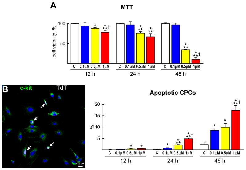
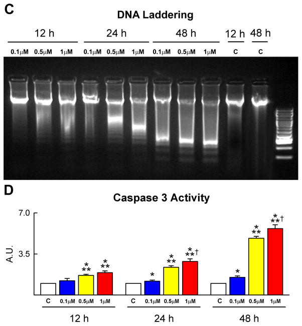
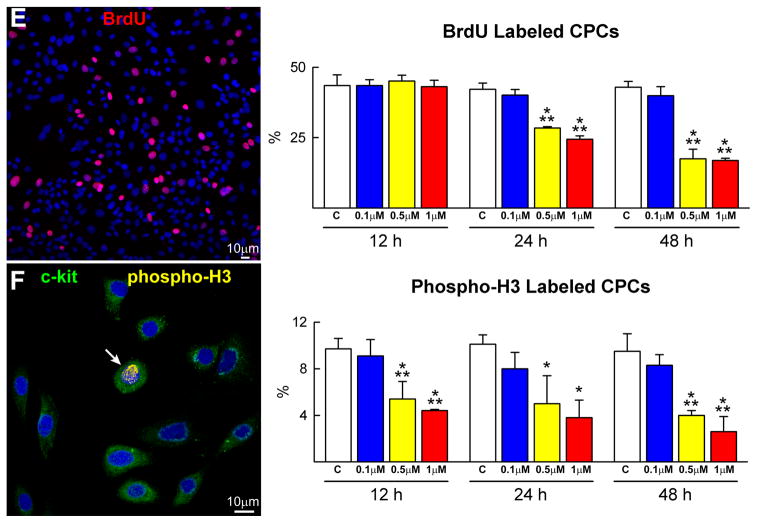
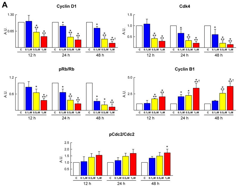
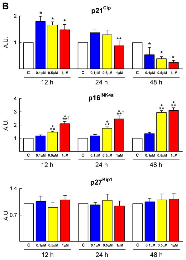
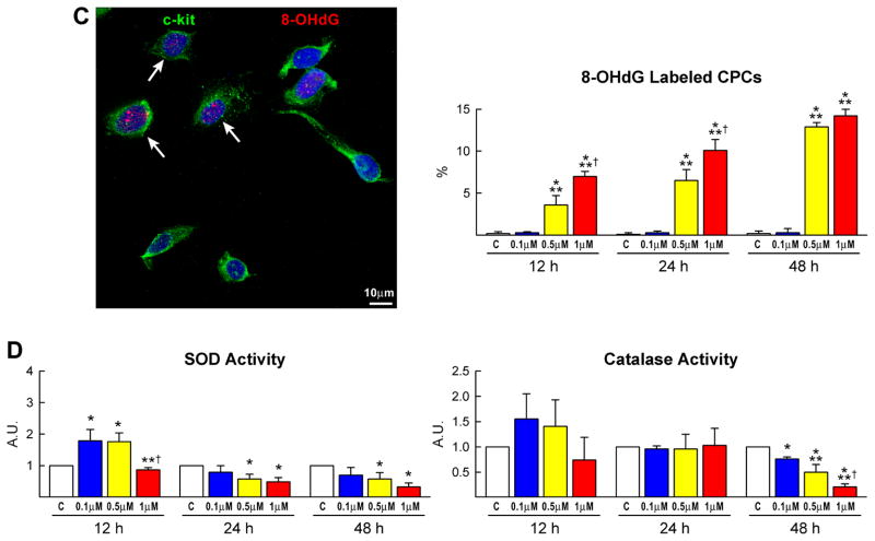
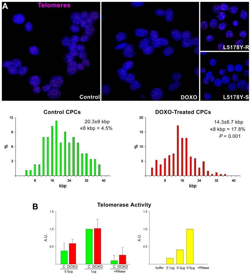
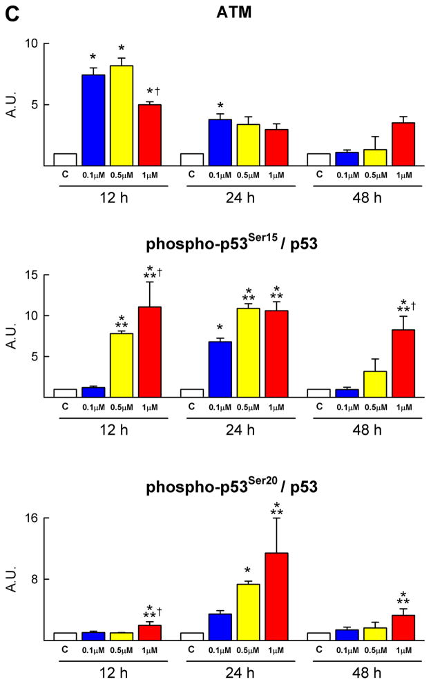
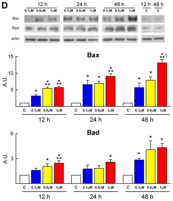
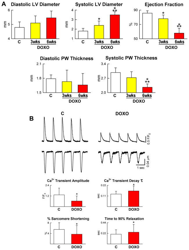
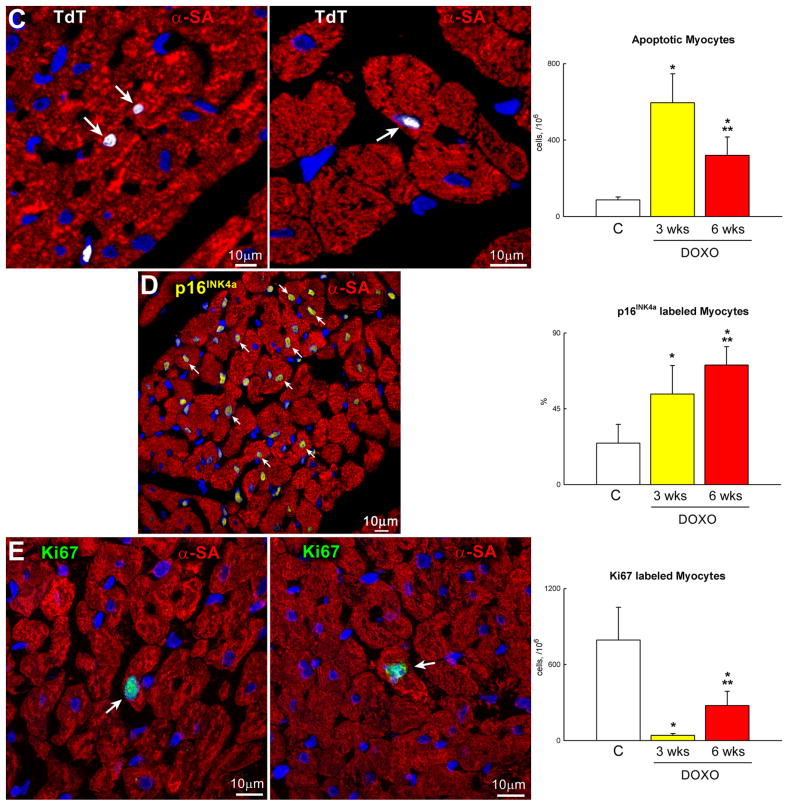
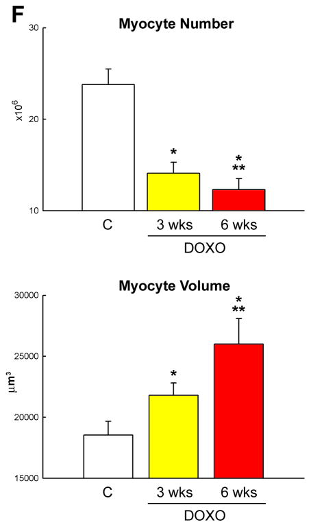
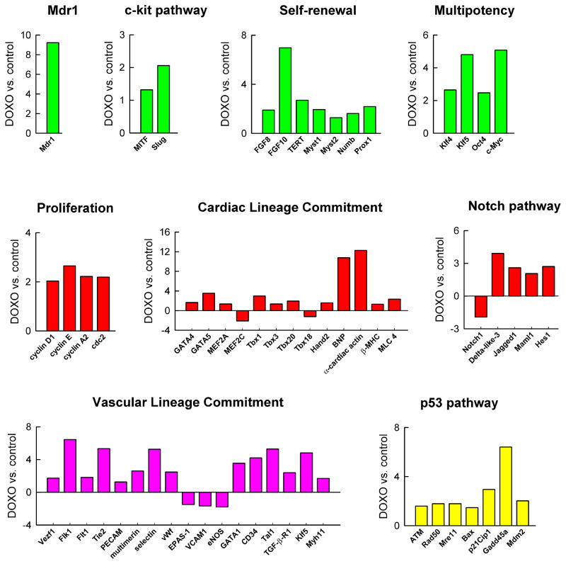
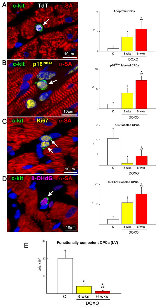
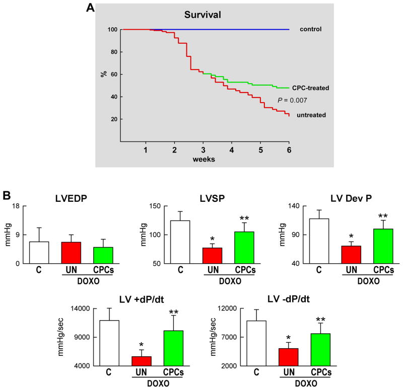
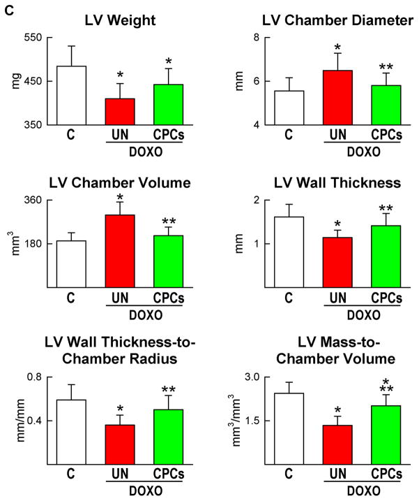
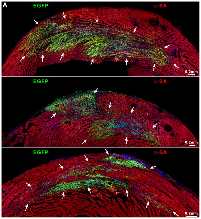
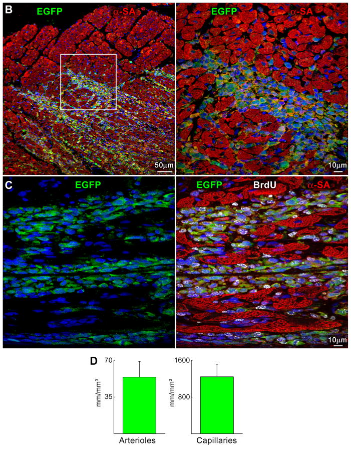
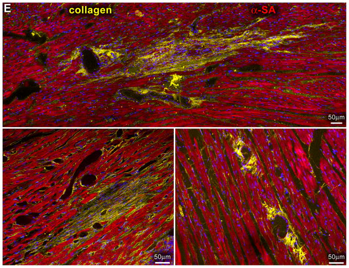
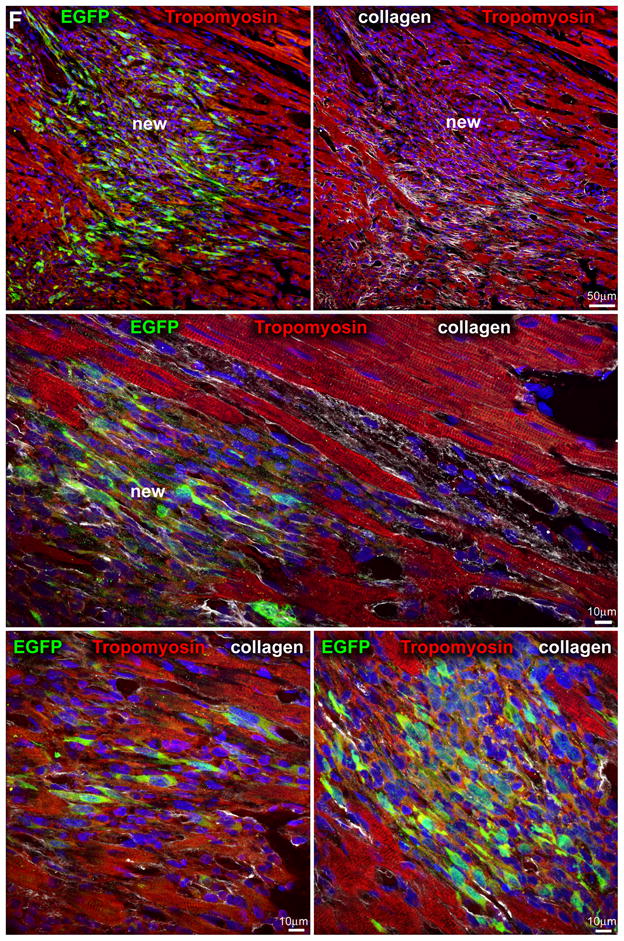
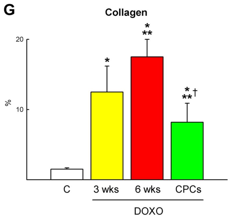
Methods and results: Administration of doxorubicin in rats led to a dilated myopathy, heart failure, and death. To test whether the effects of doxorubicin on cardiac anatomy and function were mediated by alterations in cardiac progenitor cells (CPCs), these cells were exposed to the anthracycline, which increased the formation of reactive oxygen species and caused increases in DNA damage, expression of p53, telomere attrition, and apoptosis. Additionally, doxorubicin resulted in cell-cycle arrest at the G2/M transition, which led to a significant decrease in CPC growth. Doxorubicin elicited multiple molecular adaptations; the massive apoptotic death that occurred in CPCs in the presence of anthracycline imposed on the surviving CPC pool the activation of several pathways aimed at preservation of the primitive state, cell division, lineage differentiation, and repair of damaged DNA. To establish whether delivery of syngeneic progenitor cells opposed the progression of doxorubicin cardiotoxicity, enhanced green fluorescent protein-labeled CPCs were injected in the failing myocardium; this treatment promoted regeneration of cardiomyocytes and vascular structures, which improved ventricular performance and rate of animal survival.

Conclusions: Our results raise the possibility that autologous CPCs can be obtained before antineoplastic drugs are given to cancer patients and subsequently administered to individuals who are particularly sensitive to the cardiotoxicity of these agents for prevention or management of heart failure.


Figures

Figure 1
Figure 1
CPC death and growth. A, The viability of CPCs is negatively affected by dose and time of exposure to DOXO. B–D, CPC apoptosis measured by TdT assay (B), DNA laddering (C) and caspase-3 activity (D) increases with the concentration of DOXO and from 12 to 48 h. The expression of active caspase-3 is shown as fold changes with respect to control (c). TdT labeling (white) of apoptotic CPCs (c-kit, green) is shown by immunolabeling in panel B. E and F, BrdU (E, red) and phospho-H3 (F, yellow) labeling of CPCs decreases progressively with DOXO. *,**,† P<0.05 vs. c, 0.1 μM and 0.5 μM DOXO, respectively.
Figure 1
Figure 1
CPC death and growth. A, The viability of CPCs is negatively affected by dose and time of exposure to DOXO. B–D, CPC apoptosis measured by TdT assay (B), DNA laddering (C) and caspase-3 activity (D) increases with the concentration of DOXO and from 12 to 48 h. The expression of active caspase-3 is shown as fold changes with respect to control (c). TdT labeling (white) of apoptotic CPCs (c-kit, green) is shown by immunolabeling in panel B. E and F, BrdU (E, red) and phospho-H3 (F, yellow) labeling of CPCs decreases progressively with DOXO. *,**,† P<0.05 vs. c, 0.1 μM and 0.5 μM DOXO, respectively.
Figure 2
Figure 2
Cell cycle regulators and oxidative stress. A–D, DOXO affects the expression of cyclins, cyclin-dependent kinases, Rb (A) and cyclin-dependent kinase inhibitors (B), increases DNA damage (C: 8-OH-dG, red) and decreases the activity of SOD and catalase (D) in CPCs (C: c-kit, green).
Figure 2
Figure 2
Cell cycle regulators and oxidative stress. A–D, DOXO affects the expression of cyclins, cyclin-dependent kinases, Rb (A) and cyclin-dependent kinase inhibitors (B), increases DNA damage (C: 8-OH-dG, red) and decreases the activity of SOD and catalase (D) in CPCs (C: c-kit, green).
Figure 3
Figure 3
Telomere-telomerase and p53 function. A, Telomere shortening in DOXO-treated CPCs: nuclei (blue) were stained with a telomere probe (magenta). Lymphoma cells with long (L5178Y-R, 48 kbp) and short (L5178Y-S, 7 kbp) telomeres are shown for comparison. The average telomere length is indicated together with the degree of telomeric shortening and the fraction of cells with telomeres equal/shorter than 8 kbp. B, Telomerase activity was comparable in control and DOXO-treated CPCs. Samples treated with RNase were used as negative and HeLa cells as positive control. C and D, DOXO activates the DNA-damage response in CPCs. Immunoblotting for Bax and Bad are shown in the upper part of panel D. Protein expression is shown as fold changes with respect to c. See Figure 1 for symbols.
Figure 3
Figure 3
Telomere-telomerase and p53 function. A, Telomere shortening in DOXO-treated CPCs: nuclei (blue) were stained with a telomere probe (magenta). Lymphoma cells with long (L5178Y-R, 48 kbp) and short (L5178Y-S, 7 kbp) telomeres are shown for comparison. The average telomere length is indicated together with the degree of telomeric shortening and the fraction of cells with telomeres equal/shorter than 8 kbp. B, Telomerase activity was comparable in control and DOXO-treated CPCs. Samples treated with RNase were used as negative and HeLa cells as positive control. C and D, DOXO activates the DNA-damage response in CPCs. Immunoblotting for Bax and Bad are shown in the upper part of panel D. Protein expression is shown as fold changes with respect to c. See Figure 1 for symbols.
Figure 4
Figure 4
DOXO-induced cardiomyopathy. A, Echocardiography in control (C) and at 3 and 6 weeks (wks) after the first injection of DOXO. B, Defects in myocyte mechanics and calcium transient in cells isolated from DOXO-treated hearts at 3 weeks. C–E, DOXO administration enhances apoptosis (C: TdT, white) and senescence (D: p16INK4a, yellow) and decreases proliferation (E: Ki67, green) of cardiomyocytes (α-sarcomeric actin: α-SA, red) in vivo. F, Myocyte number and volume in control and DOXO-treated rats. *,** P <0.05 vs. C and 3 wks, respectively.
Figure 4
Figure 4
DOXO-induced cardiomyopathy. A, Echocardiography in control (C) and at 3 and 6 weeks (wks) after the first injection of DOXO. B, Defects in myocyte mechanics and calcium transient in cells isolated from DOXO-treated hearts at 3 weeks. C–E, DOXO administration enhances apoptosis (C: TdT, white) and senescence (D: p16INK4a, yellow) and decreases proliferation (E: Ki67, green) of cardiomyocytes (α-sarcomeric actin: α-SA, red) in vivo. F, Myocyte number and volume in control and DOXO-treated rats. *,** P <0.05 vs. C and 3 wks, respectively.
Figure 5
Figure 5
Doxorubicin and gene expression in CPCs. Transcriptional profiling of CPCs in the absence and presence of DOXO by quantitative RT-PCR array. Transcript expression is shown as fold changes with respect to control.
Figure 6
Figure 6
CPC death and growth. A–C, DOXO administration enhances apoptosis (A: TdT, white), senescence (B: p16INK4a, yellow) and decreases proliferation (C: Ki67, yellow) of CPCs (c-kit, green) in vivo at 3 and 6 weeks after the first injection of anthracycline. D, Oxidative damage (8-OH-dG, magenta) in CPCs increases after DOXO treatment in vivo. E, Number of functionally competent CPCs in control and DOXO-treated rats at 3 and 6 weeks. *,** P< 0.05 vs. C and 3 wks, respectively.
Figure 7
Figure 7
CPC treatment, ventricular function and animal survival. A–C, Mortality (A), ventricular hemodynamics (B) and cardiac anatomy (C) of untreated and CPC-treated animals at 6 weeks.
Figure 7
Figure 7
CPC treatment, ventricular function and animal survival. A–C, Mortality (A), ventricular hemodynamics (B) and cardiac anatomy (C) of untreated and CPC-treated animals at 6 weeks.
Figure 8
Figure 8
Myocardial regeneration and DOXO-induced cardiomyopathy. A, Low-power view of transverse sections of the LV wall of DOXO-treated rats 3 weeks after CPC injection. Arrows define areas of myocardial regeneration (green). Newly formed myocytes express EGFP (green) and α-SA (red). B, The area of myocardial regeneration included in the rectangle is shown at higher magnification in the adjacent panel. Small myocytes in the cluster express EGFP (green) and α-SA (red). C, Newly formed EGFP-positive (green) myocytes are labeled by BrdU (white) and express α-SA (red). D, Number of newly formed capillaries and arterioles. E, Foci of collagen (yellow) accumulation at times surrounding small vessels. F, Regenerated myocytes (new) are positive for EGFP (green) and tropomyosin (red) and replace areas of fibrosis (collagen, white). G, Fraction of collagen within the myocardium. *,**,† P <0.05 vs. c, 3 wks and 6 wks, respectively.
Figure 8
Figure 8
Myocardial regeneration and DOXO-induced cardiomyopathy. A, Low-power view of transverse sections of the LV wall of DOXO-treated rats 3 weeks after CPC injection. Arrows define areas of myocardial regeneration (green). Newly formed myocytes express EGFP (green) and α-SA (red). B, The area of myocardial regeneration included in the rectangle is shown at higher magnification in the adjacent panel. Small myocytes in the cluster express EGFP (green) and α-SA (red). C, Newly formed EGFP-positive (green) myocytes are labeled by BrdU (white) and express α-SA (red). D, Number of newly formed capillaries and arterioles. E, Foci of collagen (yellow) accumulation at times surrounding small vessels. F, Regenerated myocytes (new) are positive for EGFP (green) and tropomyosin (red) and replace areas of fibrosis (collagen, white). G, Fraction of collagen within the myocardium. *,**,† P <0.05 vs. c, 3 wks and 6 wks, respectively.

Similar articles

Cited by

References

    1. van Dalen EC, Caron HN, Dickinson HO, Kremer LC. Cardioprotective interventions for cancer patients receiving anthracyclines. Cochrane Database Syst Rev. 2008:CD003917. - PubMed
    1. Takemura G, Fujiwara H. Doxorubicin-induced cardiomyopathy from the cardiotoxic mechanisms to management. Prog Cardiovasc Dis. 2007;49:330–352. - PubMed
    1. Shan K, Lincoff AM, Young JB. Anthracycline-induced cardiotoxicity. Ann Intern Med. 1996;125:47–58. - PubMed
    1. Nithipongvanitch R, Ittarat W, Cole MP, Tangpong J, Clair DK, Oberley TD. Mitochondrial and nuclear p53 localization in cardiomyocytes: redox modulation by doxorubicin (Adriamycin)? Antioxid. Redox Signal. 2007;9:1001–1008. - PubMed
    1. Beltrami AP, Barlucchi L, Torella D, Baker M, Limana F, Chimenti S, Kasahara H, Rota M, Musso E, Urbanek K, Leri A, Kajstura J, Nadal-Ginard B, Anversa P. Adult cardiac stem cells are multipotent and support myocardial regeneration. Cell. 2003;114:763–776. - PubMed

Publication types

MeSH terms