. 2010 Mar 11;115(10):2021-7.
doi: 10.1182/blood-2009-08-241182. Epub 2009 Dec 28.

Membrane remodeling during reticulocyte maturation

Affiliations

Membrane remodeling during reticulocyte maturation

Jing Liu et al. Blood. .

Abstract

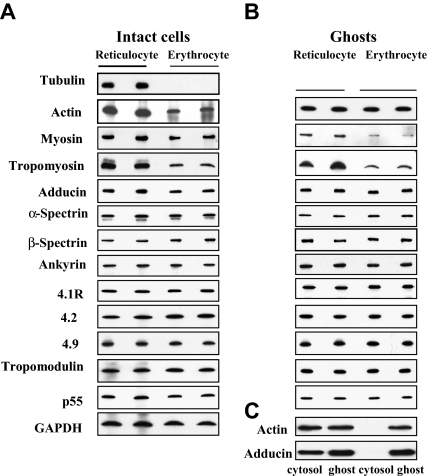
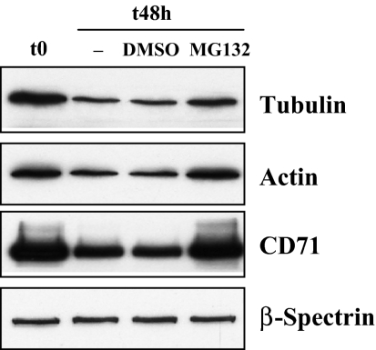
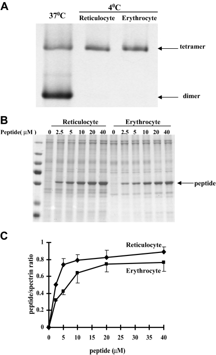
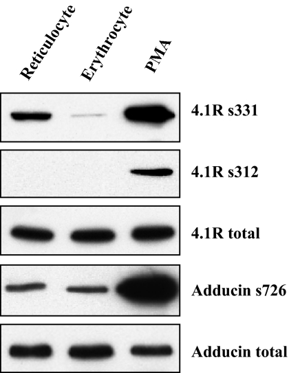
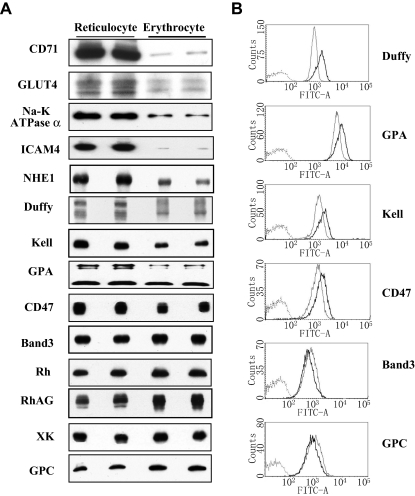
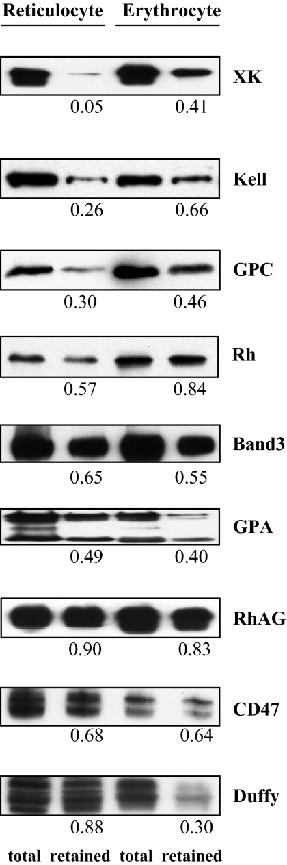
The transition of reticulocytes into erythrocytes is accompanied by extensive changes in the structure and properties of the plasma membrane. These changes include an increase in shear resistance, loss of surface area, and acquisition of a biconcave shape. The processes by which these changes are effected have remained largely undefined. Here we examine how the expression of 30 distinct membrane proteins and their interactions change during murine reticulocyte maturation. We show that tubulin and cytosolic actin are lost, whereas the membrane content of myosin, tropomyosin, intercellular adhesion molecule-4, glucose transporter-4, Na-K-ATPase, sodium/hydrogen exchanger 1, glycophorin A, CD47, Duffy, and Kell is reduced. The degradation of tubulin and actin is, at least in part, through the ubiquitin-proteasome degradation pathway. In regard to the protein-protein interactions, the formation of membrane-associated spectrin tetramers from dimers is unperturbed, whereas the interactions responsible for the formation of the membrane-skeletal junctions are weaker in reticulocytes, as is the attachment of transmembrane proteins to these structures. This weakness, in part, results from the elevated phosphorylation of 4.1R in reticulocytes, which leads to a decrease in shear resistance by reducing its interaction with spectrin and actin. These observations begin to unravel the mechanistic basis of crucial changes accompanying reticulocyte maturation.


Figures

Figure 1
Figure 1
Immunoblots of cytoskeletal proteins of reticulocytes and erythrocytes. (A) Intact cells: Western blots of SDS-PAGEs of total cellular protein prepared from reticulocytes and erythrocytes probed with antibodies against the indicated proteins. Note the absence of tubulin and the decreased expression of tropomyosin, myosin, actin and adducin in erythrocytes. (B) Ghosts: Western blots of SDS-PAGEs of ghosts prepared from reticulocytes and erythrocytes probed with antibodies against the indicated proteins. Because ghosts of both reticulocytes and erythrocytes do not contain any tubulin, there is no corresponding panel for tubulin in panel B. Note the decreased expression of tropomyosin and myosin in the erythrocyte membranes but the similar level of actin and adducin. (C) Western blots of SDS-PAGEs of the cytosol and the membrane were probed with anti-actin or anti-adducin antibody. Note actin and adducin are present in both the cytosol and the membrane fraction of reticulocytes but are only present in the membrane of erythrocytes.
Figure 2
Figure 2
Inhibition of tubulin and actin degradation by proteosome inhibitor MG132. Mouse reticulocytes were cultured for 48 hours in the presence of the proteasome inhibitor MG132 (10μM) or an equivalent volume of the solvent, DMSO. Western blots of SDS-PAGEs of total cellular protein were probed with antibodies against tubulin, actin, CD71 and β-spectrin. CD71 serves as a positive control and β-spectrin as a negative control.
Figure 3
Figure 3
Protein-protein associations in reticulocytes and erythrocytes. (A) Spectrin dimer-tetramer equilibrium in reticulocytes and mature red blood cells. Extracted spectrin was analyzed by nondenaturing gel electrophoresis. Note spectrin from both the reticulocytes and the erythrocytes is tetrameric. In panels B and C, incorporation of a β-spectrin fragment into reticulocyte and erythrocyte membranes. The Triton shells were prepared from reticulocyte or erythrocyte ghosts resealed with increasing concentrations of β-spectrin fragment 1-301. (B) Proteins retained in the Triton shells were analyzed by SDS-PAGE. (C) Quantitative analysis revealed greater incorporation of the peptide into the cytoskeleton of reticulocytes compared with that observed in erythrocytes.
Figure 4
Figure 4
Phosphorylation of 4.1R and adducin in reticulocytes and erythrocytes. Reticulocyte and erythrocyte ghosts were prepared in the presence of the phosphatase inhibitor, calyculin A. Western blots of SDS-PAGEs were probed with phospho-residue specific antibodies as indicated. PMA-treated erythrocyte ghosts were used as a positive control. Note the markedly increased phosphorylation of 4.1R at residue 331 serine (4.1R s331) in reticulocytes.
Figure 5
Figure 5
Expression of membrane proteins of reticulocytes and erythrocytes. (A) Immunoblots: Western blots of SDS-PAGEs of total cellular protein prepared from reticulocytes and erythrocytes probed with antibodies against the indicated proteins. Note the dramatic decrease of CD71, GLUT4, Na-K-ATPase α, ICAM4, and NHE1; decrease to a lesser extent of Duffy, Kell, GPA, and CD47; and increase of band 3, Rh, RhAG, XK, and GPC in erythrocytes compared with reticulocytes. (B) Flow cytometry: the ordinate measures the number of cells displaying the fluorescent intensity given by the abscissa. Dark lines indicate reticulocytes; light lines, erythrocytes. Note the decreased expression of Duffy, GPA, Kell, and CD47 and the increased expression of band 3 and GPC in erythrocytes compared with reticulocytes.
Figure 6
Figure 6
Detergent extractability of membrane proteins from reticulocytes and erythrocytes. Membrane proteins were detergent extracted and proteins retained with membrane skeletons were analyzed by SDS-PAGE and probed with antibodies as indicated. The numbers indicate the fraction retained in the pellet compared with the total protein. Note the decreased retention of XK, Kell, and GPC but the increased retention of Duffy in reticulocytes.

Similar articles

Cited by

References

    1. Bennett V. The spectrin-actin junction of erythrocyte membrane skeletons. Biochim Biophys Acta. 1989;988(1):107–121. - PubMed
    1. Bennett V, Baines AJ. Spectrin and ankyrin-based pathways: metazoan inventions for integrating cells into tissues. Physiol Rev. 2001;81(3):1353–1392. - PubMed
    1. Mohandas N, An X. New insights into function of red cell membrane proteins and their interaction with spectrin-based membrane skeleton. Transfus Clin Biol. 2006;13(1-2):29–30. - PubMed
    1. Mohandas N, Gallagher PG. Red cell membrane: past, present, and future. Blood. 2008;112(10):3939–3948. - PMC - PubMed
    1. Manno S, Takakuwa Y, Mohandas N. Modulation of erythrocyte membrane mechanical function by protein 4.1 phosphorylation. J Biol Chem. 2005;280(9):7581–7587. - PubMed

Publication types

MeSH terms