. 2010 Jan;120(1):214-22.
doi: 10.1172/JCI40231. Epub 2009 Dec 21.

CD20 deficiency in humans results in impaired T cell-independent antibody responses

Affiliations

CD20 deficiency in humans results in impaired T cell-independent antibody responses

Taco W Kuijpers et al. J Clin Invest. 2010 Jan.

Abstract

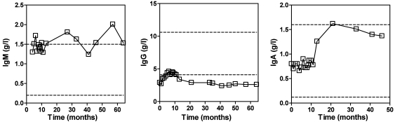
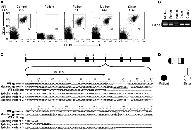
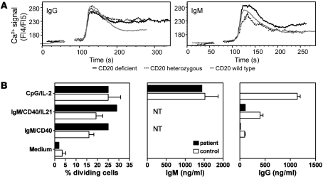
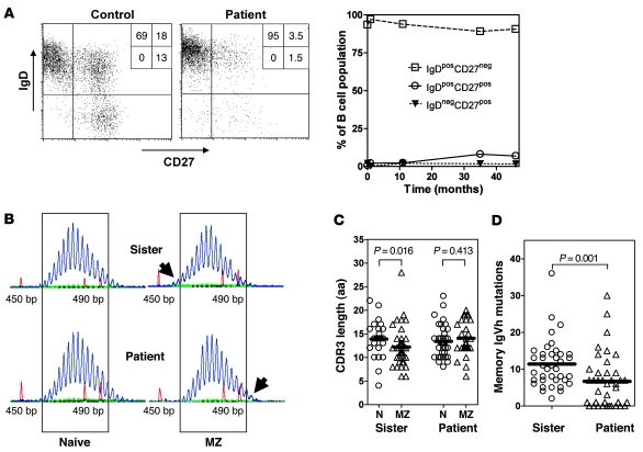
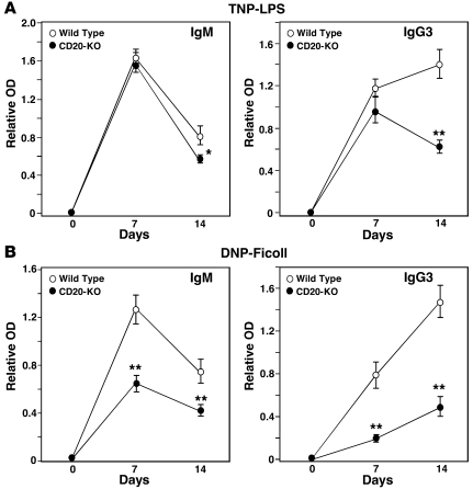
CD20 was the first B cell differentiation antigen identified, and CD20-specific mAbs are commonly used for the treatment of B cell malignancies and autoantibody-mediated autoimmune diseases. Despite this the role of CD20 in human B cell physiology has remained elusive. We describe here a juvenile patient with CD20 deficiency due to a homozygous mutation in a splice junction of the CD20 gene (also known as MS4A1) that results in "cryptic" splicing and nonfunctional mRNA species. Analysis of this patient has led us to conclude that CD20 has a central role in the generation of T cell-independent (TI) antibody responses. Key evidence to support this conclusion was provided by the observation that although antigen-independent B cells developed normally in the absence of CD20 expression, antibody formation, particularly after vaccination with TI antigens, was strongly impaired in the patient. Consistent with this, TI antipolysaccharide B cell responses were severely impeded in CD20-deficient mice. Our study therefore identifies what we believe to be a novel type of humoral immunodeficiency caused by CD20 deficiency and characterized by normal development of antigen-independent B cells, along with a reduced capacity to mount proper antibody responses.


Figures

Figure 1
Figure 1. Serum IgM, IgG, and IgA levels over time in the patient.
A turbidimetric method was used to determine serum immunoglobulin levels in serial samples. The date of first presentation of the patient at the clinic is set as 0. The upper and lower ranges of normal immunoglobulin levels are indicated by a dashed line. Intravenous immune globulin therapy was initially initiated but stopped after 6 months.
Figure 2
Figure 2. Analysis of CD20 expression and gene sequences of the patient, parents, and sibling.
(A) FACS analysis of CD20 and CD19 expression in indicated PBMC samples of normal controls and family members. MFI of CD20 staining is indicated above the plots. In a cohort of healthy children (age, 3 months to 10 years; n = 30), mean CD20 expression ranged between 874 and 1,230. (B) Agarose gel analysis of CD20 cDNA fragments of the patient, family members, and a control, revealing aberrant mRNA species in the patient and her parents. Cloning and sequencing of the cDNA fragments showed that the smallest fragment contains a complete deletion of exon 5 (not shown). The 3 fragments with increased MW contained variable parts of intron 5 sequence (see below in C). (C) CD20 gene sequences surrounding the exon 5/intron 5 boundary, as determined from cDNA and genomic fragments. The 11–base pair insertion at the noncanonical splice site is underlined in the mutated genomic sequence. The CD20 intron/exon structure is indicated on top, and the normal and aberrant sequences found in the patient cDNA clones are depicted below. The noncanonical GC splice donor site at intron 5 and the cryptic splice sites used as a result of the mutation are boxed. (D) Family pedigree indicating consanguinity of the parents. Normal and aberrant CD20 alleles are indicated by white and black, respectively.
Figure 3
Figure 3. B cell function in vitro.
(A) Cells lacking CD20 respond to IgG and IgM BCR stimulation by Ca2+ flux. B cell lines from donors expressing wild-type, intermediate, or no CD20 were cultured for 4 hours without CD40L in the presence of IL-21 before being loaded with Indo-1 AM. Cells were maintained at room temperature and incubated with a control and subsequently with anti-BCR mAbs. Fluorescence ratios of Indo-1 emission at 405/485 nm were measured by flow cytometry. Ca2+ flux curves were adjusted graphically to the same starting level fluorescence 4/fluorescence 5(FL4/FL5) ratio, and the starting point of the stimulus was set at the same time point. In general, all cells responded to BCR stimulation by showing intracellular Ca2+ fluxes. (B) CFSE-labeled PBMCs were stimulated with indicated stimuli for 6 days, and CD19+CD20+ B cells were analyzed by flow cytometry for CFSE dilution. The percentage of dividing B cells (precursor frequencies) was calculated. Supernatants were tested by ELISA for IgM and IgG secretion. NT, not tested due to stimulus. Values represent mean ± SEM and are obtained for 3–4 healthy donors.
Figure 4
Figure 4. B cell selection.
(A) FACS analysis of B cell subsets. IgD × CD27 dot plot gated on CD19+CD20+ B cells. Percentage of cells in each quadrant are indicated (left). The percentages of the patient’s naive, nonswitched, and switched memory B cells are shown over the course of approximately 4 years since initial presentation (t = 0) (right). (B) Gene scanning analyses of IgM-VH3 transcripts amplified out of IgD+CD27 mature naive and IgD+CD27+ MZ B cells of the patient and her sister show the presence of shorter MZ B cell IgM-VH3 transcripts of the sister and longer IgM-VH3 transcripts of the patient (highlighted by the top and bottom arrows on the right panel, respectively). Numbers on the x axis are lengths in base pairs, as determined using internal length markers. (C) Comparison of IgVH-CDR3 amino acid lengths of IgD+CD27 mature naive (N) and IgD+CD27+ MZ B cells. Of the patient and her sister, 25 and 39 VH3-IgM clones of the naive B cells and 42 and 28 VH3-IgM clones of the MZ B cells were sequenced, respectively. Data for the mother and a normal control were similar to those of the sister (not shown). (D) Numbers of VH3-IgVH mutations of IgG-expressing IgDCD27+ memory B cells. Of the patient and her sister, 38 and 39 VH3-IgG clones the memory B cells were sequenced, respectively. (C and D) Individual clones are indicated by the separate symbols. Mean CDR3 length and number of mutations is indicated by bars. P values of statistical analyses among groups are shown.
Figure 5
Figure 5. CD20 deficiency impairs TI antibody responses in mice.
Wild-type (n = 9/antigen) and Cd20–/– (n = 9–10/antigen) mice were immunized with either (A) TNP-LPS or (B) DNP-Ficoll on day 0, with sera collected at the indicated times. TNP- and DNP-specific IgM and IgG3 levels in all sera were assessed simultaneously by ELISA. Values represent mean ± SEM relative antigen-specific antibody levels, with 9 or more mice in each group. Significant differences between mean IgM and IgG3 levels are indicated: *P < 0.05, **P < 0.01.

References

    1. Shearer WT, Fischer A. The last 80 years in primary immunodeficiency: how far have we come, how far need we go? J Allergy Clin Immunol. 2006;117(4):748–752. doi: 10.1016/j.jaci.2006.02.029. - DOI - PubMed
    1. Conley ME, Rohrer J, Rapalus L, Boylin EC, Minegishi Y. Defects in early B-cell development: comparing the consequences of abnormalities in pre-BCR signaling in the human and the mouse. Immunol Rev. 2000;178:75–90. doi: 10.1034/j.1600-065X.2000.17809.x. - DOI - PubMed
    1. Minegishi Y, et al. Mutations in the human lambda5/14.1 gene result in B cell deficiency and agammaglobulinemia. J Exp Med. 1998;187(1):71–77. doi: 10.1084/jem.187.1.71. - DOI - PMC - PubMed
    1. Minegishi Y, et al. Mutations in Igalpha (CD79a) result in a complete block in B-cell development. . J Clin Invest. 1999;104(8):1115–1121. doi: 10.1172/JCI7696. - DOI - PMC - PubMed
    1. Tsukada S, et al. Deficient expression of a B cell cytoplasmic tyrosine kinase in human X-linked agammaglobulinemia. Cell. 1993;72(2):279–290. doi: 10.1016/0092-8674(93)90667-F. - DOI - PubMed

Publication types

Substances