. 2010 Jan;120(1):254-65.
doi: 10.1172/JCI40295. Epub 2009 Dec 21.

Cardiac fibroblasts are essential for the adaptive response of the murine heart to pressure overload

Affiliations

Cardiac fibroblasts are essential for the adaptive response of the murine heart to pressure overload

Norifumi Takeda et al. J Clin Invest. 2010 Jan.

Abstract

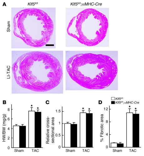
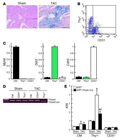
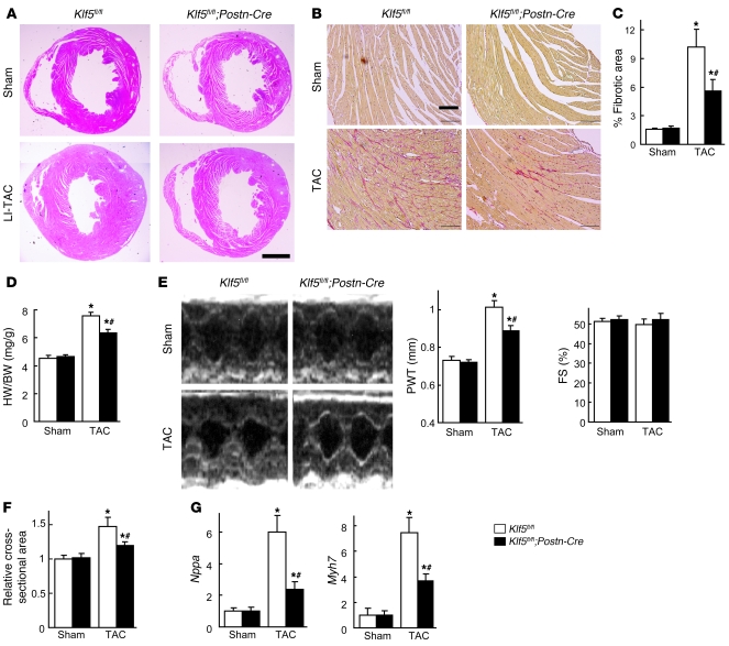
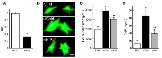
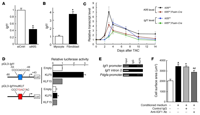
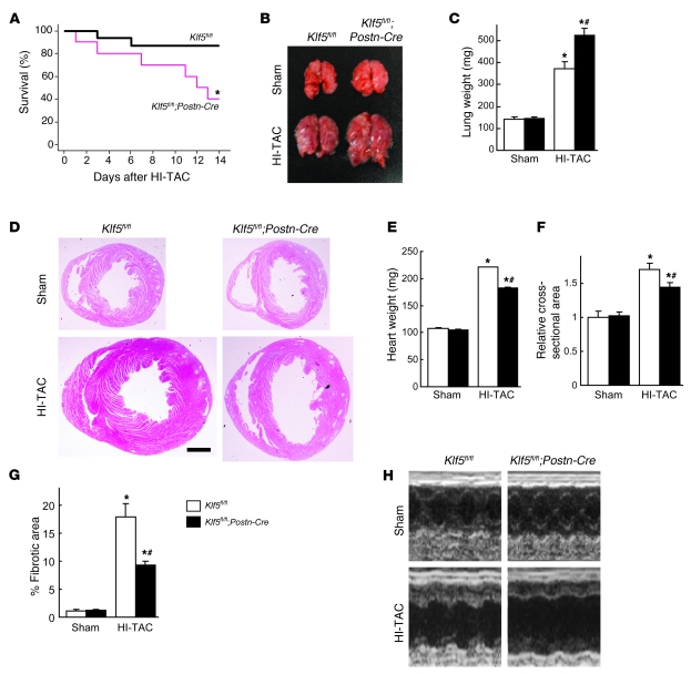
Fibroblasts, which are the most numerous cell type in the heart, interact with cardiomyocytes in vitro and affect their function; however, they are considered to play a secondary role in cardiac hypertrophy and failure. Here we have shown that cardiac fibroblasts are essential for the protective and hypertrophic myocardial responses to pressure overload in vivo in mice. Haploinsufficiency of the transcription factor-encoding gene Krüppel-like factor 5 (Klf5) suppressed cardiac fibrosis and hypertrophy elicited by moderate-intensity pressure overload, whereas cardiomyocyte-specific Klf5 deletion did not alter the hypertrophic responses. By contrast, cardiac fibroblast-specific Klf5 deletion ameliorated cardiac hypertrophy and fibrosis, indicating that KLF5 in fibroblasts is important for the response to pressure overload and that cardiac fibroblasts are required for cardiomyocyte hypertrophy. High-intensity pressure overload caused severe heart failure and early death in mice with Klf5-null fibroblasts. KLF5 transactivated Igf1 in cardiac fibroblasts, and IGF-1 subsequently acted in a paracrine fashion to induce hypertrophic responses in cardiomyocytes. Igf1 induction was essential for cardioprotective responses, as administration of a peptide inhibitor of IGF-1 severely exacerbated heart failure induced by high-intensity pressure overload. Thus, cardiac fibroblasts play a pivotal role in the myocardial adaptive response to pressure overload, and this role is partly controlled by KLF5. Modulation of cardiac fibroblast function may provide a novel strategy for treating heart failure, with KLF5 serving as an attractive target.


Figures

Figure 1
Figure 1. KLF5 is essential for pressure overload–induced hypertrophy.
(AD) Klf5+/– and wild-type mice were subjected to LI-TAC or sham operation. (A) Representative low-magnification views of H&E-stained heart sections from WT and Klf5+/– mice 2 weeks after the operations. Scale bar: 1 mm. (B and C) Heart weight/body (HW/BW) weight ratios (B) and relative cross-sectional areas of cardiomyocytes (C) from wild-type and Klf5+/– hearts. (D) Fractional areas of fibrosis in cross sections of hearts as determined by elastic picrosirius red staining. *P < 0.01 versus sham control of the same genotype; #P < 0.05 versus wild-type subjected to TAC. n = 7. (E) Expression of KLF5 in normal and hypertrophied hearts 4 days after LI-TAC. Cells were double stained for KLF5 (brown) and a cardiomyocyte marker, αMHC (red); nuclei were counterstained in blue. Scale bar: 20 μm.
Figure 2
Figure 2. Cardiomyocyte-specific deletion of Klf5 did not alter pressure overload–induced hypertrophy.
Klf5fl/fl and Klf5fl/fl;αMHC-Cre mice were subjected to LI-TAC or sham operation. (A) Representative low-magnification views of H&E-stained heart sections 2 weeks after LI-TAC. Scale bar: 1 mm. (B and C) Heart weight/body weight ratios (B) and relative cross-sectional areas of cardiomyocytes normalized to those obtained from Klf5fl/fl mice subjected to sham operations (C). (D) Fractional areas of fibrosis. *P < 0.01 versus sham control of the same genotype. n = 7.
Figure 3
Figure 3. Fibroblast-specific deletion of Klf5 in Klf5fl/fl;Postn-Cre mice.
(A) Fibroblast-specific deletion of the floxed region in Postn-Cre mice was examined using R26RstoplacZ indicator mice. LacZ expression was visualized using X-gal. Scale bars: 100 μm. (B) CD3 cells within non-myocyte-enriched cell populations isolated from adult hearts were analyzed for surface expression of the fibroblast marker Thy1 and the endothelial marker CD31. (C) Relative expression levels of cell-lineage markers in adult cardiomyocytes (CM) isolated using the Langendorff perfusion method, and in Thy1+CD31CD3 (Thy1+) and Thy1CD31+CD3 (CD31+) cells sorted from non-myocyte-enriched populations as shown in B. Myh6 (encoding αMHC), Ddr2 (encoding discoidin domain receptor 2), and Cdh5 (encoding VE-cadherin) were used as markers for cardiomyocytes, fibroblasts, and ECs, respectively. The cells were isolated from 8-week-old mice subjected to sham operations. (D) Competitive PCR analysis for quantitation of Cre-mediated recombination of the Klf5 gene region in adult cardiomyocytes, CD31+ ECs, and Thy1+ fibroblasts isolated from Klf5fl/fl and Klf5fl/fl;Postn-Cre mice 2 weeks after either the sham or LI-TAC operation. Competitive PCR was performed as shown in Supplemental Figure 6B. (E) Relative expression levels of Klf5 mRNA in adult cardiomyocytes, Thy1+ fibroblasts, and CD31+ ECs isolated from Klf5fl/fl and Klf5fl/fl;Postn-Cre mice as shown in B 5 days after either sham operation or LI-TAC. Expression levels of Klf5 mRNA were assessed using real-time PCR and normalized to those of 18s rRNA, after which they were further normalized to the levels in Thy1+ cells isolated from Klf5fl/fl mice subjected to the sham operation. *P < 0.01 versus sham control of the same genotype in the same cell lineage group; #P < 0.01 versus Klf5fl/fl mice subjected to LI-TAC in the same cell lineage group.
Figure 4
Figure 4. Fibroblast-specific deletion of Klf5 attenuates cardiac hypertrophy and fibrosis after TAC.
Klf5fl/fl and Klf5fl/fl;Postn-Cre mice were subjected to LI-TAC or sham operation. (A) Representative low-magnification views of H&E-stained heart sections 2 weeks after the operations. Scale bar: 1 mm. The bottom-left panel was composited from 2 photographs of the same section. (B) Representative elastic picrosirius red–stained sections and fibrotic areas. Scale bars: 100 μm. (C) Fibrotic areas. (D) Heart weight/body weight ratios 2 weeks after the operations. (E) Echocardiographic analysis 2 weeks after the operations. (F) Relative cross-sectional areas of cardiomyocytes. (G) Relative expression levels of Nppa and Myh7 mRNA. Expression levels of each gene were normalized to 18s ribosomal RNA levels and then further normalized with respect to those obtained with samples from Klf5fl/fl mice subjected to sham operation. *P < 0.01 versus sham control of the same genotype; #P < 0.01 versus Klf5fl/fl subjected to TAC. n = 7.
Figure 5
Figure 5. KLF5 controls expression of paracrine factors in cardiac fibroblasts that mediate cardiomyocyte hypertrophy.
(A) siRNA-mediated knockdown of Klf5 in cardiac fibroblasts. Klf5 levels were normalized to those in cells transfected with the control siRNA (siCntrl). *P < 0.01 versus siCntrl. (BD) Cultured cardiomyocytes were incubated with serum-free medium (SFM) or conditioned medium prepared from cardiac fibroblasts transfected with control or Klf5 siRNA for 48 hours. (B) Representative cardiomyocytes are shown stained for sarcomeric α-actinin (green) and nuclei (Hoechst 33258, blue). Scale bar: 10 μm. (C) Cell surface areas of 100 cells from each group. *P < 0.01 versus cells treated with SFM; #P < 0.05 versus cells treated with medium conditioned by siCntrl transfectants. (D) ANP concentrations in culture medium conditioned by cardiomyocytes. *P < 0.05 versus SFM; #P < 0.05 versus siCntrl.
Figure 6
Figure 6. KLF5 transactivates the Igf1 promoter.
(A) KLF5 knockdown reduced Igf1 expression in cardiac fibroblasts. KLF5 was knocked down as shown in Figure 5A. *P < 0.01 versus siCntrl. (B) Fibroblast-selective expression of Igf1. Igf1 mRNA levels in cultured cardiac fibroblasts were normalized to those of 18s rRNA and then further normalized with respect to those in cardiomyocytes. *P < 0.01 versus cardiomyocytes. (C) Cardiac expression of Klf5 and Igf1 mRNA after LI-TAC in Klf5fl/fl and Klf5fl/fl;Postn-Cre mice. Expression levels were normalized to those of 18s rRNA and then further normalized with respect to those in the hearts before TAC. (D) Reporter analysis of KLF5-depedent transactivation of the Igf1 promoter. Luciferase reporter constructs driven by the wild-type Igf1 promoter or a mutant promoter in which the potential KLF-binding site was mutated were cotransfected with either empty vector or a vector harboring Klf5 or Klf15. Data are representative of 3 independent experiments. (E) ChIP assays of KLF5 binding to the Igf1 and Pdgfa promoters. An intronic region of Igf1 that does not contain a KLF-binding motif served as a negative control. (F) Effects of neutralizing IGF-1 on the cardiotrophic activity of fibroblast-conditioned medium. An antibody against IGF-1 (30 μg/ml) or normal IgG was added to the conditioned medium, after which the effect of the medium on cardiomyocyte surface area was analyzed. *P < 0.01 versus cells treated with SFM; #P < 0.01 versus cells treated with fibroblast-conditioned medium.
Figure 7
Figure 7. Cardiac fibroblasts are essential for the protective response elicited by severe pressure overload.
(AH) Klf5fl/fl and Klf5fl/fl;Postn-Cre mice were subjected to HI-TAC or sham operation. (A) Kaplan-Meier survival analysis of Klf5fl/fl (n = 16) and Klf5fl/fl;Postn-Cre (n = 10) mice after HI-TAC. *P < 0.05 versus Klf5fl/fl. (B) Representative pictures of lungs 2 weeks after the operations. Note the severe lung edema in Klf5fl/fl;Postn-Cre mice subjected to HI-TAC. (C) Lung weights in Klf5fl/fl (n = 5) and Klf5fl/fl;Postn-Cre (n = 3) mice 2 weeks after the operations. (D) Representative low-magnification views of H&E-stained heart sections 2 weeks after the operations. Scale bar: 1 mm. (EG) Heart weight/body weight ratios (E), relative cross-sectional areas of cardiomyocytes (F), and fibrotic areas (G) in Klf5fl/fl (n = 5) and Klf5fl/fl;Postn-Cre (n = 3) mice 2 weeks after the HI-TAC operation. *P < 0.01 versus sham control of the same genotype; #P < 0.05 versus Klf5fl/fl mice subjected to HI-TAC. (H) M-mode echocardiographic tracings obtained 2 weeks after the operations.
Figure 8
Figure 8. An IGF-1 receptor antagonist aggravates heart failure induced by severe pressure overload.
(A) Kaplan-Meier survival analysis of wild-type mice treated with vehicle or JB1, a peptide IGF-1 receptor antagonist, after HI-TAC. n = 10 in each group. *P < 0.001 versus vehicle. (B) Representative photographs of lungs taken 1 week after the operations. Note the severe lung edema in JB1-treated mice subjected to HI-TAC. (C) Lung weights in vehicle-treated and JB1-treated groups 1 week after the operations. n = 5 in each group. (D) Representative low-magnification views of H&E-stained heart sections 1 week after the operations. Scale bars: 1 mm. (E) Heart weights. (F) Echocardiographic analysis carried out 1 week after the operation. *P < 0.05 versus sham controls in the same treatment group; #P < 0.05 versus vehicle controls subjected to HI-TAC.

Similar articles

Cited by

References

    1. Frey N, Olson EN. Cardiac hypertrophy: the good, the bad, and the ugly. Annu Rev Physiol. 2003;65:45–79. doi: 10.1146/annurev.physiol.65.092101.142243. - DOI - PubMed
    1. Baudino TA, Carver W, Giles W, Borg TK. Cardiac fibroblasts: friend or foe? Am J Physiol Heart Circ Physiol. 2006;291(3):H1015–H1026. doi: 10.1152/ajpheart.00023.2006. - DOI - PubMed
    1. Manabe I, Shindo T, Nagai R. Gene expression in fibroblasts and fibrosis: involvement in cardiac hypertrophy. Circ Res. 2002;91(12):1103–1113. doi: 10.1161/01.RES.0000046452.67724.B8. - DOI - PubMed
    1. Eghbali M, et al. Collagen chain mRNAs in isolated heart cells from young and adult rats. J Mol Cell Cardiol. 1988;20(3):267–276. doi: 10.1016/S0022-2828(88)80059-2. - DOI - PubMed
    1. Zak R. Cell proliferation during cardiac growth. Am J Cardiol. 1973;31(2):211–219. doi: 10.1016/0002-9149(73)91034-5. - DOI - PubMed

Publication types

Substances