. 2010 Jan;120(1):266-79.
doi: 10.1172/JCI39740. Epub 2009 Dec 28.

Limited forward trafficking of connexin 43 reduces cell-cell coupling in stressed human and mouse myocardium

Affiliations

Limited forward trafficking of connexin 43 reduces cell-cell coupling in stressed human and mouse myocardium

James W Smyth et al. J Clin Invest. 2010 Jan.

Abstract

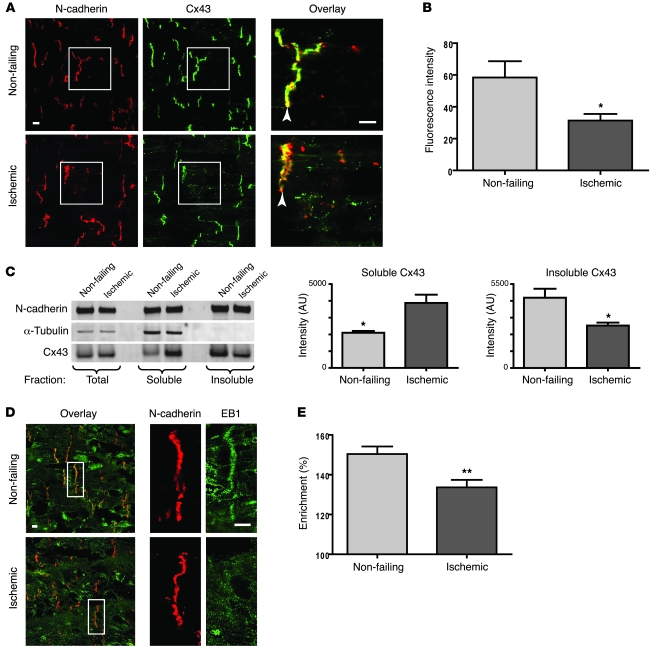
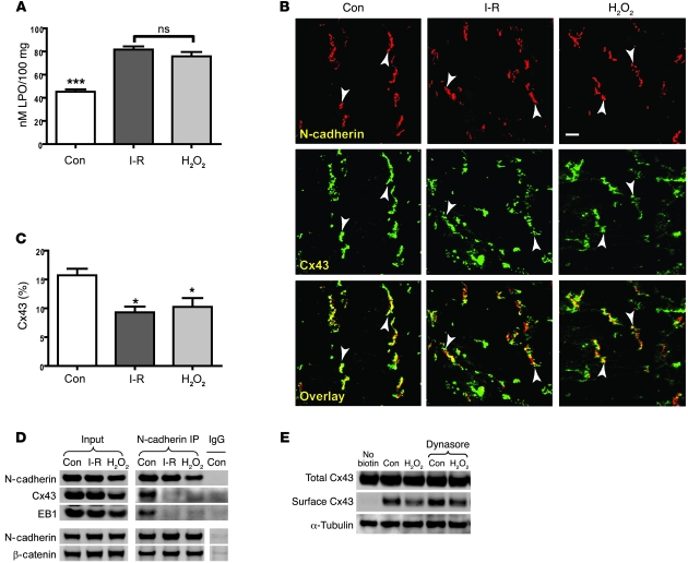
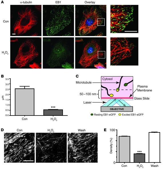
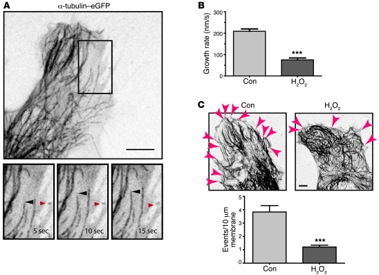
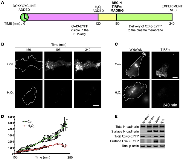
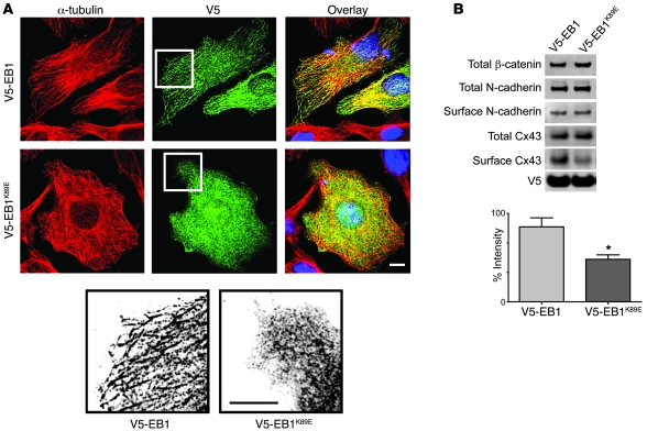
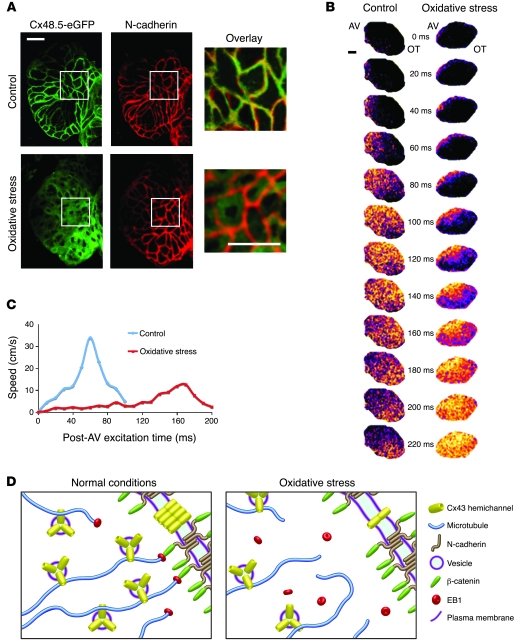
Gap junctions form electrical conduits between adjacent myocardial cells, permitting rapid spatial passage of the excitation current essential to each heartbeat. Arrhythmogenic decreases in gap junction coupling are a characteristic of stressed, failing, and aging myocardium, but the mechanisms of decreased coupling are poorly understood. We previously found that microtubules bearing gap junction hemichannels (connexons) can deliver their cargo directly to adherens junctions. The specificity of this delivery requires the microtubule plus-end tracking protein EB1. We performed this study to investigate the hypothesis that the oxidative stress that accompanies acute and chronic ischemic disease perturbs connexon forward trafficking. We found that EB1 was displaced in ischemic human hearts, stressed mouse hearts, and isolated cells subjected to oxidative stress. As a result, we observed limited microtubule interaction with adherens junctions at intercalated discs and reduced connexon delivery and gap junction coupling. A point mutation within the tubulin-binding domain of EB1 reproduced EB1 displacement and diminished connexon delivery, confirming that EB1 displacement can limit gap junction coupling. In zebrafish hearts, oxidative stress also reduced the membrane localization of connexin and slowed the spatial spread of excitation. We anticipate that protecting the microtubule-based forward delivery apparatus of connexons could improve cell-cell coupling and reduce ischemia-related cardiac arrhythmias.


Figures

Figure 1
Figure 1. Levels of Cx43 and EB1 at the intercalated disc are reduced with end-stage ischemic cardiomyopathy.
Immunofluorescence labeling of cryosections from snap-frozen non-failing and end-stage ischemic explanted human heart tissue. (A) Human heart Cx43 immunofluorescence. N-cadherin in red and Cx43 in green, with enlarged overlay images. N-cadherin was used as a marker for the intercalated disc (arrows). (B) Cx43 at intercalated discs. Quantification of Cx43 fluorescence intensity in regions also positive for N-cadherin expression. (C) Tissue fractionation. Triton X-100–based fractionation of soluble (cytoplasmic) and insoluble (junctional) protein from tissue detected by Western blot analysis, quantified in bar charts. (D) EB1 immunofluorescence. N-cadherin in red and EB1 in green with enlarged panels at right displaying comparable intercalated discs. (E) EB1 enrichment at intercalated discs. Quantification of EB1 fluorescence intensity in regions also positive for N-cadherin expression. Original magnification, ×60. Scale bars: 10 μm. Data are representative of 4 non-failing and 4 end-stage ischemic explanted hearts. Statistical analysis was performed using the Student’s unpaired t test. Values represent mean ± SEM. *P < 0.05, **P < 0.01.
Figure 2
Figure 2. The oxidative stress component of ischemia-reperfusion injury is sufficient to disrupt EB1/adherens junction association and perturb Cx43 forward trafficking.
Hearts from 6- to 8-week-old male C57BL/6 mice were maintained using a Langendorff perfusion apparatus for 30 minutes, followed either by 90 minutes of normal perfusion (control), 30 minutes of ischemia, and 60 minutes of reperfusion (I-R) or by 90 minutes of perfusion with 10 μM H2O2. (A) The lipid hydroperoxide (LPO) assay was used to quantify levels of oxidative stress induced by each treatment. (B) Immunofluorescence labeling of N-cadherin (red) and Cx43 (green) in cryosections from snap-frozen hearts. Arrows indicate intercalated discs. Original magnification, ×60. Scale bar: 10 μm. (C) Percentage of Cx43 at intercalated discs. Quantification of Cx43 fluorescence intensity in regions also positive for N-cadherin expression relative to total Cx43 expression. (D) Co-immunoprecipitation of Cx43, EB1, and β-catenin with N-cadherin detected by Western blot analysis. (E) Surface protein biotinylation of isolated adult mouse cardiomyocytes cultured for 4 hours in the presence or absence of 10 μM H2O2 detected by Western blot analysis. Dynasore was used to inhibit endocytosis. Con, control. Statistical analysis was performed using a 1-way ANOVA with Bonferroni post-hoc correction. Values represent 3 hearts from each condition; mean ± SEM. *P < 0.05 compared with control. ***P < 0.001 compared with I-R or H2O2.
Figure 3
Figure 3. EB1 is displaced off of microtubule plus ends and interacts less with the plasma membrane during oxidative stress.
(A) HeLa cells exposed to 200 μM H2O2 for 4 hours were fixed in ice-cold methanol, and confocal immunofluorescence detection of EB1 (green) and α-tubulin (red) was performed with nuclei counterstained using TO-PRO-3 (blue). Enlargement of a cell-cell border region (scale bar: 10 μm) shows a reduction in EB1 at the microtubule plus end. Original magnification, ×60. (B) Quantification of EB1 comet length. (C) Schematic representation of TIRFm detection of EB1-EGFP at dynamic microtubule plus ends interacting with the plasma membrane. (D) EB1 movement visualized by TIRFm. HeLa cells were transfected with EB1-EGFP 24 hours prior to imaging. Maximum-intensity projections of compiled images from 2-minute TIRFm acquisitions before, after 45 minutes of exposure to 200 μM H2O2, and 45 minutes following end of exposure to 200 μM H2O2. Original magnification, ×100. Scale bar: 10 μm. (E) Density of EB1 comet events detectable by TIRFm. Data are representative of 3 separate experiments. Statistical analysis was performed using the Student’s unpaired t test (B) and a 1-way ANOVA with Bonferroni post-hoc correction (E). Data represent mean ± SEM. ***P < 0.001 compared with control.
Figure 4
Figure 4. Microtubule dynamics are decreased and fewer microtubules approach the cell cortex during oxidative stress.
HeLa cells were transfected with α-tubulin–EGFP and visualized by spinning-disk confocal microscopy to capture 5-minute time courses 18 hours after transfection. When appropriate, H2O2 was added to a final concentration of 200 μM 45 minutes prior to imaging. (A) Individual microtubules were tracked using ImageJ and the MTrackJ plug-in. Red arrowheads represent a static point of reference; black arrowheads represent a dynamic microtubule plus end. (B) Quantification of microtubule growth rate. (C) Arrows point to microtubules approaching cell cortex during a 5-minute acquisition time. Graph illustrates quantification of the numbers of microtubules approaching the cell cortex/10 μm membrane. Original magnification, ×100. Scale bars: 10 μm. Results are representative of 3 separate experiments. Statistical analysis was performed using the Student’s unpaired t test. Values represent mean ± SEM. ***P < 0.001.
Figure 5
Figure 5. Forward trafficking of Cx43 to the plasma membrane is reduced during oxidative stress.
(A) Timeline of Tet-inducible Cx43-EYFP trafficking assay. A tetracycline-inducible clonal HeLa cell line expressing Cx43-EYFP was induced with 2 μg/ml doxycycline 2.5 hours prior to imaging with TIRFm in the presence or absence of 200 μM H2O2, which was added 30 minutes prior to imaging. (B) TIRFm visualization of Cx43-EYFP delivery. TIRFm detection of Cx43-EYFP at 150, 195, and 240 minutes after the addition of doxycycline in the presence or absence of H2O2. White lines outline cell cortex. (C) Comparison of widefield epifluorescence and TIRFm detection of Cx43-EYFP at 240 minutes after the addition of doxycycline, showing that cells contain comparable levels of Cx43-EYFP. (D) Quantification of TIRFm-detectable Cx43-EYFP surface intensity. Error bars represent SEM. (E) Total and surface N-cadherin and Cx43-EYFP levels following total surface protein biotinylation and pulldown through neutravidin. Input lysates and pulldowns of biotinylated surface proteins were subjected to SDS-PAGE on the same gel. Original magnification, ×100. Scale bars: 10 μm. Values represent mean ± SEM.
Figure 6
Figure 6. EB1 displacement alone is sufficient to lower Cx43 levels at the plasma membrane.
A point mutation (K89E) was introduced within the tubulin-binding domain of EB1, and a stable HeLa clonal cell line expressing Cx43 was transfected with either N-terminal V5-tagged wild-type EB1 or EB1K89E. (A) Overexpression of EB1 and EB1K89E in HeLa cells. Immunofluorescence labeling of α-tubulin in red and V5 tag in green with overlay images including the nuclear counterstain DAPI in blue. Enlarged regions were inverted and presented in grayscale to facilitate visualization of protein localization. Original magnification, ×60. Scale bar: 10 μm. (B) Surface biotinylation was performed, and input lysates and pulldowns of biotinylated surface proteins were subjected to SDS-PAGE on the same gel. Endocytosis was inhibited using 80 μM Dynasore. Results are representative of 3 separate experiments. Statistical analysis was performed using the Student’s unpaired t test. Error bars represent mean ± SEM. *P < 0.05.
Figure 7
Figure 7. Oxidative stress decreases gap junction coupling, slowing cardiac excitation in the zebrafish embryo.
(A) Zebrafish transgenic connexin and N-cadherin expression. Three-dimensional reconstruction by ×40 confocal microscopy at 48–60 hpf of Tg(cmlc2:Cx48.5-EGFP)s882 zebrafish hearts after 24 hours of control medium with or without 100 nM H2O2. Cx48.5-EGFP is presented in green and N-cadherin in red. Enlargement of the white boxes, presented as overlay images, shows cytoplasmic Cx48.5-EGFP during oxidative stress. (B) Optical mapping with pseudocolor of calcium transients in the ventricles of separate Tg(cmlc2:gCaMP)s878 hearts exposed to normal or oxidative stress conditions. The signal first crossed the AV node at t = 0. AV, atrioventricular node; OT, outflow tract. (C) Conduction velocity from the leading edge of the excitation wavefronts obtained from isochronal maps of the data in B. Original magnification, ×40. Scale bar: 20 μm. (D) Schematic representation of the proposed mechanism of failed Cx43 delivery during oxidative stress.

Comment in

Similar articles

Cited by

References

    1. Shaw RM, Rudy Y. Ionic mechanisms of propagation in cardiac tissue. Roles of the sodium and L-type calcium currents during reduced excitability and decreased gap junction coupling. Circ Res. 1997;81(5):727–741. - PubMed
    1. Gros DB, Jongsma HJ. Connexins in mammalian heart function. Bioessays. 1996;18(9):719–730. doi: 10.1002/bies.950180907. - DOI - PubMed
    1. Luke RA, Saffitz JE. Remodeling of ventricular conduction pathways in healed canine infarct border zones. J Clin Invest. 1991;87(5):1594–1602. doi: 10.1172/JCI115173. - DOI - PMC - PubMed
    1. Beardslee MA, et al. Dephosphorylation and intracellular redistribution of ventricular connexin43 during electrical uncoupling induced by ischemia. Circ Res. 2000;87(8):656–662. - PubMed
    1. Akar FG, et al. Dynamic changes in conduction velocity and gap junction properties during development of pacing-induced heart failure. Am J Physiol Heart Circ Physiol. 2007;293(2):H1223–H1230. doi: 10.1152/ajpheart.00079.2007. - DOI - PubMed

Publication types