. 2009 Dec;8(24):2406-16.
doi: 10.4161/cbt.8.24.10824. Epub 2009 Dec 2.

Sorafenib inhibits ERK1/2 and MCL-1(L) phosphorylation levels resulting in caspase-independent cell death in malignant pleural mesothelioma

Affiliations

Sorafenib inhibits ERK1/2 and MCL-1(L) phosphorylation levels resulting in caspase-independent cell death in malignant pleural mesothelioma

Sharyn I Katz et al. Cancer Biol Ther. 2009 Dec.

Abstract

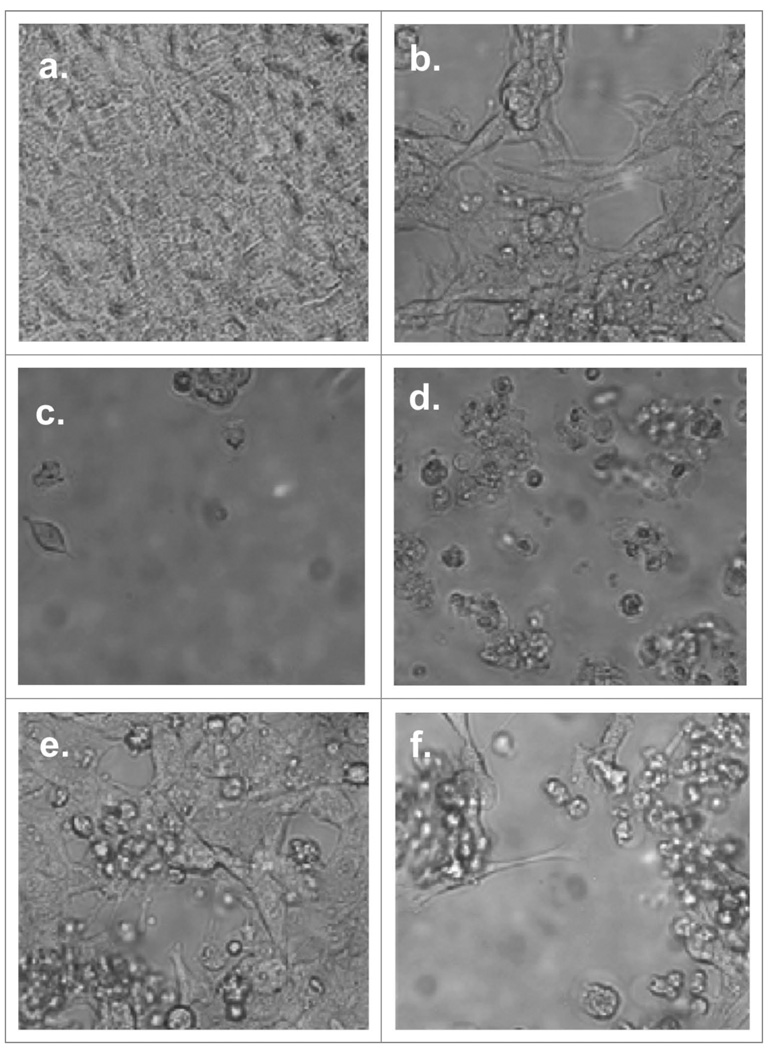
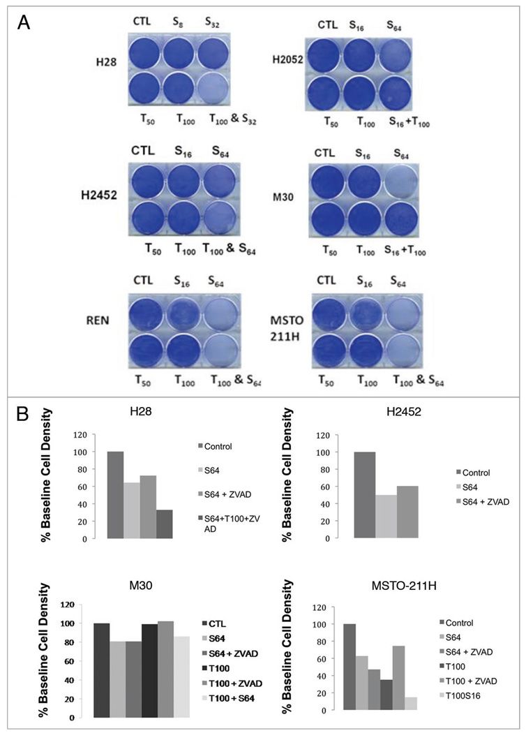
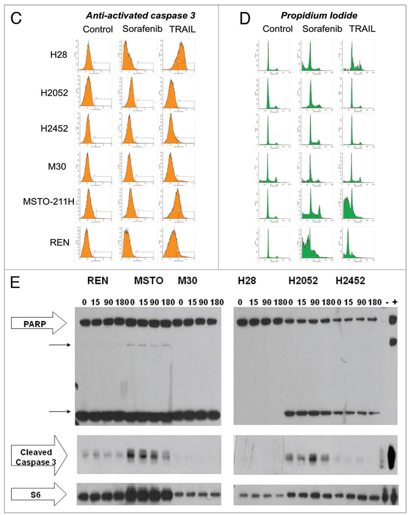
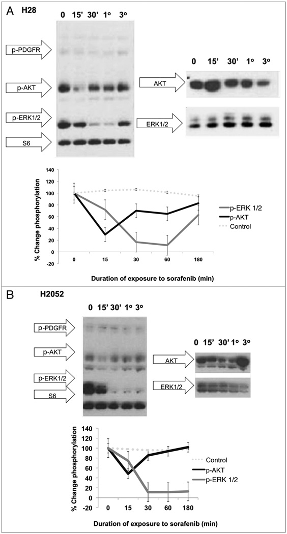
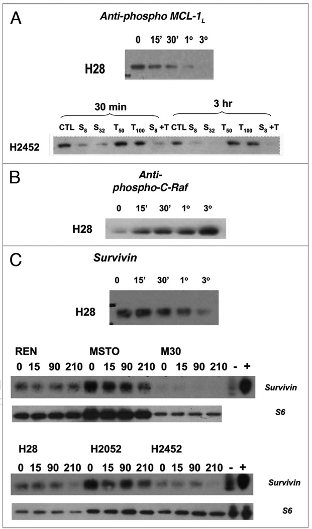
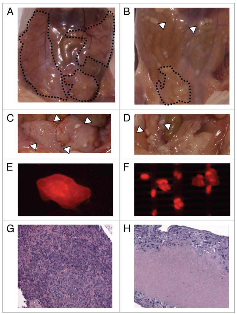
Malignant pleural mesothelioma (MPM) is an aggressive, rapidly progressive malignancy without effective therapy. We evaluate sorafenib efficacy and impact on the cellular pro-survival machinery in vitro, efficacy of sorafenib as monotherapy and in combination with the naturally occurring death receptor agonist, TRAIL using human MPM cell lines, MSTO-211H, M30, REN, H28, H2052 and H2452. In vitro studies of the six MPM lines demonstrated single agent sensitivity to the multikinase inhibitor sorafenib and resistance to TRAIL. H28 and H2452 demonstrated augmented apoptosis with the addition of TRAIL to sorafenib in vitro. Treated cell lines demonstrated sorafenib-induced rapid dephosphorylation of AKT followed shortly by near complete dephosphorylation of the constitutively phosphorylated ERK1/2. Sorafenib therapy also decreased phosphorylation of B-Raf and mTOR in several cell lines. Within 3 h of sorafenib treatment, a number of known pro-survival molecules were dephosphorylated and/or downregulated in expression including MCL-1(L), c-FLIP(L), survivin and cIAP(1). These changes and eventual cell death did not elicit significant caspase-3 activation or PARP cleavage and pretreatment with the pan-caspase inhibitor, Z-VAD-FMK, did not block sorafenib efficacy but did block the effect of TRAIL monotherapy. Pre-treatment with Z-VAD-FMK did not block the synergistic effect of TRAIL and sorafenib in H28. In summary, single agent treatment with sorafenib results in widespread inhibition of the pro-survival machinery in vitro leading to cell death via a primarily caspase-independent mechanism. Combining sorafenib therapy with TRAIL, may be useful in order to provide a more efficient death signal and this synergistic effect appears to be caspase-independent. Pilot in vivo data demonstrates promising evidence of therapeutic efficacy in human tumor bearing xenograft nu/nu mice. We document single agent activity of sorafenib against MPM, unravel novel effects of sorafenib on anti-apoptotic signaling mediators, and suggest the combination of sorafenib plus TRAIL as possible therapy for clinical testing in MPM.


Figures

Figure 1
Figure 1
Effects of incubation of MSTO-211H with TRAIL and sorafenib. Cells were allowed to adhere in sterile tissue culture plates for 16–24 h at 5% CO2, 37°C. Sorafenib or vehicle (0.1% DMSO) were added to the plates and incubated for additional 16–22 h. TRAIL was added to designated wells and wells were then imaged with bright field microscopy. Chemotherapy assignment and concentrations are as follows: (a) control, (b) Sorafenib (16 µM), (c) Sorafenib (64 µM), (d) Sorafenib (64 µM) & TRAIL (100 ng/mL), (e) TRAIL (50 ng/mL) and (f) TRAIL (100 ng/mL). Images demonstrate a dose-dependent response of MSTO-211H to sorafenib and TRAIL with a marked sensitivity to sorafenib and to the combination of TRAIL and sorafenib.
Figure 2
Figure 2
In vitro exposure of MPM cell lines to sorafenib demonstrate cytotoxicity without significant induction of caspase-3 activation or PA RP cleavage products. All six MPM cell lines, REN, MSTO-211H, M30, H28, H2052 and H2452, were allowed 24 h to adhere to tissue culture plates. (A) Coomassie blue staining demonstrating the sensitivity profile of the six cell lines to TRAIL and sorafenib. (B) Coomassie blue staining of MPM cell lines with and without pre-treatment with the irreversible pan-caspase inhibitor, Z-VAD-FMK prior to sorafenib and/or TRAIL therapy. Immunoblotting was performed S6 loading control (bottom); cleaved caspase 3; PA RP (large arrow) and PA RP cleavage products (two smaller arrows) (Cell Signaling, Danvers, MA). Control lysate, purchased from Cell Signaling, Danvers, MA, for apoptosis were also added as the (−) and (+) lanes, representing Jurkat cells untreated or treated with etoposide respectively. (C) Anti-activated caspase FACS histograms of MPM cell lines treated with sorafenib or TRAIL, fixed and stained with PE-conjugated anti-activated caspase 3 antibody. (D) Propidium iodide FACS histogram of MPM cell lines treated with sorafenib or TRAIL, fixed and stained with propidium iodide. (E) Western blot analysis of MPM cell lysates following treatment with sorafenib 64 µM or vehicle (0.1% DMSO) for a 3 h time course. Immunoblotting was performed S6 loading control (bottom); cleaved caspase 3; PA RP (large arrow) and PARP cleavage products (two smaller arrows) (Cell Signaling, Danvers, MA). Control lysate, purchased from Cell Signaling, Danvers, MA, for apoptosis were also added as the (−) and (+) lanes, representing Jurkat cells untreated or treated with etoposide respectively.
Figure 2
Figure 2
In vitro exposure of MPM cell lines to sorafenib demonstrate cytotoxicity without significant induction of caspase-3 activation or PA RP cleavage products. All six MPM cell lines, REN, MSTO-211H, M30, H28, H2052 and H2452, were allowed 24 h to adhere to tissue culture plates. (A) Coomassie blue staining demonstrating the sensitivity profile of the six cell lines to TRAIL and sorafenib. (B) Coomassie blue staining of MPM cell lines with and without pre-treatment with the irreversible pan-caspase inhibitor, Z-VAD-FMK prior to sorafenib and/or TRAIL therapy. Immunoblotting was performed S6 loading control (bottom); cleaved caspase 3; PA RP (large arrow) and PA RP cleavage products (two smaller arrows) (Cell Signaling, Danvers, MA). Control lysate, purchased from Cell Signaling, Danvers, MA, for apoptosis were also added as the (−) and (+) lanes, representing Jurkat cells untreated or treated with etoposide respectively. (C) Anti-activated caspase FACS histograms of MPM cell lines treated with sorafenib or TRAIL, fixed and stained with PE-conjugated anti-activated caspase 3 antibody. (D) Propidium iodide FACS histogram of MPM cell lines treated with sorafenib or TRAIL, fixed and stained with propidium iodide. (E) Western blot analysis of MPM cell lysates following treatment with sorafenib 64 µM or vehicle (0.1% DMSO) for a 3 h time course. Immunoblotting was performed S6 loading control (bottom); cleaved caspase 3; PA RP (large arrow) and PARP cleavage products (two smaller arrows) (Cell Signaling, Danvers, MA). Control lysate, purchased from Cell Signaling, Danvers, MA, for apoptosis were also added as the (−) and (+) lanes, representing Jurkat cells untreated or treated with etoposide respectively.
Figure 3
Figure 3
Time course of treatment of human MPM cell lines with Sorafenib (64 µM) results in an early decrease in phosphorylation in AKT followed by a marked decrease in ERK 1/2 phosphorylation in whole cell lysates. In vitro cell cultures of H28 (A) and H2052 (B) were treated with sorafenib at a concentration of 64 µM at 37°C, 5% CO2 for 0 (no sorafenib), 15 min (15'), 30 min (30'), 1 h (1°) and 3 h (3°). Cells were then washed with dPBS and lysed in 1X LDS sample buffer with 2-ME, boiled and run on a 4–12% pre-cast Nupage gel followed by immunoblotting with the following primary antibodies: PDGFR pathway activation antibody cocktail (Cell Signaling) with antibodies to phospho-PDGFR, phosphor-SHP 2, phospho-AKT, phosphor-ERK 1/2 and S6 loading control. These data reveal an early marked decrease in the phosphorylation levels of AKT in response to treatment with sorafenib with near recovery to baseline levels by 3 h of sorafenib exposure. This is followed temporally by a sharp, sustained dephosphorylation of ERK1/2 that begins to recover by 3 h of sorafenib therapy.
Figure 4
Figure 4
Time course of treatment of human MPM cell lines with Sorafenib (64 µM) results in negative regulation of anti-apoptotic resistance proteins. MPM in vitro cell cultures were treated with sorafenib at a concentration of 64 µM at 37°C, 5% CO2 for 0 (no sorafenib), 15 min (15'), 30 min (30'), 1 h (1°) and 3 h (3°). Cells were then washed with dPBS and lysed in 1X LDS sample buffer with 2-ME, boiled and run on a 4–12% pre-cast Nupage gel followed by immunoblotting with the following primary antibodies: (A) phospho-MCL-1L (B) C-Raf (C) survivin (Cell Signaling). Sorafenib therapy resulted in dephosphorylation of MCL1 followed by decreased protein levels of MCL1 (data not shown) and decreased protein levels of survivin.
Figure 5
Figure 5
Pilot evaluation of human MPM cell lines in a xenograft mouse models suggests sorafenib efficacy. nu/nu mice were injected IP with human malignant pleural mesothelioma cell line, MSTO-211H cell surface labeled with a fluorescent label, CellVue Maroon. Images above demonstrate (A) untreated (3 w daily IP vehicle) (B) treated with sorafenib 60 mg/Kg daily for 3 w (C) exposed untreated tumor with peritoneum resected (D) exposed treated tumor with peritoneum resected (E) large soft tissue IP tumors (F) and smaller tumor lesions demonstrate fluorescence in the expected excitation/absorption spectrum for CellVue Maroon cell surface labeled MSTO-211H utilized for these IP injected tumors. Immediately after sacrifice, xenografted tumor tissues were dissected, fixed in 4% formalin, paraffin-embedded, sectioned in 6 µm slices and stained with hematoxylin and eosin. Treated tumor nodules demonstrated marked hyalinization and reduce tumor viability (H) compared with untreated tumor (G).

Similar articles

Cited by

References

    1. Montanaro F, Rosato R, Gangemi M, et al. Survival of pleural malignant mesothelioma in Italy: a population-based study. Int J Cancer. 2009;124:201–207. - PubMed
    1. Kazan-Allen L. Asbestos and mesothelioma: worldwide trends. Lung Cancer. 2005;49:3–8. - PubMed
    1. Goudar RK. Review of pemetrexed in combination with cisplatin for the treatment of malignant pleural mesothelioma. Ther Clin Risk Manag. 2008;4:205–211. - PMC - PubMed
    1. Kindler HL. Systemic treatments for mesothelioma: standard and novel. Curr Treat Options Oncol. 2008;9:171–179. - PMC - PubMed
    1. Rippo MR, Moretti S, Vescovi S, et al. FLIP overexpression inhibits death receptor-induced apoptosis in malignant mesothelial cells. Oncogene. 2004;23:7753–7760. - PubMed

MeSH terms