. 2009 Fall;6(3):211-22.
doi: 10.1900/RDS.2009.6.211. Epub 2009 Nov 10.

Sequential abnormalities in type 1 diabetic encephalopathy and the effects of C-Peptide

Affiliations

Sequential abnormalities in type 1 diabetic encephalopathy and the effects of C-Peptide

Anders A F Sima et al. Rev Diabet Stud. 2009 Fall.

Abstract

Diabetic encephalopathy is a recently recognized complication in type 1 diabetes. In this review, we summarize a series of experimental results obtained longitudinally in the spontaneously type 1 diabetic BB/Wor-rat, and bringing out the beneficial effects of C-peptide replacement. It is increasingly clear that lack of insulin and C-peptide, and perturbations of their signaling cascades in type 1 diabetes are detrimental to the regulation of neurotrophic factors and their receptors. Other consequences of such deficits and perturbations are innate inflammatory responses with effects on synaptogenesis, neurite degeneration, and early behavioral abnormalities. Replacement of C-peptide, which does not effect hyperglycemia, has beneficial effects on a variety of pro-apoptotic stressors, oxidative stressors, and finally on apoptosis. Eventually, this cascade of events leads to neuronal loss and decreased densities of white matter myelinating cells, with more profound deficits in behavioral and cognitive function. Such changes are likely to underlie gray and white matter atrophy in type 1 diabetes, and are significantly prevented by full C-peptide replacement. Present data demonstrate that C-peptide replacement has beneficial effects on numerous sequential and partly interrelated pathogenetic mechanisms, resulting in prevention of neuronal and oligodendroglial cell loss, with significant prevention of neurobehavioral and cognitive functions.


Figures

Figure 1
Figure 1. Results from behavioral testing of 2-mo control, diabetic, and C-peptide replaced diabetic BB/Wor-rats using the radial arm paradigm
A: Total latencies were not altered in diabetic or C-peptide-treated diabetic rats as compared to age-matched control animals. Type I (B) type II (C) errors were significantly more common in non-treated diabetic rats, signifying impaired reference and working memory, respectively. These error types were not recorded in C-peptide-treated diabetic rats (B and C).
Figure 2
Figure 2. Micro-PET data from three 3-month diabetic BB/Wor-rats (A) and age-matched control rats (B)
The uptake rate constant of the FDA tracer (K-cplx) was decreased by 22-32% in diabetic rats as compared to control rats (C and Table). Despite this, the metabolic rate of glucose (rMRGlc) was increased approximately 3-fold in cerebral cortex, hippocampus, and whole brain (Table).
Figure 3
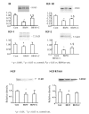
Figure 3. Neurotrophic profiles in hippocami of 2-month diabetic and C-peptide treated rats (IR, IGF-R, IGF-1 and IGF-2)
Note the significant suppression of the expression of these factors and receptors in diabetic rats, abnormalities that were significantly prevented by C-peptide replacement. Also, NGF and its receptor Trk A showed decreased expression in hippocampi of 8-month diabetic BB/Wor-rat with modest prevention in C-peptide treated rats. Equal loading of lanes was evaluated by staining of membranes with Ponceau reagent.
Figure 4
Figure 4
A: The expression of hippocampal presynaptic synaptophysin was significantly decreased in 4-month diabetic rats, and was prevented by C-peptide replacement from onset of diabetes. B: This corresponded to a decreased density of synaptophysin stained synapses in the CA1 region of diabetic rat hippocampi, suggesting presynaptic degeneration. The normal density of presynaptic buttons was preserved in C-peptide treated rats.
Figure 5
Figure 5. Latencies in the Morris water maze set up (right upper corner) in control and diabetic BB/Wor-rats at 2 (A); 4 (B); 6 (C), and 8 months (D) of diabetes
Q1-Q4 signify the four quadrants of the water maze. Note, significantly prolonged latencies are seen only after 6 months of diabetes (C). Diabetic rats treated with C-peptide from onset of diabetes showed significant prevention in the latencies at 8 months (D), suggesting prevention of spatial memory and long-term memory deficits.
Figure 6
Figure 6
Expression of post-synaptic excitatory GluR2 and GluR4 in hippocampus were both significantly increased in 7-month diabetic BB/Wor-rats. C-peptide substitution from diabetes onset fully prevented the more abundant GluR2 subunit, whereas the effect was less striking with respect to the less abundant GluR4.
Figure 7
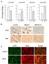
Figure 7. White matter changes in control, diabetic, and C-peptide replaced diabetic rats
A: Already in 2-month diabetic rats, there is a significant decrease in the number of oligodendroglial cells (identified by the oligo marker CAII) in temporal white matter, which is associated with increased proliferation of astrocytes. These differnces are sustained and increased in 8-month diabetic rats. B: Immunocytochemical identification of astrocytes (GFAP), which are increased in 4-month diabetic rats, which simultaneously show decreased stainability for the oligo marker myelin basic protein (MBP). There is close to normal stainability of astrocytes and oligodendroglia cells in C-peptide treated animals. C: RAGE is markedly upregulated in the white matter of 8-month diabetic rats, and colocalizes with GFAP-positive astrocytes.
Figure 8
Figure 8. Conceptual depiction of the temporal inter-relationships between pathogenetic mechanisms emanating from hyperglycemia and insulin deficiency in type 1 diabetic encephalopathy
Hyperglycemia leads to activation of the polyol pathway and AGEs, with upregulation of RAGE and activation of innate inflammatory factors. Inflammation and RAGE activation are also induced by increased expression of NF-κB secondary to impaired insulin signaling activity. Suppressed insulin signaling leads to decreased expression of IGF, NFG, and their respective receptors, with consequent neurite degeneration, and loss of presynaptic connections. This results in early (radial-arm paradigm) neuro behavioral deficits. Innate inflammatory activities and suppressed insulin signaling result in oxidative and apoptotic stresses. This results in apoptosis and cell loss of both neurons and oligodendroglial cells, with consequent gray and white matter atrophy and cognitive deficits. As outlined in this review, full substitution of C-peptide has significant beneficial effects on several of these pathogenetic components, resulting in substantial prevention of behavioral and cognitive deficits.

Similar articles

Cited by

References

    1. Sima AA, Kamiya H, Li ZG. Insulin, C-peptide hyperglycemia and central nervous system complications in diabetes. Europ J Pharmacol. 2004;490:187–197. - PubMed
    1. Biessels GJ, Deary TJ, Ryan CM. Cognition and diabetes: a lifespan perspective. Lancet Neurol. 2008;7:184–190. - PubMed
    1. Biessels GJ. Diabetic encephalopathy. In: Veves A, Malik RA, editors. Diabetic Neuropathy - Clinical Management. Humana Press; Totowa, NJ: 2007. pp. 187–205.
    1. Sima AA. Pathobiology of diabetic encephalopathy. In: Biessels GJ, Luchsinger JA, editors. Humana Press; Totowa, NJ: 2009. pp. 405–427.
    1. Zhang W, Sima AA. The role glucose, cholesterol and insulin have in amyloidogenesis in human neuroblastoma cells. Toronto, Canada. Proc XIX in Meeting of Neurodiab; 2009. p. 21. Abstract.

LinkOut - more resources