Review
. 2010 Apr;67(7):1077-88.
doi: 10.1007/s00018-009-0234-9. Epub 2009 Dec 29.

Helix insertion into bilayers and the evolution of membrane proteins

Affiliations
Review

Helix insertion into bilayers and the evolution of membrane proteins

Robert Renthal. Cell Mol Life Sci. 2010 Apr.

Abstract

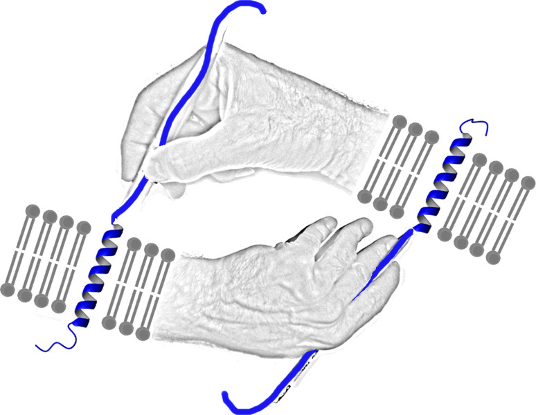
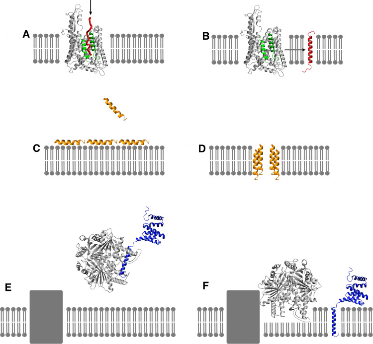
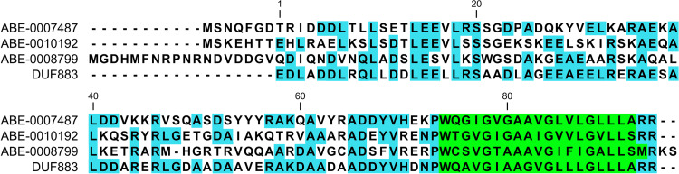
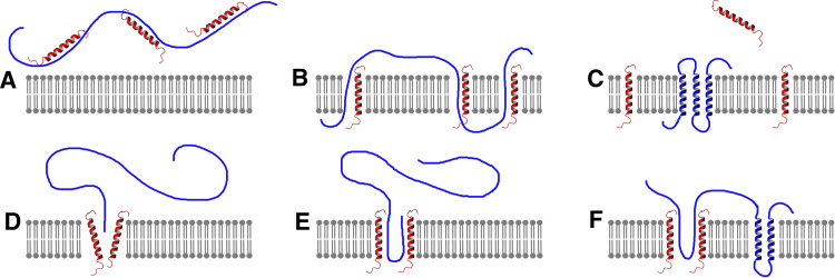
Polytopic alpha-helical membrane proteins cannot spontaneously insert into lipid bilayers without assistance from polytopic alpha-helical membrane proteins that already reside in the membrane. This raises the question of how these proteins evolved. Our current knowledge of the insertion of alpha-helices into natural and model membranes is reviewed with the goal of gaining insight into the evolution of membrane proteins. Topics include: translocon-dependent membrane protein insertion, antibiotic peptides and proteins, in vitro insertion of membrane proteins, chaperone-mediated insertion of transmembrane helices, and C-terminal tail-anchored (TA) proteins. Analysis of the E. coli genome reveals several predicted C-terminal TA proteins that may be descendents of proteins involved in pre-cellular membrane protein insertion. Mechanisms of pre-translocon polytopic alpha-helical membrane protein insertion are discussed.

Keywords: Bacteriocins; Chaperones; Tail-anchored proteins; Translocons; α-Helical membrane proteins.


Figures

Fig. 1
Fig. 1
Polytopic α-helical membrane proteins require polytopic α-helical membrane proteins for insertion into membranes. This raises the question of how these proteins evolved. Illustration after M.C. Escher’s “Drawing Hands”
Fig. 2
Fig. 2
Insertion of helical transmembrane (TM) proteins. a SecYE translocon from Thermus thermophilus [4] (Protein Data Bank 2ZJS). Nascent polypeptide chain (red) shown entering translocon channel from ribosome (arrow indicates direction of movement). Lateral gate, helices 2 and 7 (green). b TM helix (red) shown after insertion. Arrow indicates direction of motion through lateral gate. Translocon channel can accommodate two TM segments, thereby having the capability of inserting proteins with multiple TM domains and with connecting loops on both sides of membrane. c Peptide antibiotic alamethicin (yellow) (Protein Data Bank 1AMT) binds to membrane surface. d Alamethicin peptides associate to form TM channel. e Get3, a chaperone in the Asna1/TRC40 family [47] binds C-terminal tail-anchored membrane proteins and docks with receptor (gray rectangle) on target membrane (Protein Data Bank 2WOJ). TA protein (blue) modeled from water-soluble domain of Fis1 (Protein Data Bank 1PC2) and TM domain of monoamine oxidase (Protein Data Bank 2Z5Y). f After binding to receptor, chaperone inserts TM domain of TA protein into bilayer, possibly by entering bilayer outer leaflet. Protein structures drawn using MOLMOL software [90]
Fig. 3
Fig. 3
Sequence alignment of three tail-anchored proteins from E. coli. Sequences are aligned with the consensus DUF883 domain (pfam05957). Green putative C-terminal transmembrane domain, light blue sequence identities with the consensus DUF883 domain
Fig. 4
Fig. 4
Two possible mechanisms for pre-translocon insertion of polytopic helical membrane proteins. Single transmembrane (TM) helix proteins that spontaneously insert into bilayers (red) could facilitate polytopic membrane protein (blue) insertion. a–c Single TM helix proteins separately chaperone target transmembrane segments of larger protein, followed by dissociation of chaperones along with strong interaction of transmembrane helices to form folded structure. d–f Single TM helix proteins associate to form a translocon-like channel, probably involving four or more subunits (only two are shown). Binding of target protein opens lateral gate to release helical segments into bilayer. The polytopic protein in (d) and (e) is either chaperoned or cotranslationally inserted; the chaperones or ribosome are not shown

Similar articles

Cited by

References

    1. Kol S, Nouwen N, Driessen AJM. Mechanisms of YidC-mediated insertion and assembly of multimeric membrane protein complexes. J Biol Chem. 2008;283:31269–31273. doi: 10.1074/jbc.R800029200. - DOI - PubMed
    1. Bonnefoy N, Fiumera HL, Dujardin G, Fox TD. Roles of Oxa1-related inner-membrane translocases in assembly of respiratory chain complexes. Biochim Biophys Acta. 2009;1793:60–70. doi: 10.1016/j.bbamcr.2008.05.004. - DOI - PMC - PubMed
    1. Driessen AJ, Nouwen N. Protein translocation across the bacterial cytoplasmic membrane. Annu Rev Biochem. 2008;77:643–667. doi: 10.1146/annurev.biochem.77.061606.160747. - DOI - PubMed
    1. Tsukazaki T, Hori H, Fukai S, Ishitani R, Mori T, Dohmae N, Perederina A, Sugita Y, Vassylyev DG, Ito K, Nureki O. Conformational transition of Sec machinery inferred from bacterial SecYE structures. Nature. 2008;455:988–992. doi: 10.1038/nature07421. - DOI - PMC - PubMed
    1. Blobel G. Intracellular protein topogenesis. Proc Natl Acad Sci USA. 1980;77:1496–1500. doi: 10.1073/pnas.77.3.1496. - DOI - PMC - PubMed

Publication types

MeSH terms

LinkOut - more resources