. 2009 Dec;66(6):759-70.
doi: 10.1002/ana.21728.

A novel recessive Nefl mutation causes a severe, early-onset axonal neuropathy

Affiliations

A novel recessive Nefl mutation causes a severe, early-onset axonal neuropathy

Sabrina W Yum et al. Ann Neurol. 2009 Dec.

Abstract

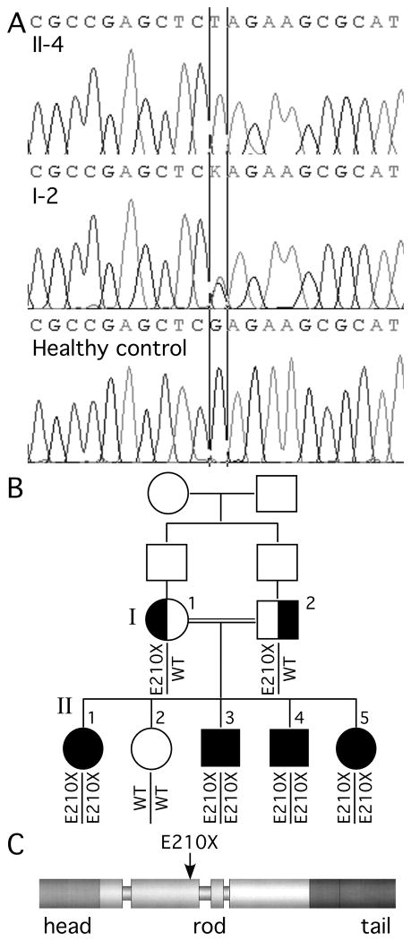
Objective: To report the first cases of a homozygous recessive mutation in NEFL, the gene that encodes the light subunit of neurofilaments.

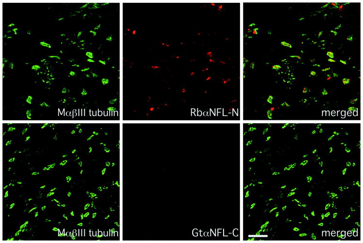
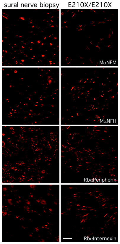
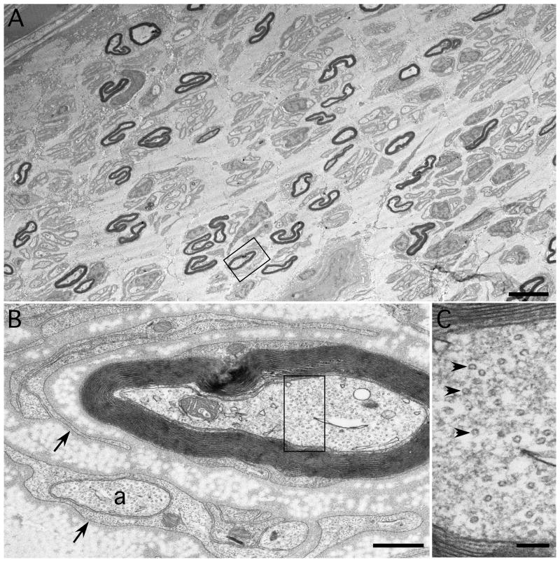
Methods: Clinical and electrophysiologic data were evaluated, and a sural nerve biopsy from one affected child was examined by immunohistochemistry and electron microscopy. The ability of the mutant protein to form filaments was characterized in an established cell culture system.

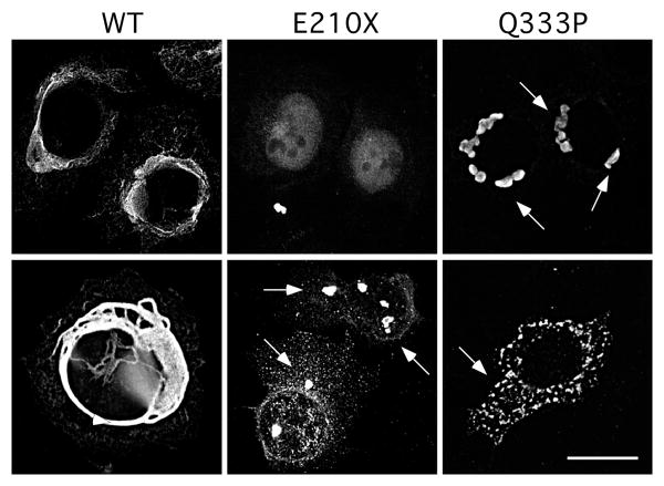
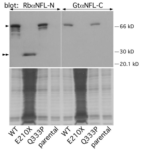
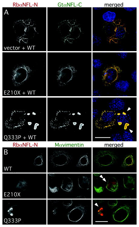
Results: Four of five siblings developed of a severe, progressive neuropathy beginning in early childhood. Serial nerve conduction studies showed progressively reduced amplitudes with age and pronounced slowing at all ages. Visual-evoked responses were slowed in three children, indicating that central nervous system axons were subclinically involved. All four affected children were homozygous for a nonsense mutation at glutamate 210 (E210X) in the NEFL gene; both parents were heterozygous carriers. A sural nerve biopsy from an affected patient showed markedly reduced numbers of myelinated axons; the remaining myelinated axons were small and lacked intermediate filaments. The E210X mutant protein did not form an intermediate filament network and did not interfere with the filament formation by wild-type human light subunit of neurofilaments in SW-13 vim(-) cells.

Interpretation: This is the first demonstration of a recessive NEFL mutation, which appears to cause a simple loss of function, resulting in a severe, early-onset axonal neuropathy with unique features. These results confirm that neurofilaments are the main determinant of axonal caliber and conduction velocity, and demonstrate for the first time that neurofilaments are required for the maintenance of myelinated peripheral nervous system axons.


Figures

Fig. 1
Fig. 1. Family pedigree and mutational analysis
Panel A shows the electrophoretograms of the DNA sequence harboring the c.628G>T mutation for individuals II-4 and I-2 as well as a healthy control (the color versions are shown in Supplemental Fig. 1). In panel B, note that the unaffected sister (II-2) does not have the mutation, all affected children (II-1, II-3, II-4, and II-5) are homozygous, and both parents (I-1 and I-2) are heterozygous for the mutation. The siblings of the parents are not shown, and their parents are not available for genetic testing. Panel C shows the predicted effect of the E210X mutation on the domain structure of the human NFL protein.
Fig. 2
Fig. 2. Altered NFL staining in a sural nerve biopsy
These are confocal images of transverse sections from the sural nerve biopsy of patient II-4 at age 16 (who is homozygous for the E210X mutation), immunostained as indicated. Note the complete lack of staining with the goat antiserum against the C-terminus of NFL, whereas the rabbit antiserum against the N-terminus labels the axons, which are βIII tubulin-positive. Scale bar: 10 μm.
Fig. 3
Fig. 3. Intermediate filament subunits in a sural nerve biopsy
These are confocal images of transverse sections of the sural nerve biopsy from patient II-4 at age 16 (right column), and an adult patient with an idiopathic neuropathy (left column), immunostained as indicated. The axons in both biopsies are immunoreactive for NFM, NFH, peripherin, and α-internexin. Scale bar: 10 μm.
Fig. 4
Fig. 4. Ultrastructure of a sural nerve biopsy
These are electron micrographs taken from a transverse section of a sural nerve biopsy from patient II-4 at age 16. In A, note that the reduced density of myelinated axons, all of which are small and many of which are irregularly shaped. The rectangle is enlarged in panel B, which shows a myelinated axon that is partially surrounded by Schwann cell processes (arrows), one of which encloses an unmyelinated axon (a). The rectangle is enlarged in panel C, which shows that the axon contains abundant microtubules (arrowheads), but no intermediate filaments. Scale bars: A, 5 μm; B, 0.5 μm; C, 0.1 μm.
Fig. 5
Fig. 5. The E210X mutant does not form filaments
These are deconvolved images of SW-13 vim- cells, transiently transfected to express WT human NFL (WT), E210X, or Q333P, immunostained with a rabbit antiserum against the N-terminus of NFL and a mouse monoclonal antibody against vimentin (not shown), and counterstained with DAPI (not shown). Note that WT NFL forms a filamentous network, Q333P forms cytoplasmic aggregates (arrowheads), and E210X forms faint, diffuse puncta and occasional larger aggregrates (arrowheads). Scale bar: 20 μm.
Fig. 6
Fig. 6. Detection of the E210X mutant protein by immunoblotting
The upper panel shows immunoblots of lysates from parental SW-13 vim- cells or cells transiently transfected to express WT human NFL (WT), E210X, or Q333P, probed with antisera against the N-terminus (left panel) or C-terminus (right panel) of NFL. Each lane contains 60 μg of lysate, except for the E210X sample, which was deliberately overloaded (∼1800 μg) to show the fainter NFL band (double arrowheads) of the truncated E210X mutant. The positions of the 20, 30, and 66 kDa size markers are shown, and full-length NFL protein is indicated by the single arrowhead. The lower panels are images of the Coomassie-stained gels after transfer.
Fig. 7
Fig. 7. The E210X mutant does not affect the ability of WT NFL or vimentin to form a network
(A) These are deconvolved images of SW-13 vim- cells, transiently transfected to express WT human NFL (WT) plus “empty vector” (vector), E210X, or Q333P, then immunostained with a rabbit antiserum against the N-terminus of NFL (red), a goat antiserum against the C-terminus of NFL (green), a mouse monoclonal antibody against vimentin (not shown), and counterstained with DAPI (blue). Note the filamentous network of NFL staining in cells expressing WT NFL plus vector or E210X, whereas cells expressing both WT NFL and Q333P form cytoplasmic aggregates (arrowheads). Scale bar: 20 μm. (B) These are deconvolved images of SW-13 vim+ cells, transiently transfected to express WT human NFL (WT), E210X, or Q333P, then immunostained with a rabbit antiserum against the N-terminus of NFL (red) and a mouse monoclonal antibody against vimentin (green). Note that vimentin and NFL staining are co-localized in cells expressing WT NFL (forming a network) or Q333P (which collapses the network and forms cytoplasmic aggregates; arrowheads), but not in cells expressing E210X, which is found in small puncta/diffuse staining that did not disrupt vimentin assembly (double arrowheads). Scale bar: 20 μm.

Comment in

Similar articles

Cited by

References

    1. Friede RL, Samorajski T. Axon caliber related to neurofilaments and microtubules in sciatic nerve fibers of rats and mice. Anat Rec. 1970;167:379–388. - PubMed
    1. Muma NA, Hoffman PN. Neurofilaments are intrinsic determinants of axonal caliber. Micron. 1993;24:677–683.
    1. Lee MK, Xu Z, Wong PC, Cleveland DW. Neurofilaments are obligate heteropolymers in vivo. J Cell Biol. 1993;122:1337–1350. - PMC - PubMed
    1. Zhu Q, Couillard-Despres S, Julien JP. Delayed maturation of regenerating myelinated axons in mice lacking neurofilaments. Exp Neurol. 1997;148:299–316. - PubMed
    1. Jacomy H, Zhu QZ, Couillard-Despres S, et al. Disrupting of type IV intermediate filament network in mice lacking the neurofilament medium and heavy subunits. J Neurochem. 1999;73:972–984. - PubMed

Publication types

MeSH terms