

Laminin-332-beta1 integrin interactions negatively regulate invadopodia
- PMID: 20039268
- PMCID: PMC3150482
- DOI: 10.1002/jcp.22018
Laminin-332-beta1 integrin interactions negatively regulate invadopodia
Abstract
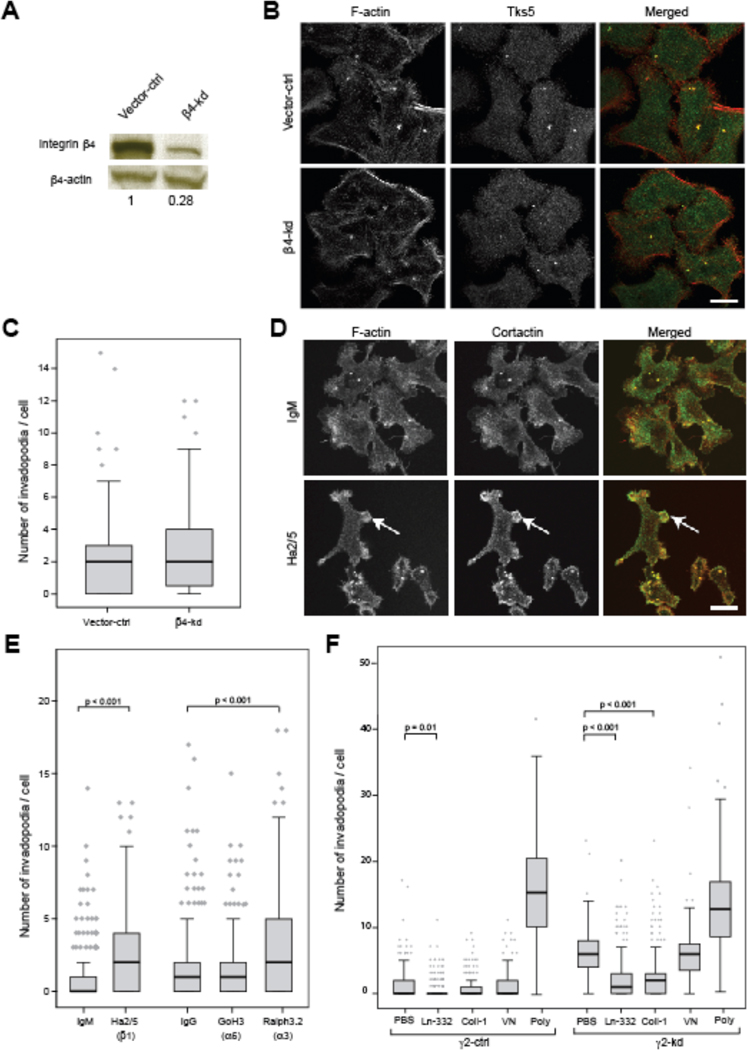
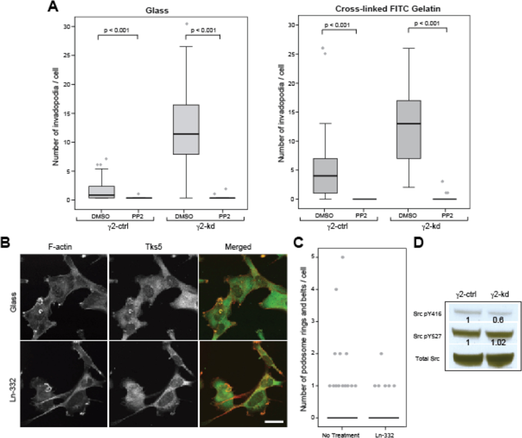
Adhesion of epithelial cells to basement membranes (BM) occurs through two major structures: actin-associated focal contacts and keratin-associated hemidesmosomes, both of which form on laminin-332 (Ln-332). In epithelial-derived cancer cells, additional actin-linked structures with putative adhesive properties, invadopodia, are frequently present and mediate BM degradation. A recent study proposed that BM invasion requires a proper combination of focal contacts and invadopodia for invading cells to gain traction through degraded BM, and suggested that these structures may compete for common molecular components such as Src kinase. In this study, we tested the role of the Ln-332 in regulating invadopodia in 804G rat bladder carcinoma cells, a cell line that secretes Ln-332 and forms all three types of adhesions. Expression of shRNA to Ln-332 gamma2 chain (gamma2-kd) led to increased numbers of invadopodia and enhanced extracellular matrix degradation. Replating gamma2-kd cells on Ln-332 or collagen-I fully recovered cell spreading and inhibition of invadopodia. Inhibition of alpha3 or beta1, but not alpha6 or beta4, phenocopied the effect of gamma2-kd, suggesting that alpha3beta1-mediated focal contacts, rather than alpha6beta4-mediated hemidesmosome pathways, intersect with invadopodia regulation. gamma2-kd cells exhibited alterations in focal contact-type structures and in activation of focal adhesion kinase (FAK) and Src kinase. Inhibition of FAK also increased invadopodia number, which was reversible with Src inhibition. These data are consistent with a model whereby actin-based adhesions can limit the availability of active Src that is capable of invadopodia initiation and identifies Ln-332-beta1 interactions as a potent upstream regulator that limits cell invasion.
J. Cell. Physiol. 223: 134-142, 2010. (c) 2009 Wiley-Liss, Inc.
Figures


Similar articles
-
Basement membrane laminin-5 is deposited in colorectal adenomas and carcinomas and serves as a ligand for alpha3beta1 integrin.APMIS. 2000 Mar;108(3):161-72. doi: 10.1034/j.1600-0463.2000.d01-40.x. APMIS. 2000. PMID: 10752684
-
Enterolobium contortisiliquum trypsin inhibitor (EcTI), a plant proteinase inhibitor, decreases in vitro cell adhesion and invasion by inhibition of Src protein-focal adhesion kinase (FAK) signaling pathways.J Biol Chem. 2012 Jan 2;287(1):170-182. doi: 10.1074/jbc.M111.263996. Epub 2011 Oct 28. J Biol Chem. 2012. PMID: 22039045 Free PMC article.
-
Co-localization of cortactin and phosphotyrosine identifies active invadopodia in human breast cancer cells.Exp Cell Res. 2006 May 1;312(8):1240-53. doi: 10.1016/j.yexcr.2005.12.012. Epub 2006 Jan 25. Exp Cell Res. 2006. PMID: 16442522
-
Defining the role of laminin-332 in carcinoma.Matrix Biol. 2009 Oct;28(8):445-55. doi: 10.1016/j.matbio.2009.07.008. Epub 2009 Aug 15. Matrix Biol. 2009. PMID: 19686849 Free PMC article. Review.
-
Laminin-5 in epithelial tumour invasion.J Mol Histol. 2004 Mar;35(3):277-86. doi: 10.1023/b:hijo.0000032359.35698.fe. J Mol Histol. 2004. PMID: 15339047 Review.
Cited by
-
Pyk2 and FAK differentially regulate invadopodia formation and function in breast cancer cells.J Cell Biol. 2018 Jan 2;217(1):375-395. doi: 10.1083/jcb.201702184. Epub 2017 Nov 13. J Cell Biol. 2018. PMID: 29133485 Free PMC article.
-
Signaling inputs to invadopodia and podosomes.J Cell Sci. 2013 Jul 15;126(Pt 14):2979-89. doi: 10.1242/jcs.079475. Epub 2013 Jul 10. J Cell Sci. 2013. PMID: 23843616 Free PMC article. Review.
-
A RhoG-mediated signaling pathway that modulates invadopodia dynamics in breast cancer cells.J Cell Sci. 2017 Mar 15;130(6):1064-1077. doi: 10.1242/jcs.195552. Epub 2017 Feb 15. J Cell Sci. 2017. PMID: 28202690 Free PMC article.
-
Adhesion rings surround invadopodia and promote maturation.Biol Open. 2012 Aug 15;1(8):711-22. doi: 10.1242/bio.20121867. Epub 2012 Jun 12. Biol Open. 2012. PMID: 23213464 Free PMC article.
-
Cdc42 and Tks5: a minimal and universal molecular signature for functional invadosomes.Cell Adh Migr. 2014;8(3):280-92. doi: 10.4161/cam.28833. Cell Adh Migr. 2014. PMID: 24840388 Free PMC article.
References
-
- Borradori L, Sonnenberg A. Structure and function of hemidesmosomes: more than simple adhesion complexes. J Invest Dermatol. 1999;112(4):411–418. - PubMed
-
- Buccione R, Orth JD, McNiven MA. Foot and mouth: podosomes, invadopodia and circular dorsal ruffles. Nature reviews. 2004;5(8):647–657. - PubMed
-
- Cance WG, Harris JE, Iacocca MV, Roche E, Yang X, Chang J, Simkins S, Xu L. Immunohistochemical analyses of focal adhesion kinase expression in benign and malignant human breast and colon tissues: correlation with preinvasive and invasive phenotypes. Clin Cancer Res. 2000;6(6):2417–2423. - PubMed
-
- Caron-Lormier G, Berry H. Amplification and oscillations in the FAK/Src kinase system during integrin signaling. Journal of theoretical biology. 2005;232(2):235–248. - PubMed
Publication types
MeSH terms
Substances
Grants and funding
LinkOut - more resources
Full Text Sources
Medical
Molecular Biology Databases
Miscellaneous
