. 2010 Jan;293(1):1-24.
doi: 10.1002/ar.21001.

The developmental remodeling of eye-specific afferents in the ferret dorsal lateral geniculate nucleus

Affiliations

The developmental remodeling of eye-specific afferents in the ferret dorsal lateral geniculate nucleus

Colenso M Speer et al. Anat Rec (Hoboken). 2010 Jan.

Abstract

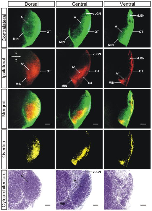
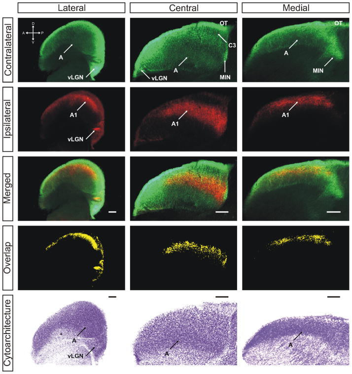
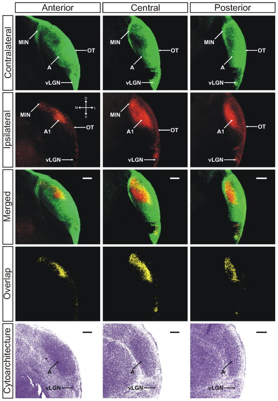
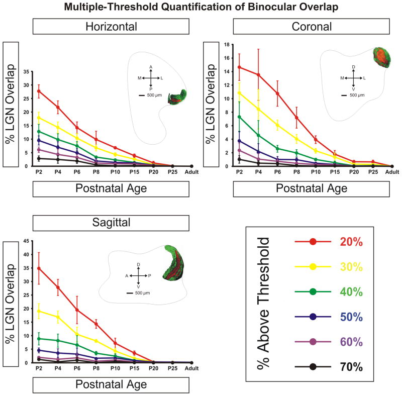
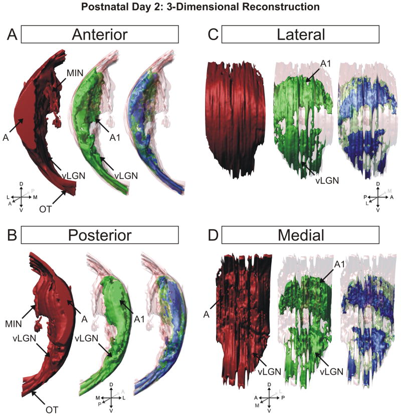
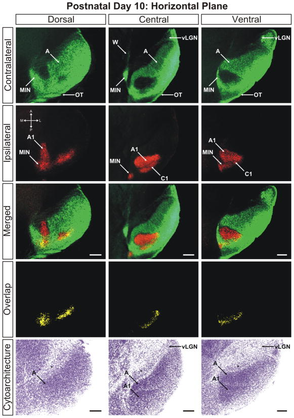
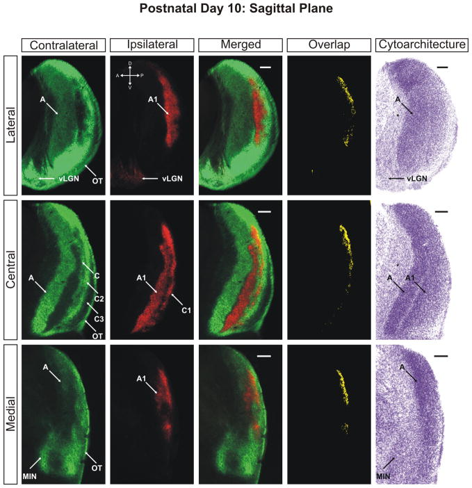
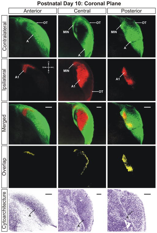
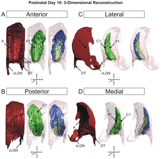
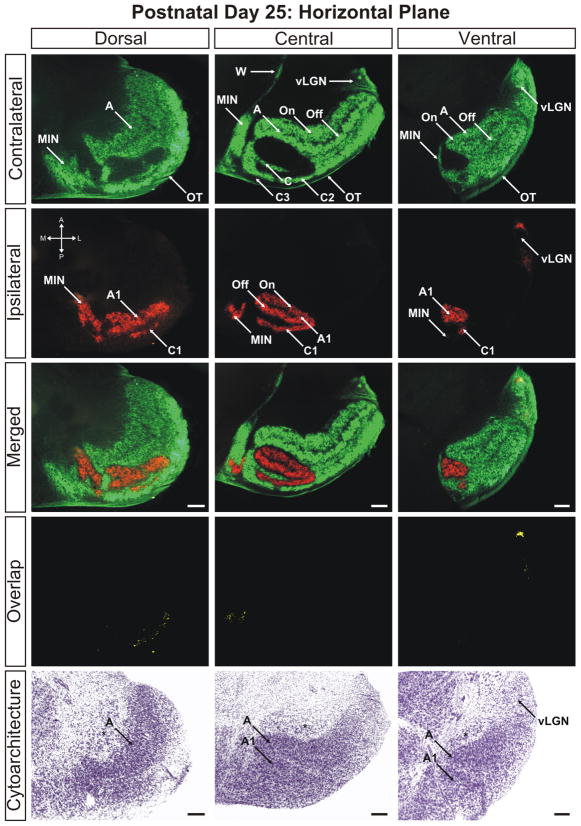
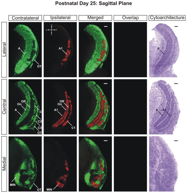
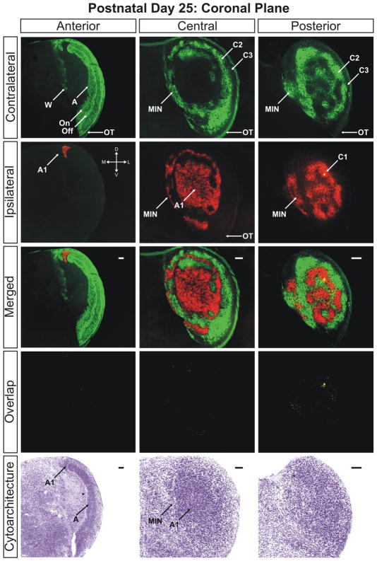
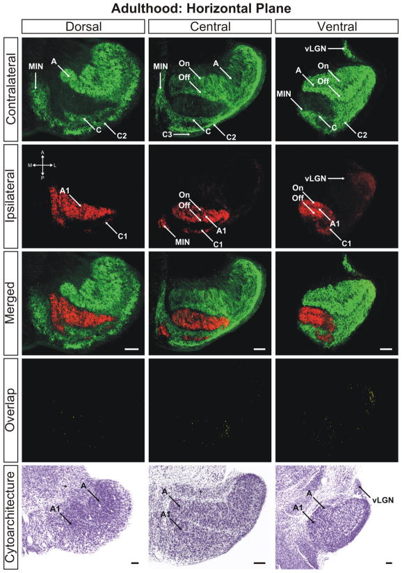
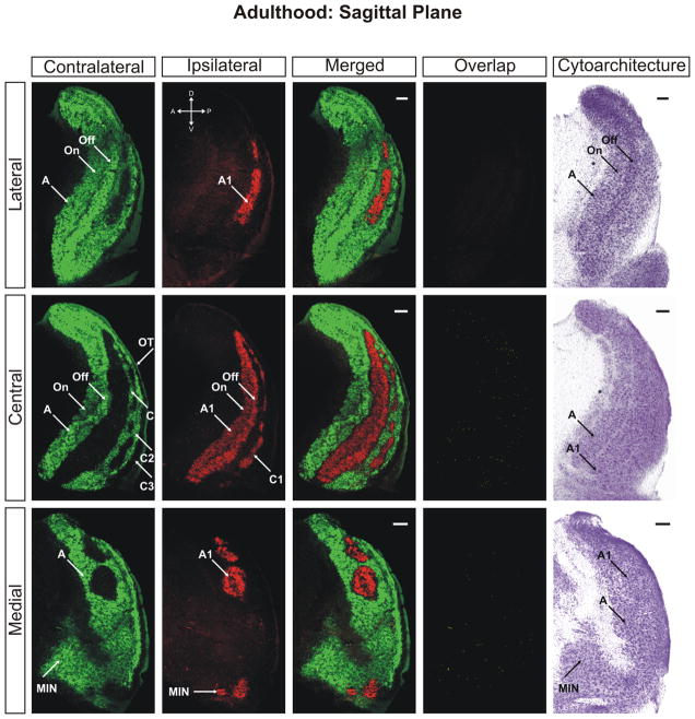
Eye-specific projections to the dorsal lateral geniculate nucleus (dLGN) serve as a model for exploring how precise patterns of circuitry form during development in the mammalian central nervous system. Using a combination of dual-label anterograde retinogeniculate tracing and Nissl-staining, we studied the patterns of eye-specific afferents and cellular laminae in the dLGN of the pigmented sable ferret at eight developmental timepoints between birth and adulthood. Each time point was investigated in the three standard orthogonal planes of section, allowing us to generate a complete anatomical map of eye-specific development in this species. We find that eye-specific retinal ganglion cell axon segregation varies according to location in the dLGN, with the principle contralateral (A) and ipsilateral layers (A1) maturing first, followed by the contralateral and ipsilateral C laminae. Cytoarchitectural lamination lags behind eye-specific segregation, except in the C laminae where underlying cellular layers never develop to accompany eye-specific afferent domains. The emergence of On/Off sublaminae occurs following eye-specific segregation in this species. On the basis of these findings, we constructed a three-dimensional map of eye-specific channels in the developing and mature ferret dLGN.


Figures

Figure 1
Figure 1
The ferret dLGN in the horizontal plane of section at postnatal day 2. Contralateral retinal inputs are green, ipsilateral inputs are red. Dorsal, central, and ventral sections through the nucleus are shown. Overlap represents multiplication of ipsilateral and contralateral signals thresholded to 30% above background. A, contralateral lamina; A1, ipsilateral lamina; OT, optic tract; vLGN, ventral lateral geniculate nucleus; MIN, medial intralaminar nucleus; *, perigeniculate nucleus. Scale bars: 200 μM
Figure 2
Figure 2
The ferret dLGN in the sagittal plane of section at postnatal day 2. Contralateral retinal inputs are green, ipsilateral inputs are red. Lateral, central, and medial sections through the nucleus are shown. Overlap represents multiplication of ipsilateral and contralateral signals thresholded to 30% above background. A, contralateral lamina; A1, ipsilateral lamina; OT, optic tract; vLGN, ventral lateral geniculate nucleus; MIN, medial intralaminar nucleus; *, perigeniculate nucleus. Scale bars: 200 μM
Figure 3
Figure 3
The ferret dLGN in the coronal plane of section at postnatal day 2. Contralateral retinal inputs are green, ipsilateral inputs are red. Anterior, central, and posterior sections through the nucleus are shown. Overlap represents multiplication of ipsilateral and contralateral signals thresholded to 30% above background. A, contralateral lamina; A1, ipsilateral lamina; OT, optic tract; vLGN, ventral lateral geniculate nucleus; MIN, medial intralaminar nucleus; *, perigeniculate nucleus. Scale bars: 200 μM
Figure 4
Figure 4
Quantification of binocular retinogeniculate afferent overlap across development. Threshold analysis results based on the methods described in Supplemental Figure 19 are shown for threshold values 20–70% above background for the three planes of section. Overlap is maximal at birth and decreases smoothly during the first 20 postnatal days at which point overlap between left/right eye inputs is absent. Each data point reflects mean value of measured overlap. Error bars reflect ± S.E.M. Photomicrograph insets are representative of the dLGN in adult animals. Note the Y-axes differ amongst the three graphs. See Methods for details on the quantification analyses performed.
Figure 5
Figure 5
3D reconstruction of the ferret dLGN at postnatal day 2 (P2). Perspectives facing the nucleus from the anterior pole (A), posterior pole (B), lateral aspect (C), and medial aspect (D) are shown. Contralateral projections are shown in red, ipsilateral projections are shown in green, overlapping projections are shown in blue for clarity. At this age, the major axis of the ipsilateral projections is oriented rostrocaudally. A, contralateral lamina; A1, ipsilateral lamina; C1, ipsilateral lamina; MIN, medial intralaminar nucleus; vLGN, ventral lateral geniculate nucleus; OT, optic tract.
Figure 6
Figure 6
The ferret dLGN in the horizontal plane of section at postnatal day 10. Dorsal, central, and ventral sections through the nucleus are shown. Overlap represents multiplication of ipsilateral and contralateral signals thresholded to 30% above background. A, contralateral lamina; A1, ipsilateral lamina; C1, ipsilateral lamina; OT, optic tract; vLGN, ventral lateral geniculate nucleus; W, wing of the geniculate; MIN, medial intralaminar nucleus; *, perigeniculate nucleus. Scale bars: 200 μm
Figure 7
Figure 7
The ferret dLGN in the sagittal plane of section at postnatal day 10. Contralateral retinal inputs are green, ipsilateral inputs are red. Lateral, central, and medial sections through the nucleus are shown. Overlap represents multiplication of ipsilateral and contralateral signals thresholded to 30% above background. A, contralateral lamina; A1, ipsilateral lamina; C, contralateral lamina; C1, ipsilateral lamina; C2, contralateral lamina; C3, retinal afferent free lamina; OT, optic tract; vLGN, ventral lateral geniculate nucleus; MIN, medial intralaminar nucleus; *, perigeniculate nucleus. Scale bars: 200 μm
Figure 8
Figure 8
The ferret dLGN in the coronal plane of section at postnatal day 10. Contralateral retinal inputs are green, ipsilateral inputs are red. Anterior, central, and posterior sections through the nucleus are shown. Overlap represents multiplication of ipsilateral and contralateral signals thresholded to 30% above background. A, contralateral lamina; A1, ipsilateral lamina; OT, optic tract; vLGN, ventral lateral geniculate nucleus; MIN, medial intralaminar nucleus; *, perigeniculate nucleus. Scale bars: 200 μm
Figure 9
Figure 9
3D reconstruction of the ferret dLGN at postnatal day 10 (P10). Perspectives facing the nucleus from the anterior pole (A), posterior pole (B), lateral aspect (C), and medial aspect (D) are shown. Contralateral projections are shown in red, ipsilateral projections are shown in green, overlapping projections are shown in blue for clarity. At this age, the major axis of the ipsilateral projections is oriented dorsoventrally. A, contralateral lamina; A1, ipsilateral lamina; C1, ipsilateral lamina; MIN, medial intralaminar nucleus; vLGN, ventral lateral geniculate nucleus; OT, optic tract.
Figure 10
Figure 10
The ferret dLGN in the horizontal plane of section at postnatal day 25. Dorsal, central, and ventral sections through the nucleus are shown. A, contralateral lamina; A1, ipsilateral lamina; On, ‘On’ center sublamina; Off, ‘Off’ center sublamina; C, contralateral lamina; C1, ipsilateral lamina; C2, contralateral lamina; C3, retinal afferent free lamina; OT, optic tract; vLGN, ventral lateral geniculate nucleus; W, wing of the geniculate; MIN, medial intralaminar nucleus; *, perigeniculate nucleus. Scale bars: 200 μm
Figure 11
Figure 11
The ferret dLGN in the sagittal plane of section at postnatal day 25. Contralateral retinal inputs are green, ipsilateral inputs are red. Lateral, central, and medial sections through the nucleus are shown. Overlap represents multiplication of ipsilateral and contralateral signals thresholded to 30% above background. A, contralateral lamina; A1, ipsilateral lamina; On, ‘On’ center sublamina; Off, ‘Off’ center sublamina; C, contralateral lamina; C1, ipsilateral lamina; C2, contralateral lamina; C3, retinal afferent free lamina; OT, optic tract; MIN, medial intralaminar nucleus; *, perigeniculate nucleus. Scale bars: 200 μm
Figure 12
Figure 12
The ferret dLGN in the coronal plane of section at postnatal day 25. Anterior, central, and posterior sections through the nucleus are shown. A, contralateral lamina; A1, ipsilateral lamina; On, ‘On’ center sublamina; Off, ‘Off’ center sublamina; C1, ipsilateral lamina; C2, contralateral lamina; C3, retinal afferent free lamina; OT, optic tract; W, wing of the geniculate; MIN, medial intralaminar nucleus; *, perigeniculate nucleus. Scale bars: 200 μm
Figure 13
Figure 13
The ferret dLGN in the horizontal plane of section at adulthood (>P100). Dorsal, central, and ventral sections through the nucleus are shown. A, contralateral lamina; A1, ipsilateral lamina; On, ‘On’ center sublamina; Off, ‘Off’ center sublamina; C, contralateral lamina; C1, ipsilateral lamina; C2, contralateral lamina; C3, retinal afferent free lamina; OT, optic tract; vLGN, ventral lateral geniculate nucleus; MIN, medial intralaminar nucleus; *, perigeniculate nucleus. Scale bars: 200 μm
Figure 14
Figure 14
The ferret dLGN in the sagittal plane of section at adulthood (>P100). Contralateral retinal inputs are green, ipsilateral inputs are red. Lateral, central, and medial sections through the nucleus are shown. Overlap represents multiplication of ipsilateral and contralateral signals thresholded to 30% above background. A, contralateral lamina; A1, ipsilateral lamina; On, ‘On’ center sublamina; Off, ‘Off’ center sublamina; C, contralateral lamina; C1, ipsilateral lamina; C2, contralateral lamina; C3, retinal afferent free lamina; OT, optic tract; MIN, medial intralaminar nucleus; *, perigeniculate nucleus. Scale bars: 200 μm

Similar articles

Cited by

References

    1. Akerman CJ, Grubb MS, Thompson ID. Spatial and temporal properties of visual responses in the thalamus of the developing ferret. J Neurosci. 2004;24:170–182. - PMC - PubMed
    1. Akerman CJ, Smyth D, Thompson ID. Visual experience before eye-opening and the development of the retinogeniculate pathway. Neuron. 2002;36:869–879. - PubMed
    1. Akerman CJ, Tolhurst DJ, Morgan JE, Baker GE, Thompson ID. Relay of visual information to the lateral geniculate nucleus and the visual cortex in albino ferrets. J Comp Neurol. 2003;461:217–235. - PubMed
    1. Angelucci A, Clasca F, Sur M. Anterograde axonal tracing with the subunit B of cholera toxin: a highly sensitive immunohistochemical protocol for revealing fine axonal morphology in adult and neonatal brains. J Neurosci Methods. 1996;65:101–112. - PubMed
    1. Bjartmar L, Huberman AD, Ullian EM, Renteria RC, Liu X, Xu W, Prezioso J, Susman MW, Stellwagen D, Stokes CC, Cho R, Worley P, Malenka RC, Ball S, Peachey NS, Copenhagen D, Chapman B, Nakamoto M, Barres BA, Perin MS. Neuronal pentraxins mediate synaptic refinement in the developing visual system. J Neurosci. 2006;26:6269–6281. - PMC - PubMed

Publication types

MeSH terms