

Biliary cystadenocarcinoma diagnosed with real-time contrast-enhanced ultrasonography: report of a case with diagnostic features
- PMID: 20039461
- PMCID: PMC2799910
- DOI: 10.3748/wjg.v16.i1.131
Biliary cystadenocarcinoma diagnosed with real-time contrast-enhanced ultrasonography: report of a case with diagnostic features
Abstract
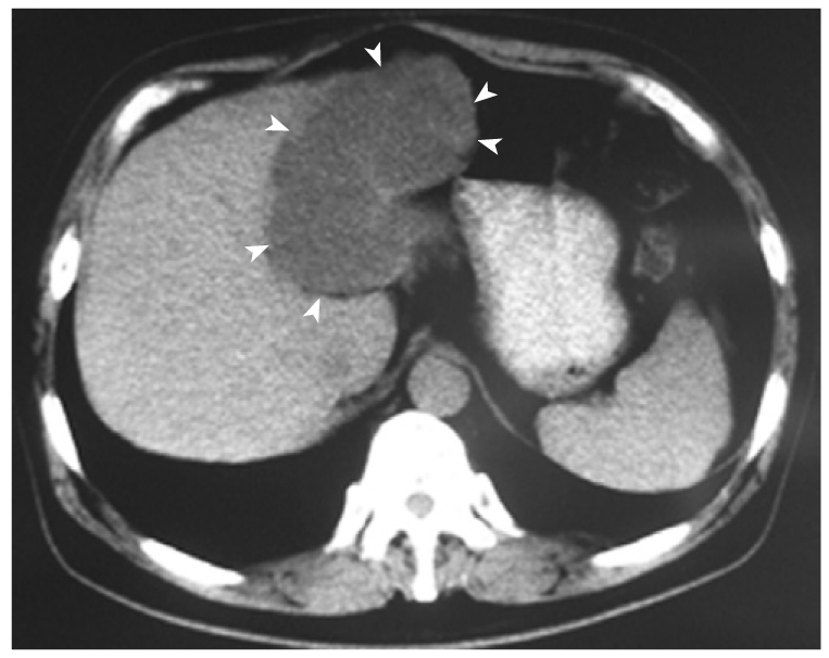
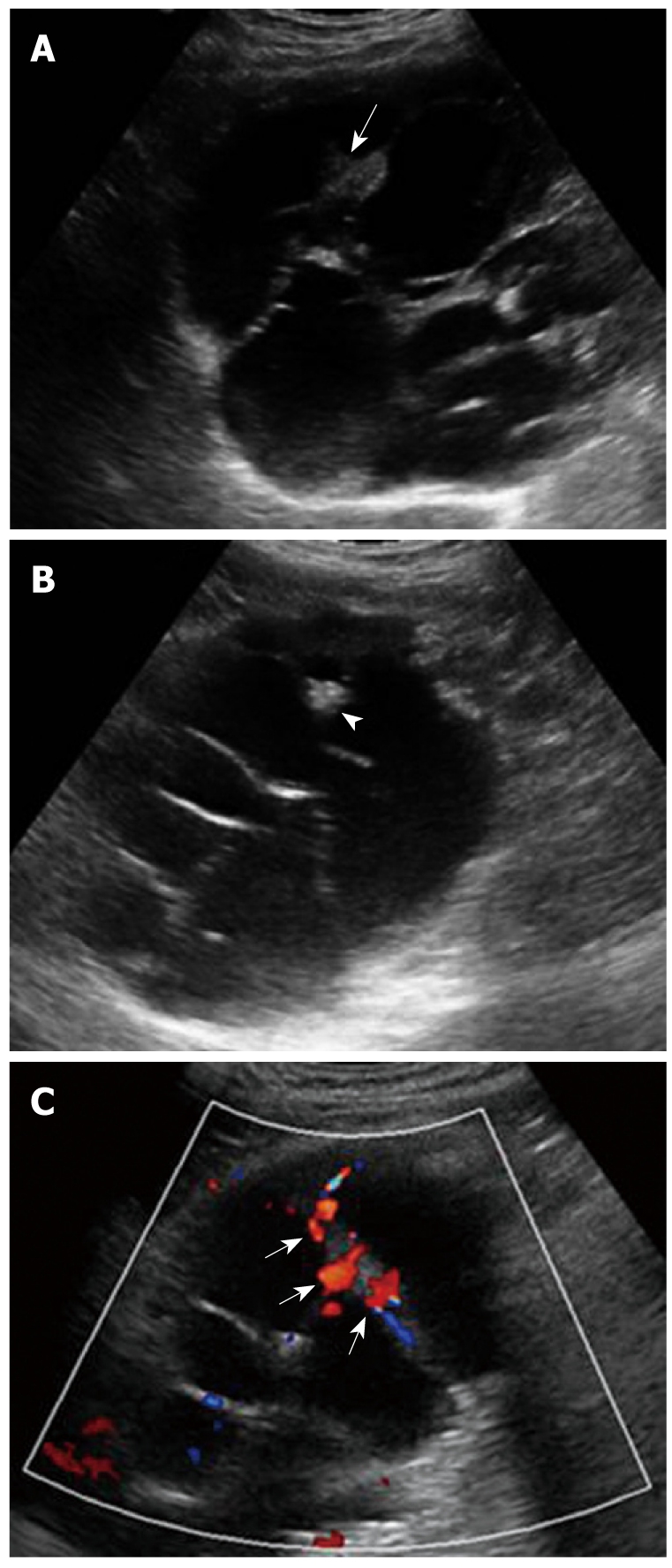
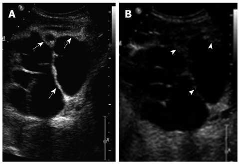
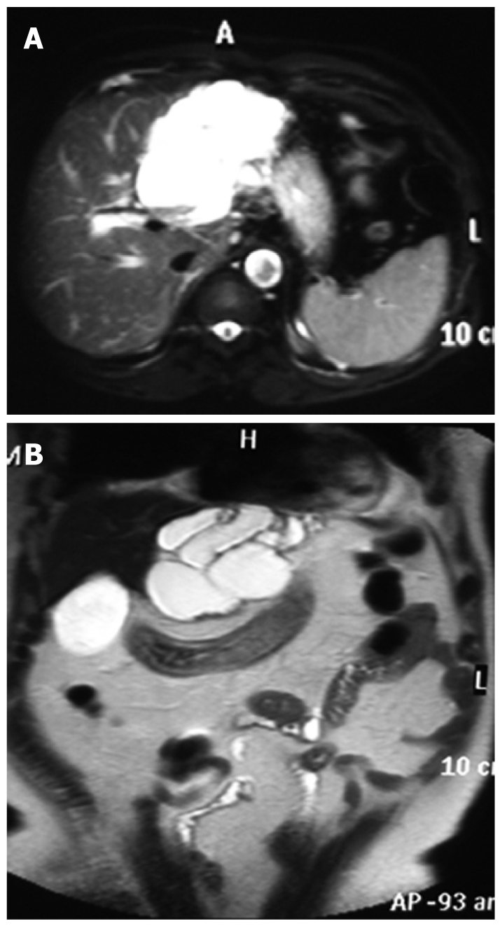
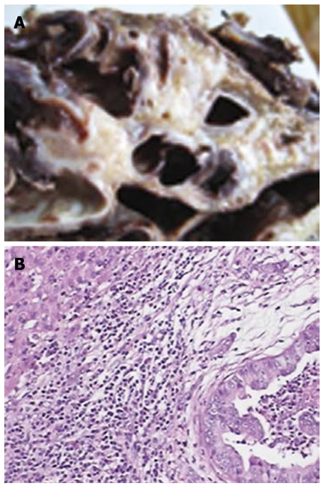
Biliary cystadenocarcinoma is a very rare malignant cystic tumor of the liver, which is often misdiagnosed due to a poor recognition of it. We report a case of a 60-year-old man with biliary cystadenocarcinoma with his real time contrast enhanced ultrasound (CEUS) characteristics compared to those of computed tomography (CT) and magnetic resonance imaging (MRI), which were correlated with the surgical and pathologic findings. Cystic wall enhancement, internal septations and intra-cystic solid portions in the arterial phase were observed on CEUS after contrast agent injection. The enhancement was washed out progressively and depicted as hypo-enhancement in the portal and late phases. CT revealed a large irregular cystic lesion in the left liver lobe with no clear septations and solid components. MRI showed an irregular cystic occupying lesion with septations.
Figures
Similar articles
-
Imaging features of intrahepatic biliary cystadenoma and cystadenocarcinoma on B-mode and contrast-enhanced ultrasound.Ultraschall Med. 2012 Dec;33(7):E241-E249. doi: 10.1055/s-0031-1299276. Epub 2012 Nov 15. Ultraschall Med. 2012. PMID: 23154870
-
Contrast enhanced ultrasound features of hepatic cystadenoma and hepatic cystadenocarcinoma.Scand J Gastroenterol. 2017 Mar;52(3):365-372. doi: 10.1080/00365521.2016.1259652. Epub 2016 Nov 25. Scand J Gastroenterol. 2017. PMID: 27887203
-
Multifocal biliary cystadenocarcinoma of the liver: CT and pathologic findings.Tumori. 2006 Jul-Aug;92(4):358-60. doi: 10.1177/030089160609200418. Tumori. 2006. PMID: 17036531
-
A case of biliary cystadenocarcinoma.Korean J Intern Med. 1997 Jan;12(1):109-13. doi: 10.3904/kjim.1997.12.1.109. Korean J Intern Med. 1997. PMID: 9159050 Free PMC article. Review.
-
Hepatobiliary cystadenomas and cystadenocarcinoma. Report of five cases.HPB Surg. 1996;9(2):83-92. doi: 10.1155/1996/36975. HPB Surg. 1996. PMID: 8871248 Free PMC article. Review.
Cited by
-
Morphological and dynamic evaluation of complex cystic focal liver lesions by contrast-enhanced ultrasound: current state of the art.J Ultrasound. 2019 Sep;22(3):251-259. doi: 10.1007/s40477-019-00385-2. Epub 2019 May 13. J Ultrasound. 2019. PMID: 31087277 Free PMC article. Review.
-
Role of contrast-enhanced ultrasound to define prognosis and predict response to biotherapy in pancreatic neuroendocrine tumors.J Endocrinol Invest. 2017 Dec;40(12):1373-1380. doi: 10.1007/s40618-017-0723-x. Epub 2017 Jun 30. J Endocrinol Invest. 2017. PMID: 28667452
-
Hepatobiliary cystadenocarcinoma.Case Reports Hepatol. 2012;2012:298957. doi: 10.1155/2012/298957. Epub 2012 Nov 1. Case Reports Hepatol. 2012. PMID: 25374705 Free PMC article.
-
A case of mucinous cystic neoplasm of the liver: description of Sonazoid-enhanced ultrasound imaging and histopathologic findings.J Med Ultrason (2001). 2013 Jul;40(3):243-50. doi: 10.1007/s10396-012-0422-3. Epub 2012 Dec 5. J Med Ultrason (2001). 2013. PMID: 27277243
-
Invasive biliary mucinous cystic neoplasm: a review.HPB (Oxford). 2012 Nov;14(11):725-40. doi: 10.1111/j.1477-2574.2012.00532.x. Epub 2012 Jul 22. HPB (Oxford). 2012. PMID: 23043661 Free PMC article. Review.
References
-
- Takayasu K, Muramatsu Y, Moriyama N, Yamada T, Hasegawa H, Hirohashi S, Ichikawa T, Ohno G. Imaging diagnosis of bile duct cystadenocarcinoma. Cancer. 1988;61:941–946. - PubMed
-
- Ishak KG, Willis GW, Cummins SD, Bullock AA. Biliary cystadenoma and cystadenocarcinoma: report of 14 cases and review of the literature. Cancer. 1977;39:322–338. - PubMed
-
- Itai Y, Araki T, Furui S, Yashiro N, Ohtomo K, Iio M. Computed tomography of primary intrahepatic biliary malignancy. Radiology. 1983;147:485–490. - PubMed
-
- Forrest ME, Cho KJ, Shields JJ, Wicks JD, Silver TM, McCormick TL. Biliary cystadenomas: sonographic-angiographic-pathologic correlations. AJR Am J Roentgenol. 1980;135:723–727. - PubMed
-
- Federle MP, Filly RA, Moss AA. Cystic hepatic neoplasms: complementary roles of CT and sonography. AJR Am J Roentgenol. 1981;136:345–348. - PubMed
Publication types
MeSH terms
Substances
LinkOut - more resources
Full Text Sources
Medical
