Review
. 2010 Feb;45(1):23-49.
doi: 10.3109/10409230903501819.

DNA interstrand crosslink repair in mammalian cells: step by step

Affiliations
Review

DNA interstrand crosslink repair in mammalian cells: step by step

Parameswary A Muniandy et al. Crit Rev Biochem Mol Biol. 2010 Feb.

Abstract

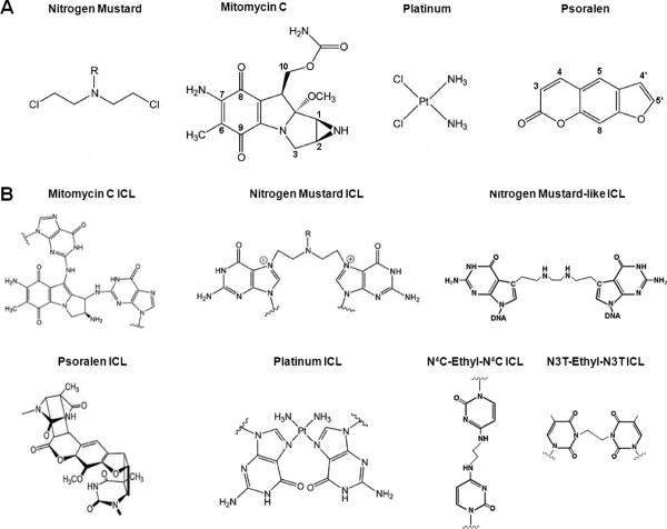
Interstrand DNA crosslinks (ICLs) are formed by natural products of metabolism and by chemotherapeutic reagents. Work in E. coli identified a two cycle repair scheme involving incisions on one strand on either side of the ICL (unhooking) producing a gapped intermediate with the incised oligonucleotide attached to the intact strand. The gap is filled by recombinational repair or lesion bypass synthesis. The remaining monoadduct is then removed by nucleotide excision repair (NER). Despite considerable effort, our understanding of each step in mammalian cells is still quite limited. In part this reflects the variety of crosslinking compounds, each with distinct structural features, used by different investigators. Also, multiple repair pathways are involved, variably operative during the cell cycle. G(1) phase repair requires functions from NER, although the mechanism of recognition has not been determined. Repair can be initiated by encounters with the transcriptional apparatus, or a replication fork. In the case of the latter, the reconstruction of a replication fork, stalled or broken by collision with an ICL, adds to the complexity of the repair process. The enzymology of unhooking, the identity of the lesion bypass polymerases required to fill the first repair gap, and the functions involved in the second repair cycle are all subjects of active inquiry. Here we will review current understanding of each step in ICL repair in mammalian cells.


Figures

Figure 1
Figure 1
DNA ICL forming agents. (A) Common crosslinking agents used in ICL experiments. (B) DNA-ICL structures including synthetic substrates.
Figure 2
Figure 2
Laser localized lesions and repair in G1 mammalian cells. Localized lesions (green stripe) formed by dig-tagged psoralen in WT and XPC deficient cells. Cells are stained with a cell cycle marker (NPAT, red spots) to show cells in G1 phase. NPAT shows 2 spots in G1 cells and four spots in S/G2 cells, and the spots may be present in different focal planes. A color version of this figure is available in the online version of this manuscript.
Figure 3
Figure 3
The `Cole' model. A schematic of ICL repair in E. coli. After the 1st cycle of incision, the gap is either filled by strand exchange or repair synthesis bypassing the adducted base. A color version of this figure is available in the online version of this manuscript.
Figure 4
Figure 4
Entry into an ICL repair pathway depends on the mode of recognition. (A) In the context of a non-replicating DNA, the distortion of the DNA helical structure caused by the lesion attracts protein(s) involved in the global damage surveillance of DNA. This process has been shown to involve proteins of the NER pathway, with XPC leading the initial recognition. The first incision step on either side of the lesion on one strand of the duplex by XPF-ERCC1 complex (and perhaps XPG) generates a gapped structure which serves as a substrate for bypass polymerases. The now flipped out monoadduct-like structure can be recognized by DDB2 and perhaps also by glycosylases such as MPG or Neil1. Again, the NER pathway will initiate the second cycle of repair and remove the remaining adduct on the opposite strand. (B) Stalling of RNA polymerases at the site of lesion during transcription can also serve as a means of ICL recognition in non-replicating DNA. (C) and (D), a stalled replication fork, either due to a single or dual fork encounter, is attractive to proteins of the Fanconi Anemia (FA) pathway and proteins such as Mus81-EME1/2, Snm1B, and MRN. Initial recognition is thought to be mediated by the FancM-FAAP24 complex, which then becomes part of the FA core complex. The FA core complex is a prerequisite to recruit the FancD2 and FancI proteins which are modified via ubiquitination and phosphorylation. The ICL is incised by XPF-ERCC1 and Mus81-EME1 on the leading strand, generating a DSB at the fork. In the case of converging forks (D), the first incision cycle may occur on either strand. The gapped structure will be filled in by lesion bypass polymerases, including polζ, polκ, polι, polN, polη and Rev1. When a single fork is stalled by the ICL polymerase(s) will extend a parental strand to fill the gap. When two forks converge on the ICL a leading daughter strand is extended to bypass the lesion. Upon removal of the remaining single adduct on the opposite strand, in an NER dependent pathway, the broken fork will be reconstructed by recombinational repair. A color version of this figure is available in the online version of this manuscript.

Similar articles

Cited by

References

    1. Adair GM, Rolig RL, Moore-Faver D, Zabelshansky M, Wilson JH, Nairn RS. Role of ERCC1 in removal of long non-homologous tails during targeted homologous recombination. EMBO J. 2000;19:5552–5561. - PMC - PubMed
    1. Ahmad A, Robinson AR, Duensing A, van DE, Beverloo HB, Weisberg DB, Hasty P, Hoeijmakers JH, Niedernhofer LJ. ERCC1-XPF endonuclease facilitates DNA double-strand break repair. Mol. Cell Biol. 2008;28:5082–5092. - PMC - PubMed
    1. Akagi J, Masutani C, Kataoka Y, Kan T, Ohashi E, Mori T, Ohmori H, Hanaoka F. Interaction with DNA polymerase eta is required for nuclear accumulation of REV1 and suppression of spontaneous mutations in human cells. DNA Repair (Amst) 2009;8:585–599. - PubMed
    1. Akkari YM, Bateman RL, Reifsteck CA, Olson SB, Grompe M. DNA replication is required to elicit cellular responses to psoralen-induced DNA interstrand cross-links. Mol. Cell Biol. 2000;20:8283–8289. - PMC - PubMed
    1. Allan JM, Engelward BP, Dreslin AJ, Wyatt MD, Tomasz M, Samson LD. Mammalian 3-methyladenine DNA glycosylase protects against the toxicity and clastogenicity of certain chemotherapeutic DNA cross-linking agents. Cancer Res. 1998;58:3965–3973. - PubMed

Publication types

LinkOut - more resources