. 2009 Dec 29;16(1):114.
doi: 10.1186/1423-0127-16-114.

Reduced Ca2+ transport across sarcolemma but enhanced spontaneous activity in cardiomyocytes isolated from left atrium-pulmonary veins tissue of myopathic hamster

Affiliations

Reduced Ca2+ transport across sarcolemma but enhanced spontaneous activity in cardiomyocytes isolated from left atrium-pulmonary veins tissue of myopathic hamster

Yue-Xia Loh et al. J Biomed Sci. .

Abstract

Background: Several lines of evidence point to a particularly important role of the left atrium (LA) in initiating and maintaining atrial fibrillation (AF). This role may be related to the location of pulmonary veins (PVs) in the LA. The aim of the present study was to investigate the action potential (AP) and ionic currents in LA-PV cardiomyocytes isolated from Bio14.6 myopathic Syrian hamsters (36-57 week-old) versus age-matched F1B healthy control hamsters.

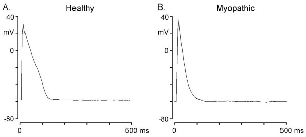
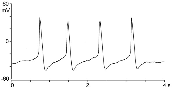
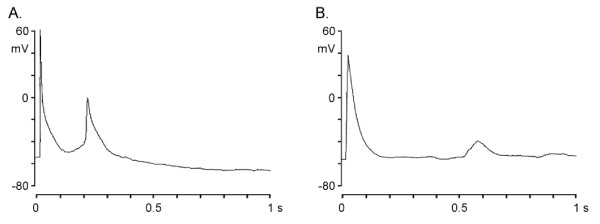
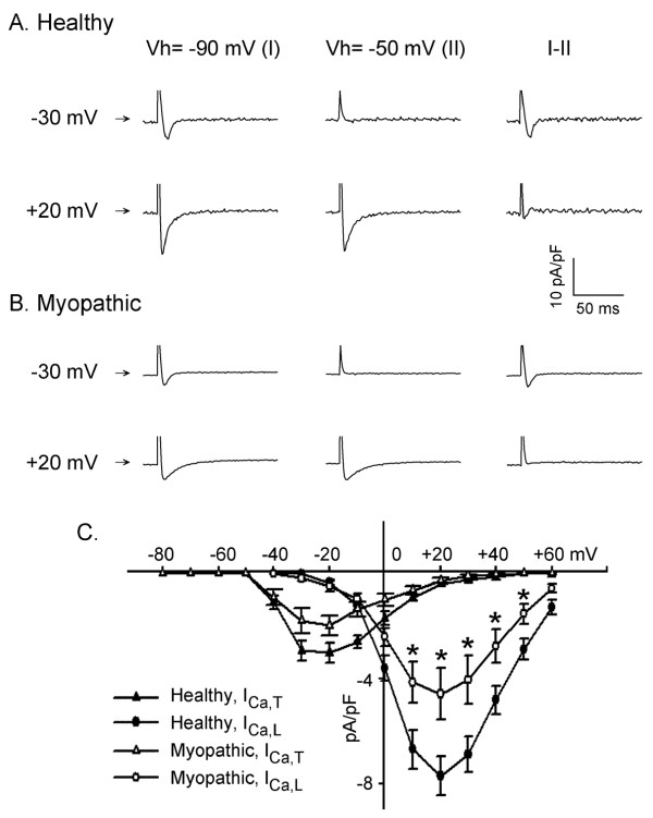
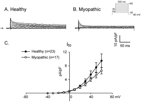
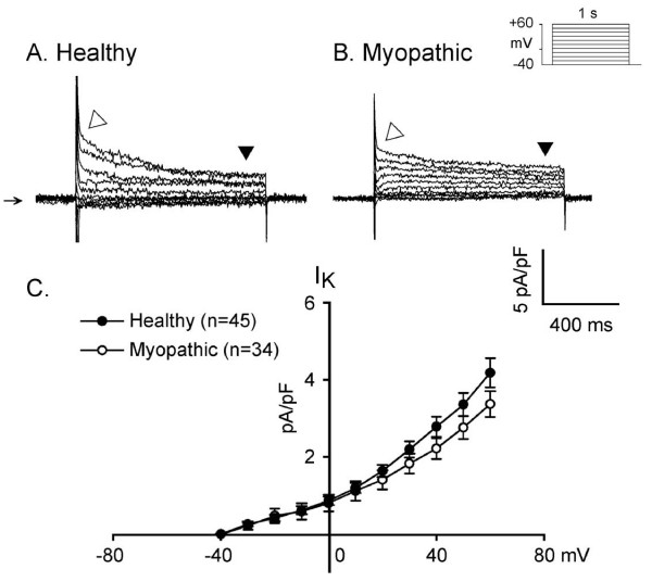
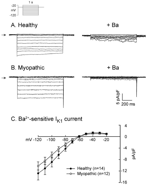
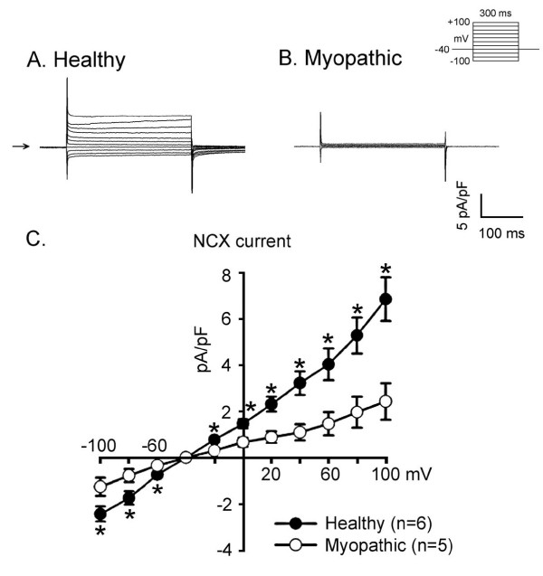
Methods and results: Whole-cell patch-clamp techniques were used to record AP in current-clamp mode and ionic currents in voltage-clamp mode. The results obtained show that in both healthy and myopathic LA-PV tissue spontaneously discharging cardiomyocytes can be found, but they are more numerous in myopathic (9/29) than in healthy hamsters (4/42, p < 0.05 by chi2 analysis). Myopathic myocytes have shorter AP duration (APD) with smaller ICa,L and INCX than the healthy control. The currents ITO, IK, IK1 and ICa,T are not significantly different in myopathic versus healthy cells.

Conclusions: Our results indicate that in myopathic Syrian hamsters LA-PV cardiomyocytes are more prone to automatic rhythms. Also, they show altered electrophysiologic properties, which may be due to abnormal Ca2+ channels and may account for contractile dysfunction.


Figures

Figure 1
Figure 1
Different configuration and duration of driven action potentials in LA-PV cardiomyocytes obtained from healthy (panel A) and myopathic (panel B) hamsters.
Figure 2
Figure 2
Spontaneously discharging action potentials in the absence of electrical stimulation recorded in a LA-PV cardiomyocyte.
Figure 3
Figure 3
Triggered rhythms in hamster LA-PV cardiomyocyte. Panel A and B show typical examples of early and delayed afterdepolarization (EAD and DAD, respectively) recorded in a myopathic myocyte stimulated at a rate of 1 Hz in normal Tyrode's solution.
Figure 4
Figure 4
Differences in two types of calcium currents recorded from healthy and myopathic LA-PV cardiomyocytes. A and B: selected membrane currents elicited by depolarizing clamps to various voltages from the holding potentials (Vh) of -90 mV (I) and -50 mV (II, ICa,L). Horizontal arrows near left margin indicate zero current. The difference current (I-II, ICa,T) was obtained from the subtraction of current elicited from -90 mV and -50 mV, respectively. C: Mean current densities of peak ICa,L and ICa,T from 10 myopathic and 12 healthy LA-PV myocytes. *P < 0.05 by group comparisons.
Figure 5
Figure 5
Transient outward K+ current (ITO) activated on depolarization in healthy (panel A) and myopathic LA-PV myocytes (panel B). Clamp protocol is shown on top of panel B. The mean current-voltage relations of ITO in 23 healthy (closed circles) and 17 myopathic myocytes (open circles) are shown in panel C.
Figure 6
Figure 6
Delayed outward K+ current (IK) activated on depolarization in LA-PV healthy (panel A) and myopathic myocytes (panel B). Clamp protocol is shown at the top of panel B. IK was measured near the end of the 1 s depolarizing pulses. Downward solid triangle indicated IK recorded at test potential of +60 mV. In contrast, open triangle indicated ITO in the same myocytes. Panel C shows the mean current-voltage relations of IK in 45 healthy (closed circles) and 34 myopathic myocytes (open circles).
Figure 7
Figure 7
Ba2+-sensitive inward rectifier K+ current, IK1. Panel A illustrates a series of IK1 elicited on hyperpolarization before (control) and after 1 mM Ba2+ superfusion in a healthy myocyte. Similar traces obtained from a myopathic myocyte are shown in panel B. Horizontal arrows near left margin indicate zero current. The difference current densities (Ba2+-sensitive K+ current, IK1) (mean ± S.E.M.) in 14 healthy myocytes and 12 myopathic myocytes are summarized in the current-voltage relations shown in panel C.
Figure 8
Figure 8
Na+-Ca2+ exchange current (NCX) in healthy (panel A) and myopathic LA-PV myocytes (panel B). The clamp protocol is shown between panels A and B. The holding potential was -40 mV. Horizontal arrow at left margin indicates zero current. For inward NCX currents the myocyte was hyperpolarized Vh -40 to -100 mV in 20 mV increments. For outward NCX currents, the myocyte was depolarized from -40 to +100 mV in 20 mV increments. Mean current-voltage relationships of NCX in 6 healthy and 5 myopathic myocytes (from 3 hamsters in each group) are summarized in panel C.

Similar articles

Cited by

References

    1. Gertz EW. Cardiomyopathic Syrian hamster: a possible model of human disease. Prog Exp Tumor Res. 1972;16:242–247. - PubMed
    1. Homburger F. Myopathy of hamster dystrophy: history and morphologic aspects. Ann NY Acad Sci. 1979;317:1–7. - PubMed
    1. Thuringer D, Deroubaix E, Coulombe A, Coraboeuf E, Mercadier JJ. Ionic basis of the action potential prolongation in ventricular myocytes from Syrian hamsters with dilated cardiomyopathy. Cardiovasc Res. 1996;31:747–757. - PubMed
    1. Kuo TH. Defective Ca2+-pumping ATPase of heart sarcolemma from cardiomyopathic hamster. Biochim Biophys Acta. 1987;900:10–16. doi: 10.1016/0005-2736(87)90272-0. - DOI - PubMed
    1. Chiesi M, Wrzosek A, Gryueninger S. The role of the sarcoplasmic reticulum in various types of cardiomyocytes. Mol Cell Biochem. 1994;130:159–171. doi: 10.1007/BF01457397. - DOI - PubMed

Publication types